. 2022 Nov 1:13:1002084.
doi: 10.3389/fmicb.2022.1002084. eCollection 2022.

Vancomycin-induced gut microbiota dysbiosis aggravates allergic rhinitis in mice by altered short-chain fatty acids

Affiliations

Vancomycin-induced gut microbiota dysbiosis aggravates allergic rhinitis in mice by altered short-chain fatty acids

Zhen Chen et al. Front Microbiol. .

Abstract

Objective: This study aims to explore how gut microbiota dysbiosis affects allergic rhinitis (AR) and whether short-chain fatty acids (SCFAs) play a role in this process.

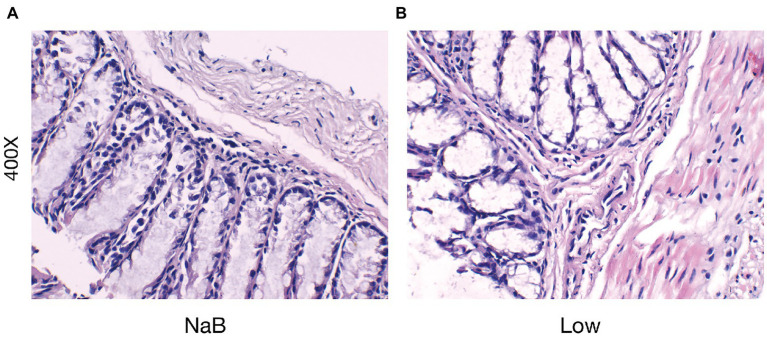
Methods: A mouse gut microbiota dysbiosis model was established by adding vancomycin to drinking water for 2 weeks before ovalbumin (OVA) sensitization. Then an OVA-alum AR mouse model was established by intraperitoneal OVA injection followed by nasal excitation. Hematoxylin and eosin (H&E) staining was performed to observe pathological changes in nasal and colon tissues of AR mice. Serum levels of total-IgE, OVA-sIgE, IL-4, IL-5, IL-10, and TGF-β1 were measured. The composition and diversity of the mouse gut microbiota were observed by 16S rDNA sequencing. Levels of SCFAs in feces were determined using SCFA-targeted metabolomics. Sodium butyrate (NaB) was added daily to mice on a low-fiber basal diet 2 weeks before the first sensitization, until the end of the study.

Results: After gut microbiota dysbiosis, serum levels of the total IgE, OVA-sIgE, IL-4, and IL-5 in AR mice were significantly increased, compared with the control group. The composition and diversity of gut microbiota were significantly altered after gut microbiota dysbiosis, with the fecal SCFAs significantly reduced as well. The reduced bacterial genera after gut microbiota dysbiosis, such as Ruminococcus and Lactobacillus, were significantly and positively correlated with SCFAs. In contrast, the increased genera in the Van group, such as Escherichia-Shigella and Klebsiella, were significantly negatively correlated with SCFAs in feces. NaB treatment significantly reduced total-IgE, OVA-sIgE, IL-4, and IL-5 levels in serum, and inflammatory infiltration of the nasal and colon mucosa. In addition, serum levels of IL-10 and TGF-β1 increased significantly after NaB treatment. Foxp3 protein in the colon was upregulated considerably after NaB intervention.

Conclusion: Vancomycin-induced gut microbiota dysbiosis increased susceptibility and severity of AR, which is significantly related to reduced SCFA-producing bacteria, fecal SCFAs, and specific bacterial taxa. In addition, it was found that NaB alleviated low dietary fiber base-fed symptoms and immune status in AR mice.

Keywords: allergic rhinitis; butyrate; gut microbiota dysbiosis; short-chain fatty acids; tregs.


Conflict of interest statement

The authors declare that the research was conducted in the absence of any commercial or financial relationships that could be construed as a potential conflict of interest.

Figures

Figure 1
Figure 1
The protocol of treatment of mice in each group. (A) Establishment of Vancomycin-induced gut microbiota dysbiosis AR model. (B) Establishment of AR mice model fed a low dietary fiber basal diet or Sodium Butyrate (NaB).
Figure 2
Figure 2
Vancomycin aggravates inflammatory cell infiltration and mucus secretion of nasal mucosa in the OVA-induced AR model. H&E staining of the nasal mucosa (400×). (A) the Control group; (B) the AR group; (C) the Van group.
Figure 3
Figure 3
Vancomycin elevated serum cytokine and IgE levels in the OVA-induced AR model. (A) ELISA detection of OVA-sIgE in serum. (B) ELISA detection of total IgE in serum. (C) ELISA detection of IL-4 in serum. (D) ELISA detection of IL-5 in serum. p < 0.05, ∗∗p < 0.01, ∗∗∗p < 0.001, ∗∗∗∗p < 0.0001.
Figure 4
Figure 4
Vancomycin reduced the richness and diversity of gut microbiota in AR mice. Chao1 index (A) and Shannon index (B) at the overall level of OUT; ∗∗∗p < 0.001, ∗∗∗∗p < 0.0001.
Figure 5
Figure 5
PCA analysis plot. The dots with different colors represent different sample grouping. The abscissa represents the first principal component, the ordinate represents the second principal component, and the percentage represents the contribution value to the samples difference. The more similar the community composition of the sample is, the closer their distances in the PCA map are. C, the control group; AR, the AR group; Van, the Van group.
Figure 6
Figure 6
Vancomycin-induced gut microbiota dysbiosis altered the proportion of gut microbiota in the OVA-induced AR model. (A) Histogram of species composition analysis at the phylum level. (B) Histogram of species composition analysis at the genus level.
Figure 7
Figure 7
Discriminative taxa among the three groups. LDA (Linear discriminant analysis) image shows the differential bacterial genus in each group. The length of the column represents the effect of differential species on this sample (an LDA effect value of more than 4).
Figure 8
Figure 8
Determination of short-chain fatty acids in mouse feces based on metabolomics. (A) Histogram shows the average proportion value of short-chain fatty acids, (B–H) changes in short-chain fatty acids in feces of mice in the control group (Con), AR model group (AR), and vancomycin-induced gut microbiota dysbiosis AR model group (Van). **p < 0.01, ***p < 0.001, ****p < 0.0001.
Figure 9
Figure 9
Correlation analysis heatmap. (A) Heat map of association between differential phyla and SCFAs. (B) Heat map of association between differential genera and SCFAs. acetic acid (AA), butyrate (BA), and propionic acid (PA), Isobutyric acid (IBA), isovaleric acid (IVA), valeric acid (VA), and caproic acid (CA). *p < 0.05, **p < 0.01, ***p < 0.001.
Figure 10
Figure 10
Vancomycin-induced gut microbiota dysbiosis aggravates intestinal barrier inflammation and injury. H&E staining of colonic mucosa (400×). (A) the Control group; (B) the AR group; (C) the Van group.
Figure 11
Figure 11
Butyrate alleviated symptoms in AR mice fed a low dietary fiber basal diet. (A,B) ELISA detection of OVA-sIgE and total IgE in serum. (C–F) ELISA detection of serum cytokines (IL-4, IL-5, IL-10, and TGF-β1). p < 0.05, ∗∗p < 0.01.
Figure 12
Figure 12
Sodium butyrate alleviates the infiltration of inflammatory cells in the nasal mucosa in the OVA-induced AR model. H&E staining of the nasal mucosa (400×). (A) the NaB group; (B) the Low group.
Figure 13
Figure 13
Butyrate reduced the severity of intestinal barrier inflammation and injury. H&E staining of colonic mucosa (400×) (A) the NaB group; (B) the Low group.
Figure 14
Figure 14
The protein expression of Foxp3 was detected by western blotting. (A) Protein banding. (B) Histogram. ****p < 0.0001.

References

    1. Agus A., Denizot J., Thévenot J., Martinez-Medina M., Massier S., Sauvanet P., et al. (2016). Western diet induces a shift in microbiota composition enhancing susceptibility to adherent-Invasive E. coli infection and intestinal inflammation. Sci. Rep. 6:19032. doi: 10.1038/srep19032, PMID: - DOI - PMC - PubMed
    1. Andreassen M., Rudi K., Angell I. L., Dirven H., Nygaard U. C. (2018). Allergen immunization induces major changes in microbiota composition and short-chain fatty acid production in different gut segments in a mouse model of lupine food allergy. Int. Arch. Allergy Immunol. 177, 311–323. doi: 10.1159/000492006, PMID: - DOI - PubMed
    1. Bae J. S., Kim J. H., Kim E. H., Mo J. H. (2017). The role of IL-17 in a lipopolysaccharide-induced rhinitis model. Allergy Asthma Immunol. Res. 9, 169–176. doi: 10.4168/aair.2017.9.2.169, PMID: - DOI - PMC - PubMed
    1. Bianchi F., Dall Asta M., Del Rio D., Mangia A., Musci M., Scazzina F. (2011). Development of a headspace solid-phase microextraction gas chromatography–mass spectrometric method for the determination of short-chain fatty acids from intestinal fermentation. Food Chem. 129, 200–205. doi: 10.1016/j.foodchem.2011.04.022 - DOI
    1. Cait A., Hughes M. R., Antignano F., Cait J., Dimitriu P. A., Maas K. R., et al. (2018). Microbiome-driven allergic lung inflammation is ameliorated by short-chain fatty acids. Mucosal Immunol. 11, 785–795. doi: 10.1038/mi.2017.75, PMID: - DOI - PubMed