. 2022 Oct 17;18(16):6052-6067.
doi: 10.7150/ijbs.75188. eCollection 2022.

Intermittent Fasting Attenuates Hallmark Vascular and Neuronal Pathologies in a Mouse Model of Vascular Cognitive Impairment

Affiliations

Intermittent Fasting Attenuates Hallmark Vascular and Neuronal Pathologies in a Mouse Model of Vascular Cognitive Impairment

Vismitha Rajeev et al. Int J Biol Sci. .

Abstract

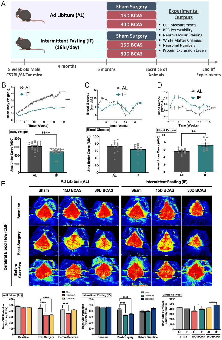
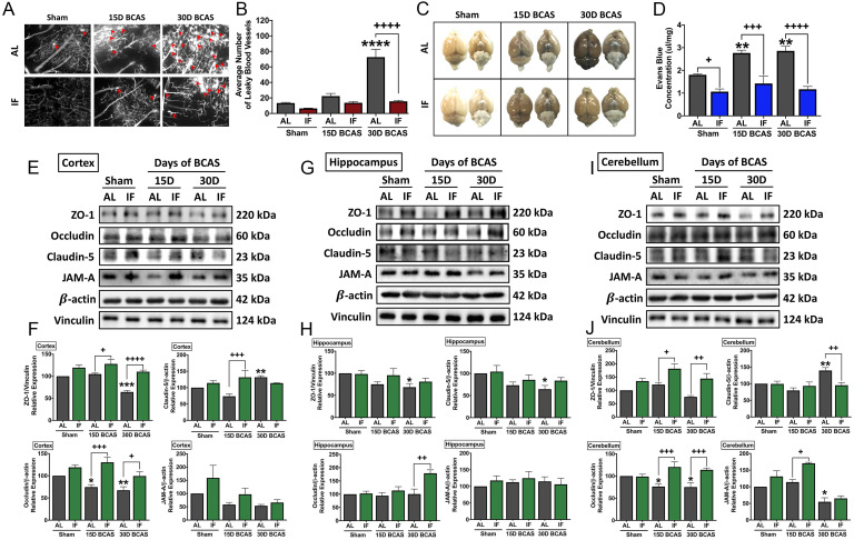
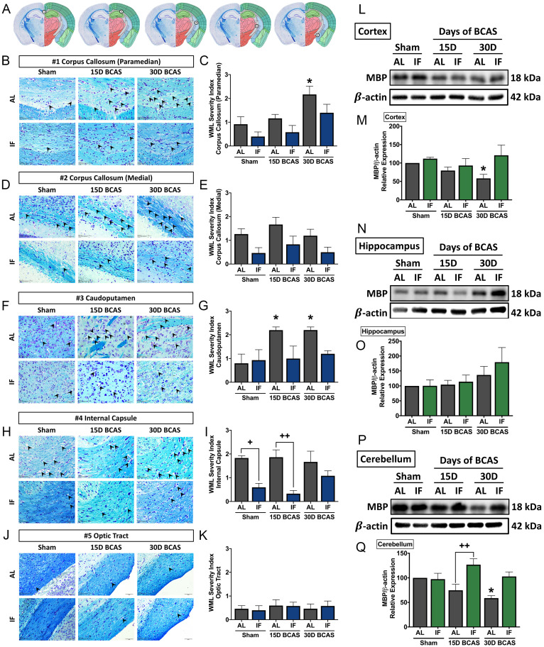
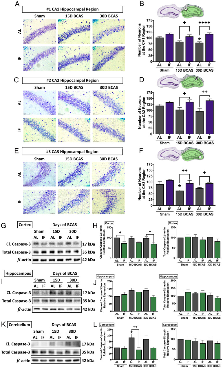
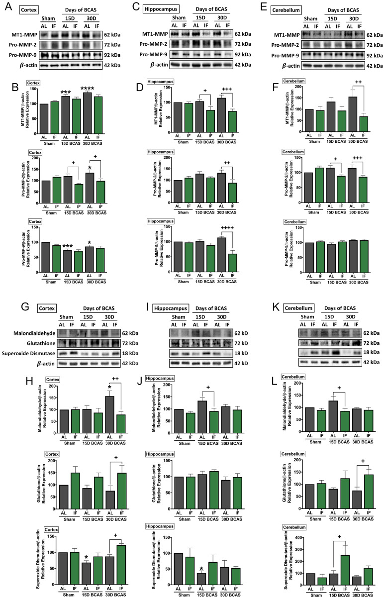
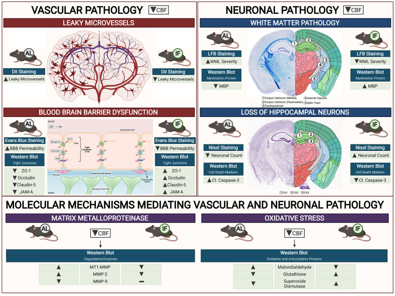
Background - Chronic cerebral hypoperfusion (CCH) is an important pathophysiological mechanism of vascular cognitive impairment (VCI). The heterogeneous effects of CCH complicate establishing single target therapies against VCI and its more severe form, vascular dementia (VaD). Intermittent fasting (IF) has multiple targets and is neuroprotective across a range of disease conditions including stroke, but its effects against CCH-induced neurovascular pathologies remain to be elucidated. We therefore assessed the effect of IF against CCH-associated neurovascular pathologies and investigated its underlying mechanisms. Methods - Male C57BL/6NTac mice were subjected to either ad libitum feeding (AL) or IF (16 hours of fasting per day) for 4 months. In both groups, CCH was experimentally induced by the bilateral common carotid artery stenosis (BCAS) method. Sham operated groups were used as controls. Measures of leaky microvessels, blood-brain barrier (BBB) permeability, protein expression of tight junctions, extracellular matrix components and white matter changes were determined to investigate the effect of IF against CCH-induced neurovascular pathologies. Results - IF alleviated CCH-induced neurovascular pathologies by reducing the number of leaky microvessels, BBB breakdown and loss of tight junctional proteins. In addition, IF mitigated the severity of white matter lesions, and maintained myelin basic protein levels, while concurrently reducing hippocampal neuronal cell death. Furthermore, IF reduced the CCH-induced increase in levels of matrix metalloproteinase (MMP)-2 and its upstream activator MT1-MMP, which are involved in the breakdown of the extracellular matrix that is a core component of the BBB. Additionally, we observed that IF reduced CCH-induced increase in the oxidative stress marker malondialdehyde, and increased antioxidant markers glutathione and superoxide dismutase. Overall, our data suggest that IF attenuates neurovascular damage, metalloproteinase and oxidative stress-associated pathways, and cell death in the brain following CCH in a mouse model of VCI. Conclusion - Although IF has yet to be assessed in human patients with VaD, our data suggest that IF may be an effective means of preventing the onset or suppressing the development of neurovascular pathologies in VCI and VaD.

Keywords: Blood brain barrier breakdown, Chronic cerebral hypoperfusion; Intermittent fasting; Neuronal death; Vascular cognitive impairment; Vascular dementia; White matter lesions.


Conflict of interest statement

Competing Interests: The authors have declared that no competing interest exists.

Figures

Figure 1
Figure 1
Effect of intermittent fasting on physiological measurements and blood flow in the brain following BCAS in a mouse model of VCI. A. Schematic representation of the experimental design and outputs performed. B. Mean body weight measurements over 25 weeks. ***P<0.001, ****P<0.0001 compared with AL. C. Blood glucose measurements over 25 weeks. D. Blood ketone measurements over 25 weeks. **P<0.01, ***P<0.001 compared with AL. E. Representative contrast images and quantification of CBF at baseline, effective blood flow reduction post-surgery, and the final level of cerebral blood flow before sacrifice at each individual end point demonstrated significant changes in cerebral blood flow following BCAS. Histographs illustrating the comparison of CBF levels at the sacrifice timepoint. The rate of blood flow was expressed in perfusion units (PU) using the PeriMed Software. *P<0.05, ***P<0.001 compared with corresponding AL BCAS; ****P<0.0001 compared with corresponding Sham. Data are represented as mean ± standard error of the mean of n=10-12 mice in each experimental group. Abbreviations: AL, ad libitum; BBB, blood-brain barrier; BCAS, bilateral common carotid artery stenosis; CBF, cerebral blood flow; IF, intermittent fasting; VCI, vascular cognitive impairment.
Figure 2
Figure 2
Effect of intermittent fasting on leaky blood vessel incidence and blood-brain barrier integrity following BCAS. A-B. Representative DiI staining images and quantification illustrating leaky blood vessel incidence in AL and IF mice following BCAS. Arrowheads point to leaky areas of the vessels. Data are represented as mean ± standard error of the mean of n=5 mice in each experimental group. ****P<0.0001 compared with AL Sham; ++++P<0.0001 compared with AL BCAS. C-D. Representative dorsal and ventral views of mouse brains injected with Evans Blue dye and respective quantification. Data are represented as mean ± standard error of the mean of n=5-7 mice in each experimental group. **P<0.01 compared with AL Sham; +P<0.05, +++P<0.001, ++++P<0.0001 compared to corresponding AL group. E-J. Representative immunoblots and quantification of tight junction proteins zonula occludens (ZO)-1, occludin, claudin-5 and junctional adhesion molecule (JAM)-A in the cortex (E-F), hippocampus (G-H) and cerebellum (I-J). Data are expressed as mean ± standard error of the mean. n=4-7 mice in each experimental group. β-actin or vinculin was used as a loading control. *P<0.05, **P<0.01, ***P<0.001 compared with AL Sham; +P<0.05, ++P<0.01, +++P<0.001, ++++P<0.0001, compared with corresponding AL BCAS. Abbreviations: AL, ad libitum; BCAS, bilateral common carotid artery stenosis; IF, intermittent fasting; VCI, vascular cognitive impairment.
Figure 3
Figure 3
Effect of intermittent fasting on white matter integrity in the brain following BCAS. A. Schematic diagrams illustrating areas where white matter severity was measured, namely the corpus callosum (medial), corpus callosum (paramedian), caudoputamen, internal capsule and optic tract. B-K. Representative Luxol fast blue stained images and quantification illustrating white matter changes at the corpus callosum (paramedian) (B-C), corpus callosum (medial) (D-E), caudoputamen (F-G), internal capsule (H-I) and optic tract (J-K). The severity of white matter damage was graded as follows: Grade 0=no disruptions, Grade 1=disarrangement of nerve fibres, Grade 2=formation of marked vacuoles, and Grade 3=disappearance of myelinated fibres. Arrowheads point only to marked vacuoles throughout the image. Magnification x60. Scale bar, 20 μm. Images were taken under identical exposures and conditions. Data are represented as mean ± standard error of the mean. n=5 mice in each experimental group. *P<0.05 compared with AL Sham; +P<0.05, ++P<0.01 compared with corresponding AL group. L-Q. Representative immunoblots and quantification of myelin basic protein (MBP) expression following BCAS in the cortex (L-M), hippocampus (N-O) and cerebellum (P-Q). Data are represented as mean ± standard error of the mean. n=4-7 mice in each experimental group. β-actin was used as a loading control. *P<0.05 compared with AL Sham; ++P<0.01 compared with corresponding AL BCAS. Abbreviations: AL, ad libitum; BCAS, bilateral common carotid artery stenosis; IF, intermittent fasting; VCI, vascular cognitive impairment.
Figure 4
Figure 4
Effect of intermittent fasting on hippocampal neuronal loss and apoptotic death in the brain following BCAS A-F. Representative cresyl violet images and quantification illustrating Nissl positively stained neurons in hippocampal CA1 (A-B), CA2 (C-D) and CA3 (E-F) regions. Magnification x60. Scale bar, 20 μm. Images were taken under identical exposures and conditions. Data are represented as mean ± standard error of the mean. n=5 mice in each experimental group. *P<0.05 compared with AL Sham; +P<0.05, ++P<0.01, ++++P<0.0001 compared with corresponding AL BCAS. G-L. Representative immunoblots and quantification illustrating apoptosis marker cleaved caspase-3 at the cortex (G-H), hippocampus (I-J) and cerebellum (K-L). Data are represented as mean ± standard error of the mean. n=4-7 mice in each experimental group. β-actin was used as a loading control. *P<0.05 compared with AL Sham; +P<0.05, ++P<0.01 compared with corresponding AL group. Abbreviations: AL, ad libitum; BCAS, bilateral common carotid artery stenosis; Cl, cleaved; IF, intermittent fasting; VCI, vascular cognitive impairment.
Figure 5
Figure 5
Effect of intermittent fasting on matrix metalloproteinase and oxidative stress in the brain following BCAS. A-F. Representative immunoblots and quantification illustrating matrix metalloproteinase (MMP)-2, its upstream membrane type (MT)-1 MMP (MT1MMP) and MMP-9 levels in the cortex (A-B), hippocampus (C-D) and cerebellum (E-F). Data are represented as mean ± standard error of the mean. n=5-7 mice in each experimental group. β-actin was used as a loading control. *P<0.05, ***P<0.001, ****P<0.0001 compared with AL Sham; +P<0.05, ++P<0.01, +++P<0.001, ++++P<0.0001 compared with corresponding AL BCAS. G-L. Representative immunoblots and quantification illustrating oxidative stress marker, malondialdehyde, and anti-oxidant markers, glutathione and superoxide dismutase in the cortex (G-H), hippocampus (I-J) and cerebellum (K-L). Data are expressed as mean ± standard error of the mean. n=4-7 mice in each experimental group. β-actin was used as a loading control. *P<0.05 compared with AL Sham; +P<0.05, ++P<0.01 compared with corresponding AL BCAS. Abbreviations: AL, ad libitum; BCAS, bilateral common carotid artery stenosis; IF, intermittent fasting; VCI, vascular cognitive impairment.
Figure 6
Figure 6
Schematic diagram illustrating the effects of intermittent fasting on the molecular mechanisms mediating vascular and neuronal pathology in the brain following BCAS in a mouse model of VCI. Vascular pathology in the brain following decreased cerebral blood flow (CBF) is defined as changes in the number of leaky microvessels and damage to the blood brain barrier (BBB). DiI staining was used to stain the vasculature, and IF was found to decrease the number of leaky microvessels in the brain following CCH. Evans blue staining was used to assess the extent of BBB damage in the brain. IF was found to decrease BBB permeability through increased tight junction proteins zonula-occludens (ZO)-1, Occludin, Claudin-5 and junctional adhesion molecule (JAM)-A. Vascular pathology in the brain following decreased CBF is defined as changes in the white matter pathology and loss of hippocampal neurons. Luxol fast blue (LFB) staining method was used to stain and image 5 different white matter regions (corpus callosum medial, corpus callosum paramedian, caudoputamen, internal capsule and optic tract). IF decreased the severity of white matter damage in the brain following CCH, through maintenance of myelin basic protein (MBP) levels. Nissl staining was used to visualise the hippocampal neurons in the brain, and IF was found to be able to increase neuronal counts through reducing apoptotic death indicated by the levels of cleaved caspase-3 in the brain. IF was found to reduce membrane-type 1 MMP (MT1-MMP) and MMP2 levels in the brain following CCH. However, MMP9 was not involved in breaking down the extracellular matrix following CCH. IF reduced oxidative stress as indicated through markers of lipid oxidation such as malondialdehyde, and antioxidative glutathione, and superoxide dismutase levels.

References

    1. Rizzi L, Rosset I, Roriz-Cruz M. Global epidemiology of dementia: Alzheimer's and vascular types. Biomed Res Int. 2014. 2014. - PMC - PubMed
    1. Roman G, Sachdev P, Royall D, Bullock R, Orgogozo J-M, López-Pousa S. et al. Vascular cognitive disorder: A new diagnostic category updating vascular cognitive impairment and vascular dementia. J Neurol Sci. 2004 Nov 15;226:81–7. - PubMed
    1. Akinyemi R, Mukaetova-Ladinska E, Attems J, Ihara M, Kalaria R. Vascular risk factors and neurodegeneration in ageing related dementias: Alzheimer's disease and vascular dementia. Curr Alzheimer Res [Internet] 2013;10(6):642–53. Available from: https://pubmed.ncbi.nlm.nih.gov/23627757/ - PubMed
    1. Cheng Y-W, Chiu M-J, Chen Y-F, Cheng T-W, Lai Y-M, Chen T-F. The contribution of vascular risk factors in neurodegenerative disorders: from mild cognitive impairment to Alzheimer's disease. Alzheimer's Res Ther 2020 121 [Internet] 2020;12(1):1–10. Available from: https://alzres.biomedcentral.com/articles/10.1186/s13195-020-00658-7. - PMC - PubMed
    1. Gyanwali B, Shaik M, Tan B, Venketasubramanian N, Chen C, Hilal S. Risk Factors for and Clinical Relevance of Incident and Progression of Cerebral Small Vessel Disease Markers in an Asian Memory Clinic Population. J Alzheimers Dis [Internet] 2019;67(4):1209–19. Available from: https://pubmed.ncbi.nlm.nih.gov/30714960/ - PubMed

Publication types