Review
. 2023 Mar;314(1):158-180.
doi: 10.1111/imr.13173. Epub 2022 Nov 28.

The formation and function of the neutrophil phagosome

Affiliations
Review

The formation and function of the neutrophil phagosome

Emily Naish et al. Immunol Rev. 2023 Mar.

Abstract

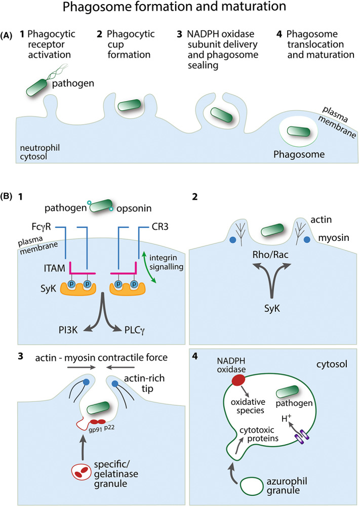
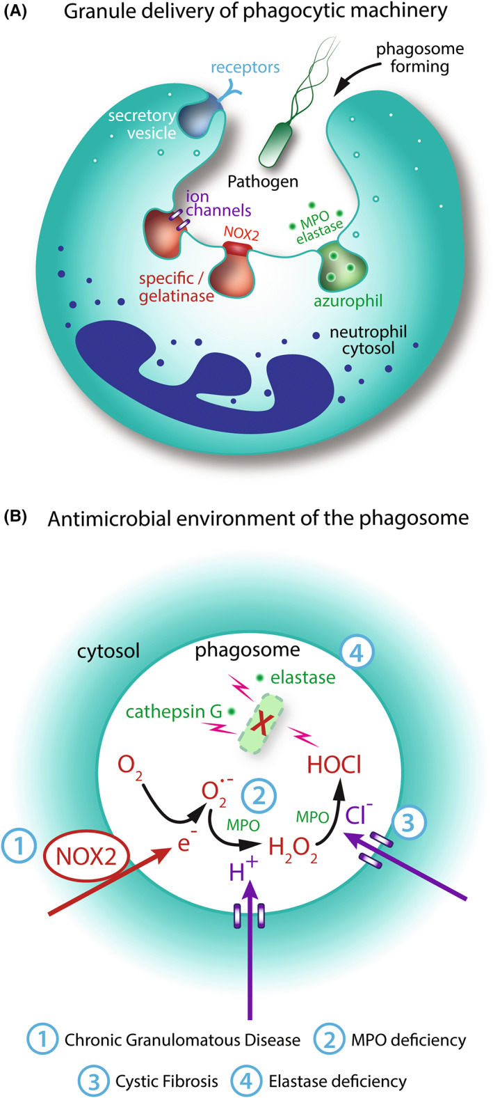
Neutrophils are the most abundant circulating leukocyte and are crucial to the initial innate immune response to infection. One of their key pathogen-eliminating mechanisms is phagocytosis, the process of particle engulfment into a vacuole-like structure called the phagosome. The antimicrobial activity of the phagocytic process results from a collaboration of multiple systems and mechanisms within this organelle, where a complex interplay of ion fluxes, pH, reactive oxygen species, and antimicrobial proteins creates a dynamic antimicrobial environment. This complexity, combined with the difficulties of studying neutrophils ex vivo, has led to gaps in our knowledge of how the neutrophil phagosome optimizes pathogen killing. In particular, controversy has arisen regarding the relative contribution and integration of nicotinamide adenine dinucleotide phosphate (NADPH) oxidase-derived antimicrobial agents and granule-delivered antimicrobial proteins. Clinical syndromes arising from dysfunction in these systems in humans allow useful insight into these mechanisms, but their redundancy and synergy add to the complexity. In this article, we review the current knowledge regarding the formation and function of the neutrophil phagosome, examine new insights into the phagosomal environment that have been permitted by technological advances in recent years, and discuss aspects of the phagocytic process that are still under debate.

Keywords: neutrophil; phagocytosis; phagosome.


Conflict of interest statement

Andrew Conway Morris is a member of the scientific advisory board of Cambridge Infection Diagnostics Ltd and reports speaking fees from Boston Scientific.

Figures

FIGURE 1
FIGURE 1
Phagosome formation and maturation. (A) Overview of the major steps in neutrophil phagosome formation following pathogen detection. Events 1–4 are depicted in further detail in panel B. (B) 1: An opsonized pathogen engages Fc receptors (FcγR) or complement receptors (e.g., CR3) to initiate phagocytosis. Both FcγR and CR3 can employ immunoreceptor signaling pathways: SH2‐domain‐bearing proteins (e.g., Syk) associate with phosphorylated ITAM, signaling downstream through phosphatidylinositol 3‐kinase (PI3K) and/or phospholipase Cγ (PLCγ). CR3 also employs independent inside‐out and outside‐in integrin signaling pathways. 2: Phagocytic receptor signaling induces regulation of the actin cytoskeleton via Rac and/or Rho. Myosin motor control of actin rearrangement drives extending pseudopod protrusions from the plasma membrane to form the phagocytic cup around the pathogen. 3: Cytosolic specific/gelatinase granules deliver proteins to the membrane of the forming phagosome, for example, the membrane‐bound subunits of NADPH oxidase, gp91phox (NOX2), and p22phox. Actin polymerization at the pseudopod tips facilitates membrane sealing to complete the phagocytic vacuole around the pathogen. 4: The formed pathogen‐containing phagosome translocates toward the granule‐rich centriole within the neutrophil cytosol. NADPH oxidase generates antimicrobial reactive oxygen species inside the phagosome. The negative charge generated by this process is compensated by an influx of protons. Cytosolic azurophil granules, containing cytotoxic proteins, for example, elastase, fuse with the phagosome membrane to deliver their contents to the lumen of the phagosome
FIGURE 2
FIGURE 2
Maturation of phagosomes formed by human peripheral blood neutrophils ingesting Staphylococcus aureus. (A) Neutrophil that has not encountered bacteria (Blue: Nucleus labeled with Hoescht, White: Actin labeled with Alexa Fluor(AF)647‐phalloidin). (B) Early ingestion of S. aureus (Green: AF488‐labeled S. aureus bioparticles), with limited change in phagosomal pH as assessed by co‐localized lysotracker signal (Red: Lyso Tracker Red DND‐99, Yellow: co‐localization with AF488 S. aureus). (C): Mid‐maturation with mixture of low pH (mature) and high pH (immature) phagosomes. (D): Late maturation with the majority of phagosomes demonstrating low pH. (E) Early endosomal antigen 1 (EEA1) staining on the surface of S. aureus containing phagosomes. Note the penumbral rather than co‐localized signature indicating membrane distribution (Red: EEA1, mouse anti‐human EEA1 with secondary anti‐mouse AF568 phalloidin staining omitted for clarity)
FIGURE 3
FIGURE 3
Role of granules and reactive oxygen species in phagosome formation and function. (A): Granules deliver phagocytic machinery to the plasma and phagosome membranes during phagocytosis. Secretory vesicles supply phagocytic receptors to the plasma membrane. Specific and gelatinase granules deliver NADPH oxidase components and ion channels to the phagosome membrane. Azurophil granules supply myeloperoxidase (MPO) and cytotoxic proteins and proteases, including elastase. (B) Reactive oxygen species (ROS) and granules contribute synergistically to microbial killing. NADPH oxidase translocates electrons into the phagosome which react with molecular oxygen to form superoxide (O2.−). Electrogenic charge is compensated by proton (H+) influx, predominantly through Hv1 channels. MPO catalyzes the reaction of O2.− and H+ to form hydrogen peroxide (H2O2). Chloride (Cl) enters the phagosome, for example, through CFTR channels. MPO oxidizes Cl to form HOCl which is likely to be directly microbicidal. Non‐oxidative proteins and proteases, for example, elastase and cathepsin G, are also directly microbicidal. Numbers in blue indicate clinical syndromes associated with mutations in various steps of the pathway (Table 1)

References

    1. Rabinovitch M. Professional and non‐professional phagocytes: an introduction. Trends Cell Biol. 1995;5(3):85‐87. - PubMed
    1. Uribe‐Querol E, Rosales C. Phagocytosis: our current understanding of a universal biological process. Front Immunol. 2020;11:1066. - PMC - PubMed
    1. Matsushima H, Geng S, Lu R, et al. Neutrophil differentiation into a unique hybrid population exhibiting dual phenotype and functionality of neutrophils and dendritic cells. Blood. 2013;121(10):1677‐1689. - PMC - PubMed
    1. Levin R, Grinstein S, Canton J. The life cycle of phagosomes: formation, maturation, and resolution. Immunol Rev. 2016;273(1):156‐179. - PubMed
    1. Mócsai A. Diverse novel functions of neutrophils in immunity, inflammation, and beyond. J Exp Med. 2013;210(7):1283‐1299. - PMC - PubMed

Publication types

Substances