

Generating and Imaging Mouse and Human Epithelial Organoids from Normal and Tumor Mammary Tissue Without Passaging
- PMID: 36440890
- PMCID: PMC10149050
- DOI: 10.3791/64626
Generating and Imaging Mouse and Human Epithelial Organoids from Normal and Tumor Mammary Tissue Without Passaging
Abstract
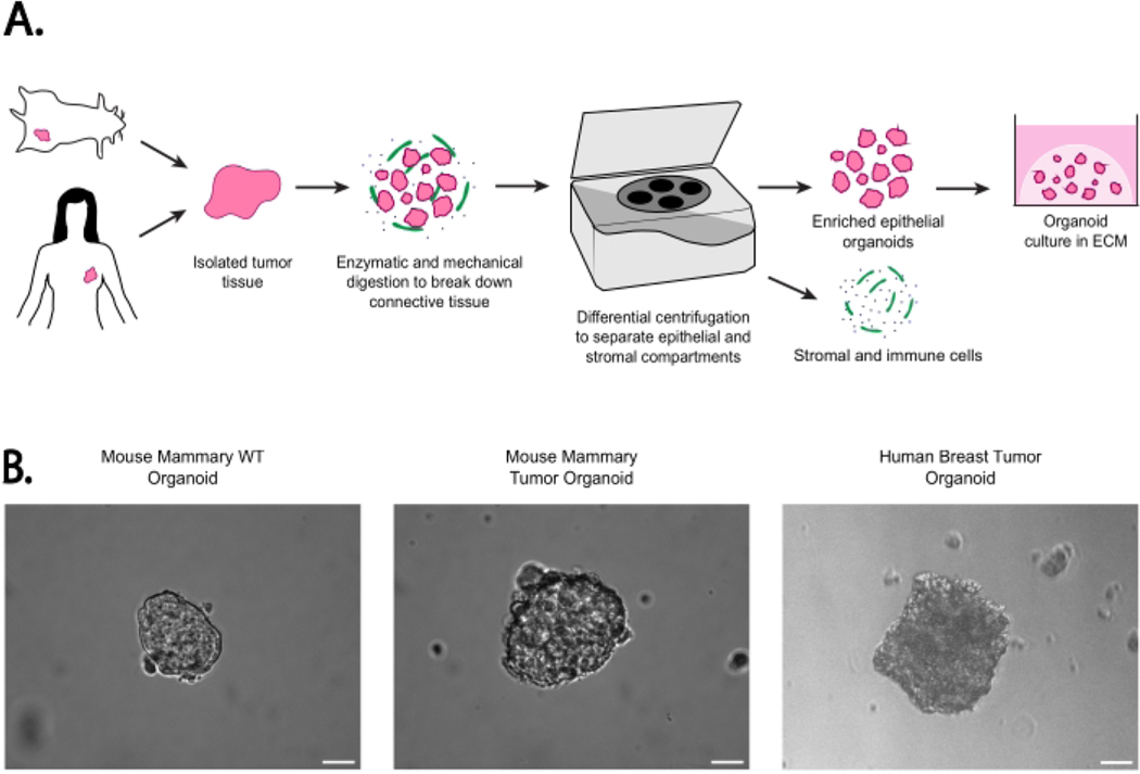
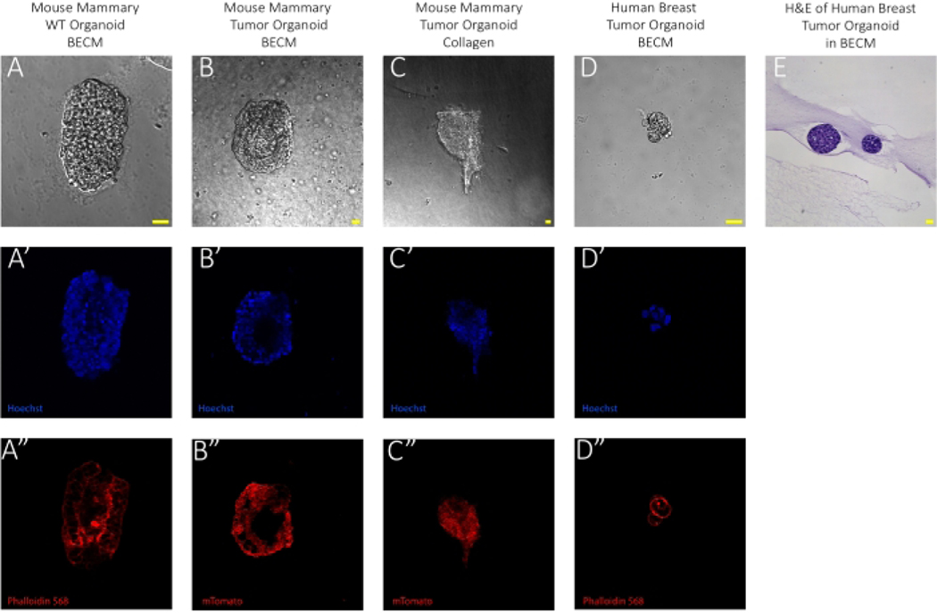
Organoids are a reliable method for modeling organ tissue due to their self-organizing properties and retention of function and architecture after propagation from primary tissue or stem cells. This method of organoid generation forgoes single-cell differentiation through multiple passages and instead uses differential centrifugation to isolate mammary epithelial organoids from mechanically and enzymatically dissociated tissues. This protocol provides a streamlined technique for rapidly producing small and large epithelial organoids from both mouse and human mammary tissue in addition to techniques for organoid embedding in collagen and basement extracellular matrix. Furthermore, instructions for in-gel fixation and immunofluorescent staining are provided for the purpose of visualizing organoid morphology and density. These methodologies are suitable for myriad downstream analyses, such as co-culturing with immune cells and ex vivo metastasis modeling via collagen invasion assay. These analyses serve to better elucidate cell-cell behavior and create a more complete understanding of interactions within the tumor microenvironment.
Conflict of interest statement
Disclosures
The authors declare no conflicts of interest.
Figures
Similar articles
-
Triple-negative breast cancer cells invade adipocyte/preadipocyte-encapsulating geometrically inverted mammary organoids.Integr Biol (Camb). 2023 Apr 11;15:zyad004. doi: 10.1093/intbio/zyad004. Integr Biol (Camb). 2023. PMID: 37015816 Free PMC article.
-
3D bioprinted mammary organoids and tumoroids in human mammary derived ECM hydrogels.Acta Biomater. 2019 Sep 1;95:201-213. doi: 10.1016/j.actbio.2019.06.017. Epub 2019 Jun 21. Acta Biomater. 2019. PMID: 31233891 Free PMC article.
-
Single Organoids Droplet-Based Staining Method for High-End 3D Imaging of Mammary Organoids.Methods Mol Biol. 2022;2471:259-269. doi: 10.1007/978-1-0716-2193-6_14. Methods Mol Biol. 2022. PMID: 35175602
-
Mammary Organoids and 3D Cell Cultures: Old Dogs with New Tricks.J Mammary Gland Biol Neoplasia. 2020 Dec;25(4):273-288. doi: 10.1007/s10911-020-09468-x. Epub 2020 Nov 18. J Mammary Gland Biol Neoplasia. 2020. PMID: 33210256 Review.
-
Organoid Cultures for the Study of Mammary Biology and Breast Cancer: The Promise and Challenges.Cold Spring Harb Perspect Med. 2024 Jul 1;14(7):a041661. doi: 10.1101/cshperspect.a041661. Cold Spring Harb Perspect Med. 2024. PMID: 38110241 Review.
Cited by
-
Digital droplet PCR analysis of organoids generated from mouse mammary tumors demonstrates proof-of-concept capture of tumor heterogeneity.Front Cell Dev Biol. 2024 May 15;12:1358583. doi: 10.3389/fcell.2024.1358583. eCollection 2024. Front Cell Dev Biol. 2024. PMID: 38827528 Free PMC article.
-
Antibody-drug conjugates in breast cancer: overcoming resistance and boosting immune response.J Clin Invest. 2023 Sep 15;133(18):e172156. doi: 10.1172/JCI172156. J Clin Invest. 2023. PMID: 37712425 Free PMC article. Review.
References
-
- Hanahan D.Hallmarks of cancer: New dimensions. Cancer Discovery. 12 (1), 31–46 (2022). - PubMed
Publication types
MeSH terms
Substances
Grants and funding
LinkOut - more resources
Full Text Sources
Medical
