. 2023 Jan 1;80(1):30-39.
doi: 10.1001/jamaneurol.2022.4251.

Autoimmune Encephalitis Misdiagnosis in Adults

Affiliations

Autoimmune Encephalitis Misdiagnosis in Adults

Eoin P Flanagan et al. JAMA Neurol. .

Abstract

Importance: Autoimmune encephalitis misdiagnosis can lead to harm.

Objective: To determine the diseases misdiagnosed as autoimmune encephalitis and potential reasons for misdiagnosis.

Design, setting, and participants: This retrospective multicenter study took place from January 1, 2014, to December 31, 2020, at autoimmune encephalitis subspecialty outpatient clinics including Mayo Clinic (n = 44), University of Oxford (n = 18), University of Texas Southwestern (n = 18), University of California, San Francisco (n = 17), University of Washington in St Louis (n = 6), and University of Utah (n = 4). Inclusion criteria were adults (age ≥18 years) with a prior autoimmune encephalitis diagnosis at a participating center or other medical facility and a subsequent alternative diagnosis at a participating center. A total of 393 patients were referred with an autoimmune encephalitis diagnosis, and of those, 286 patients with true autoimmune encephalitis were excluded.

Main outcomes and measures: Data were collected on clinical features, investigations, fulfillment of autoimmune encephalitis criteria, alternative diagnoses, potential contributors to misdiagnosis, and immunotherapy adverse reactions.

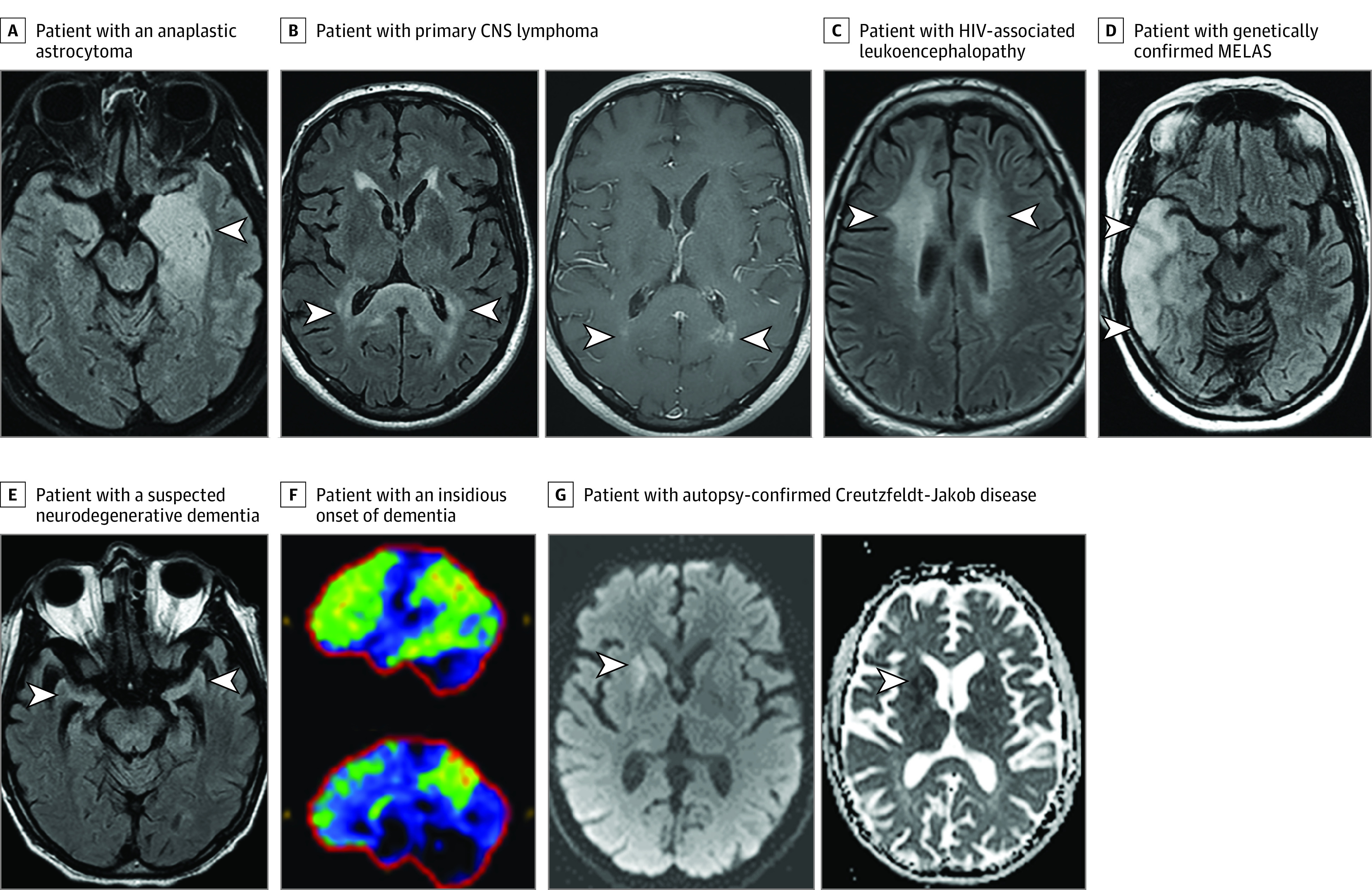
Results: A total of 107 patients were misdiagnosed with autoimmune encephalitis, and 77 (72%) did not fulfill diagnostic criteria for autoimmune encephalitis. The median (IQR) age was 48 (35.5-60.5) years and 65 (61%) were female. Correct diagnoses included functional neurologic disorder (27 [25%]), neurodegenerative disease (22 [20.5%]), primary psychiatric disease (19 [18%]), cognitive deficits from comorbidities (11 [10%]), cerebral neoplasm (10 [9.5%]), and other (18 [17%]). Onset was acute/subacute in 56 (52%) or insidious (>3 months) in 51 (48%). Magnetic resonance imaging of the brain was suggestive of encephalitis in 19 of 104 patients (18%) and cerebrospinal fluid (CSF) pleocytosis occurred in 16 of 84 patients (19%). Thyroid peroxidase antibodies were elevated in 24 of 62 patients (39%). Positive neural autoantibodies were more frequent in serum than CSF (48 of 105 [46%] vs 7 of 91 [8%]) and included 1 or more of GAD65 (n = 14), voltage-gated potassium channel complex (LGI1 and CASPR2 negative) (n = 10), N-methyl-d-aspartate receptor by cell-based assay only (n = 10; 6 negative in CSF), and other (n = 18). Adverse reactions from immunotherapies occurred in 17 of 84 patients (20%). Potential contributors to misdiagnosis included overinterpretation of positive serum antibodies (53 [50%]), misinterpretation of functional/psychiatric, or nonspecific cognitive dysfunction as encephalopathy (41 [38%]).

Conclusions and relevance: When evaluating for autoimmune encephalitis, a broad differential diagnosis should be considered and misdiagnosis occurs in many settings including at specialized centers. In this study, red flags suggesting alternative diagnoses included an insidious onset, positive nonspecific serum antibody, and failure to fulfill autoimmune encephalitis diagnostic criteria. Autoimmune encephalitis misdiagnosis leads to morbidity from unnecessary immunotherapies and delayed treatment of the correct diagnosis.


Conflict of interest statement

Conflict of Interest Disclosures: Dr Flanagan has served on advisory boards for Alexion, Genentech, UCB, and Horizon Therapeutics outside the submitted work; has a patent for DACH1-IgG as a biomarker of paraneoplastic autoimmunity pending; has received speaker honoraria from Pharmacy Times; has received royalties from UpToDate; was a site primary investigator in a randomized clinical trial on inebilizumab in neuromyelitis optica spectrum disorder run by Medimmune/Viela-Bio/Horizon Therapeutics; has received funding from the National Institutes of Health (grant R01NS113828); is a member of the medical advisory board of the MOG project; and is an editorial board member of the Journal of the Neurological Sciences and Neuroimmunology Reports. Dr Geschwind reported grants from the National Institute on Aging (grants R01 AG031189, R56 AG055619, and R01 AG062562) and research support from Michael J. Homer Family Fund during the conduct of the study; personal fees from MedConnect Pro LLC Medical Legal, Clarion, Blade Therapeutics, Clearview Healthcare Partners, LifeSci Capital LLC, Ascel Health LLC, Teledoc Health Inc, Microvention Terumo, Reata Pharmaceuticals, Wolters Kluwer, Maupin Cox, Wallace & Milsap LLC, Trinity Partners LLC, Anderson Boutwell Traylor, and Adept Field; nonfinancial support from Ionis Pharmaceuticals; has consulted for Best Doctors Inc, Biohaven Pharma Inc, Bioscience Pharma Partners, LLC, First Thought Consulting, Grand Rounds Inc/UCSF Second Opinion Inc, Quest Diagnostics, and Smith & Hennessey LLC; has received speaking honoraria from Oakstone Publishing; has received research support from Alliance Biosecure, CurePSP, the Tau Consortium, Quest Diagnostics, and the National Institutes of Health; and serves on the board of directors for San Francisco Bay Area Physicians for Social Responsibility and on the editorial board of Dementia & Neuropsychologia. Dr Lopez-Chiriboga has served on advisory boards for Genentech and Horizon Therapeutics. Dr Blackburn reported personal fees from Genentech, grants from Siegel Rare Neuroimmune Association outside the submitted work. Dr Binks reported grants from Wellcome Trust during the conduct of the study and had a patent for Ref. JA94536P.GBA pending (diagnostic strategy to improve specificity of CASPR2 antibody detection). Dr Gelfand reported grants from Genentech/Roche for research support to University of California, San Francisco for clinical trials; service on trial steering committees and grants from Vigil Neuroscience for research support to University of California, San Francisco for clinical research study; and personal fees from Biogen for consulting outside the submitted work. Dr Day reported grants from National Institute on Aging (grant K23AG064029) during the conduct of the study; personal fees from PeerView Media, Continuing Education, DynaMed, and Parabon Nanolabs outside the submitted work; is co–principal investigator of the ExTINGUISH Trial (1U01NS120901); owns stock (>$10 000) in ANI Pharmaceuticals; and is the clinical director of the Anti-NMDA Receptor Encephalitis Foundation (uncompensated). Dr Clardy reported being site investigator for an Alexion clinical trial; grants from National Institute of Neurological Disorders and Stroke for the ExTINGUISH Trial, Western Institute for Veteran Research, and Sumaira Foundation for NMO; research support from Siegel Rare Neuroimmune Association Funding and Barbara Gural Steinmetz Foundation Funding; personal fees from American Academy of Neurology (section editor, Neurology Podcast and Neurology Minute), from Alexion, VielaBio/Horizon, Genentech/Roche, Guidepoint, ExpertConnect, and Clarion Healthcare (majority fees to University of Utah); and funding from Viela Bio/Horizon and Alexion/AstraZeneca outside the submitted work. Dr Solomon reported research funding from Bristol Myers Squibb; consulting and nonpromotional speaking for EMD Serono; personal fees from Genentech, Biogen, Alexion, Celgene, Greenwich Bioscience, and Octave Biosciences; expert witness testimony for Jacob D. Fuchsberg Law Firm and Koskoff, Koskoff, and Bieder; served on advisory board of Genentech, Biogen, Alexion, Celgene, Greenwich Biosciences, and TG Therapeutics; and conducted contract research for Sanofi, Biogen, Novartis, Actelion, and Genetech outside the submitted work. Dr Pittock reported grants, personal fees, and nonfinancial support from Alexion and MedImmune/Viela Bio/Horizon (all compensation is paid directly to the Mayo Clinic); grants from the National Institutes of Health, Grifols, NovelMed, and F. Hoffmann-LaRoche/Roche/Genentech (all compensation is paid directly to Mayo Clinic); consulting for Astellas (compensation to Mayo Clinic and personal compensation); personal fees from Sage Therapeutics, UCB, and F. Hoffmann-LaRoche/Roche/Genentech; and had patent #8,889,102 issued, patent #9,891,219B2 issued, and a patent for GFAP-IgG; Septin-5-IgG; MAP1B-IgG; Kelch-like protein 11; PDE10A pending. Dr McKeon reported grants from the National Institutes of Health (grants RO1NS126227 and U01NS120901) during the conduct of the study; consulting fees from Janssen and Roche (all paid to Mayo Clinic) outside the submitted work; and had a patent for MAP1B antibody issued, a patent for Septins 5, 7, GFAP, PDE10A, KLCHL11 antibodies pending, a patent for Septin antibodies licensed, and a patent for MAP1B antibodies with royalties paid. Dr Dubey reported a patent for KLHL11 pending, a patent for LUZP4 pending, and a patent for CAVIN4 pending; and has consulted for UCB, Astellas, Argenx, Immunovant and Arialys pharmaceuticals (all compensation paid directly to Mayo Clinic). Dr Zekeridou reported grants from Roche/Genentech outside the submitted work and had a patent for DACH1-IgG as biomarker of neurological autoimmunity pending and a patent for PDE10A-IgG as biomarker of neurological autoimmunity pending. Dr Vernino has served as a consultant for Alterity, Argenx, Catalyst, Genentech, and Sage Therapeutics and has received research support from Dysautonomia International, BioHaven, Grifols, and Quest Diagnostics (through a licensing contract). Dr Irani reported grants from UCB, CSL Behring, and ONO Pharmaceuticals outside the submitted work; had a patent for LGI1/Caspr2 antibodies with royalties paid from EIAG, a patent for Autoantibody diagnostics issued, and a patent for Relapse predictions pending; and honoraria/research support from UCB, Immunovant, MedImmun, Roche, Janssen, Cerebral therapeutics, ADC therapeutics, Brain, CSL Behring, and ONO Pharmaceuticals. No other disclosures were reported.

Figures

Figure.
Figure.. Imaging Examples of Patients Who Were Initially Thought to Have Autoimmune Encephalitis but Later Had an Alternative Diagnosis Made
A T2-weighted axial fluid-attenuated inversion recovery (T2-FLAIR) image reveals a left mesial temporal lobe T2-hyperintensity and swelling (A, arrowhead) in a patient with an anaplastic astrocytoma. Note in retrospect the fullness/enlargement of the affected region, possibly suggesting some mass effect. Axial T2-FLAIR image reveals bilateral splenium T2-hyperintensity (B, left panel, arrowheads) with multifocal punctate enhancement (B, right panel, arrowheads) in a patient with primary central nervous system (CNS) lymphoma. An axial T2-FLAIR image reveals bilateral confluent T2-hyperintensity in the subcortical white matter (C, arrowheads) in a patient with HIV-associated leukoencephalopathy. Axial T2-FLAIR image reveals right temporal cortical swelling and T2-hyperintensity (D, arrowheads) in a patient with genetically confirmed mitochondrial encephalomyopathy, lactic acidosis, and stroke-like episodes (MELAS). An axial T2-FLAIR image shows disproportionate bilateral hippocampal atrophy (E, arrowheads) in a patient with a suspected neurodegenerative dementia with features potentially consistent with mixed Alzheimer disease and dementia with Lewy bodies. 18F-Fluorodeoxyglucose positron emission tomography reveals reduced uptake of glucose (normal, dark blue/black; mildly reduced, green; moderately reduced, yellow; severely reduced, red) in the frontotemporoparietal region, precuneus and posterior cingulate (F) most suspicious for underlying Alzheimer disease in a patient with an insidious onset of dementia and elevated cerebrospinal fluid phospho-Tau and low cerebrospinal fluid amyloid-β 42 also suggestive of this diagnosis. Axial diffusion weighted hyperintensity (G, left panel) and apparent diffusion coefficient hypointensity (G, right panel) consistent with restricted diffusion in the right caudate and putamen in a patient in whom autopsy later confirmed Creutzfeldt-Jakob disease.

Comment in

References

    1. Graus F, Titulaer MJ, Balu R, et al. . A clinical approach to diagnosis of autoimmune encephalitis. Lancet Neurol. 2016;15(4):391-404. doi:10.1016/S1474-4422(15)00401-9 - DOI - PMC - PubMed
    1. Dubey D, Pittock SJ, Kelly CR, et al. . Autoimmune encephalitis epidemiology and a comparison to infectious encephalitis. Ann Neurol. 2018;83(1):166-177. doi:10.1002/ana.25131 - DOI - PMC - PubMed
    1. Abboud H, Probasco JC, Irani S, et al. ; Autoimmune Encephalitis Alliance Clinicians Network . Autoimmune encephalitis: proposed best practice recommendations for diagnosis and acute management. J Neurol Neurosurg Psychiatry. 2021;92(7):757-768. doi:10.1136/jnnp-2020-325300 - DOI - PMC - PubMed
    1. Budhram A, Dubey D, Sechi E, et al. . Neural antibody testing in patients with suspected autoimmune encephalitis. Clin Chem. 2020;66(12):1496-1509. doi:10.1093/clinchem/hvaa254 - DOI - PubMed
    1. Valencia-Sanchez C, Pittock SJ, Mead-Harvey C, et al. . Brain dysfunction and thyroid antibodies: autoimmune diagnosis and misdiagnosis. Brain Commun. 2021;3(2):fcaa233. doi:10.1093/braincomms/fcaa233 - DOI - PMC - PubMed

Publication types

Supplementary concepts