Review
. 2022 Nov 22;4(1):obac042.
doi: 10.1093/iob/obac042. eCollection 2022.

Roles of Mono- and Bi-articular Muscles in Human Limbs: Two-joint Link Model and Applications

Affiliations
Review

Roles of Mono- and Bi-articular Muscles in Human Limbs: Two-joint Link Model and Applications

Tsutomu Miyake et al. Integr Org Biol. .

Abstract

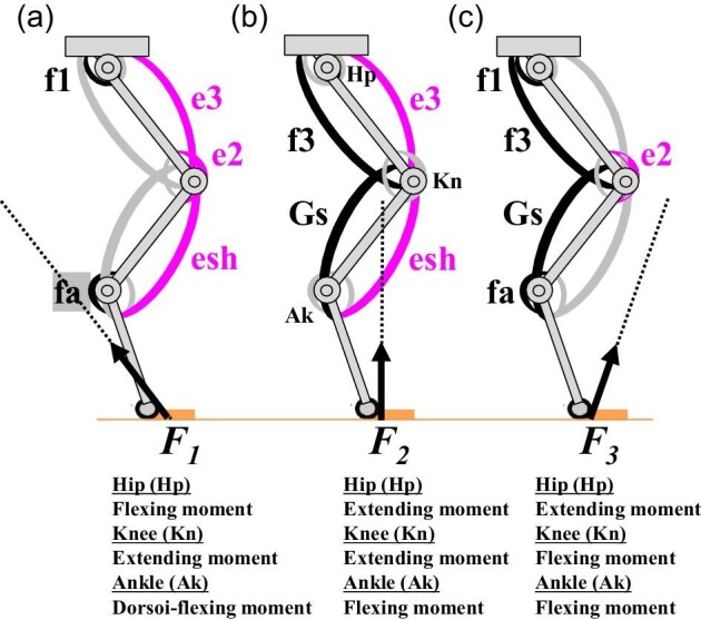
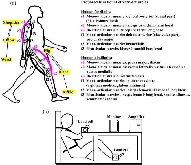
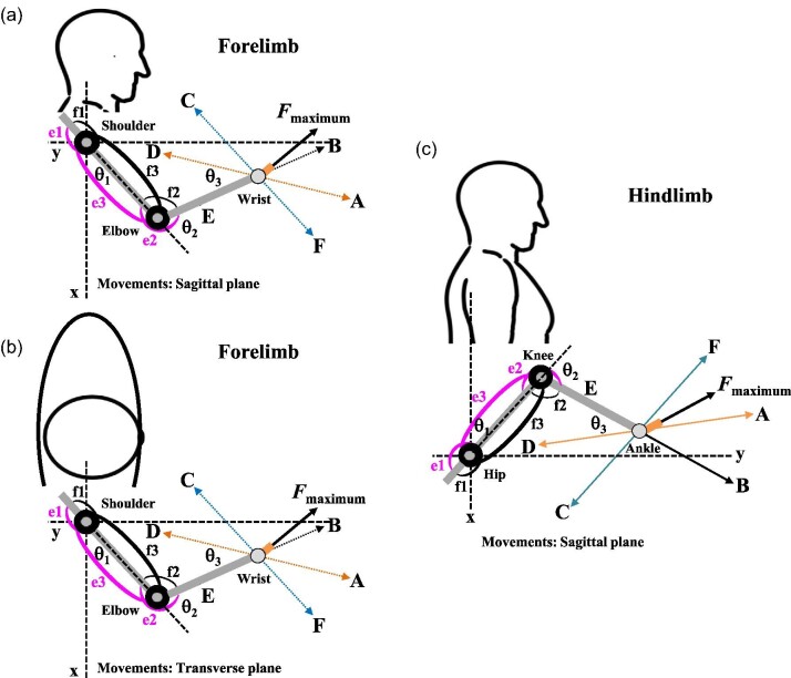
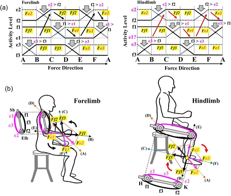
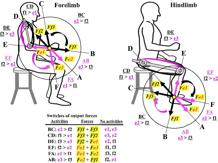
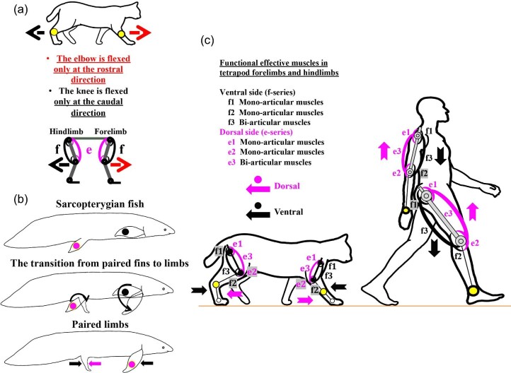
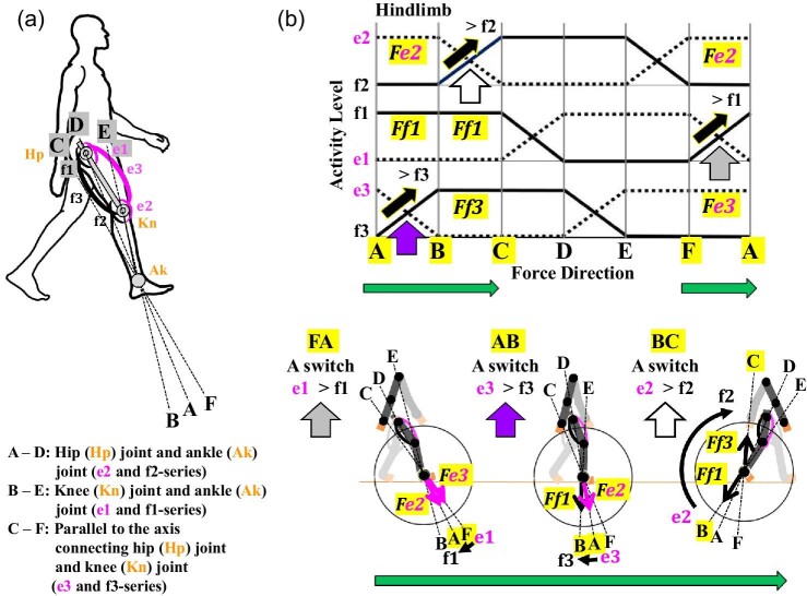
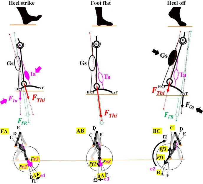
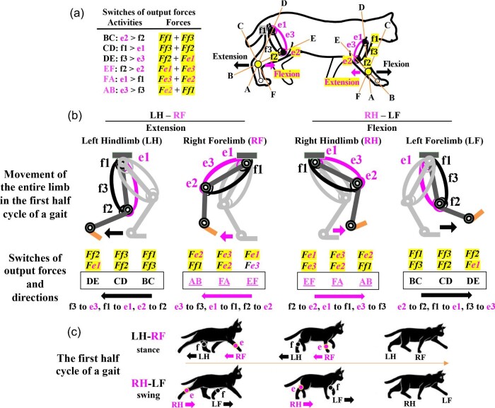
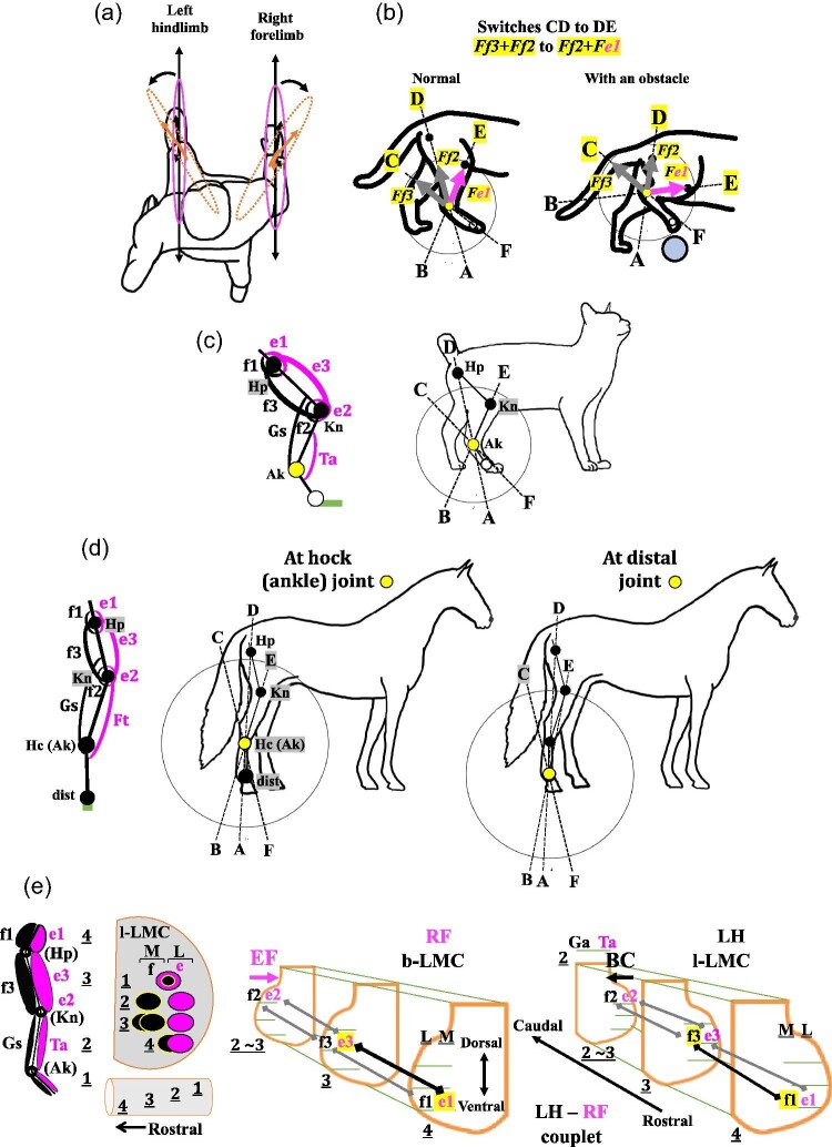
We review the two-joint link model of mono- and bi-articular muscles in the human branchium and thigh for applications related to biomechanical studies of tetrapod locomotion including gait analyses of humans and non-human tetrapods. This model has been proposed to elucidate functional roles of human mono- and bi-articular muscles by analyzing human limb movements biomechanically and testing the results both theoretically and mechanically using robotic arms and legs. However, the model has not yet been applied to biomechanical studies of tetrapod locomotion, in part since it was established based mainly on mechanical engineering analyses and because it has been applied mostly to robotics, fields of mechanical engineering, and to rehabilitation sciences. When we discovered and published the identical pairs of mono- and bi-articular muscles in pectoral fins of the coelacanth fish Latimeria chalumnae to those of humans, we recognized the significant roles of mono- and bi-articular muscles in evolution of tetrapod limbs from paired fins and tetrapod limb locomotion. Therefore, we have been reviewing the theoretical background and mechanical parameters of the model in order to analyze functional roles of mono- and bi-articular muscles in tetrapod limb locomotion. Herein, we present re-defined biological parameters including 3 axes among 3 joints of forelimbs or hindlimbs that the model has formulated and provide biological and analytical tools and examples to facilitate applicable power of the model to our on-going gait analyses of humans and tetrapods.

本総説は、ヒトやヒト以外の四肢動物における歩容解析を含む四肢運動を生体工学研究へ応用することを目的として、ヒトの上腕や大腿の一関節筋・二関節筋リンクモデルを概説したものである。このモデルは元来、ヒトの四肢運動から得られた生体力学的解析結果を、ロボットアームやロボットレッグを用いて理論的・力学的に検証するというアプローチで提唱された概念である。機械工学的な解析に基づいて確立された研究背景から、このモデルは主に、ロボット工学や機械工学、リハビリテーション科学の分野への応用が進んだが、四肢動物の四肢運動に関する生体力学への応用は進まなかった。同じ頃、我々は、シーラカンスLatimeria chalumnaeの胸鰭にヒト上腕と相当の対をなす一関節筋・二関節筋が存在することを発見し、鰭から四肢への進化や四肢運動において、これらの筋が重要な役割を果たす可能性を見出していた。この発見を起点として、我々は、四肢動物の四肢運動における一関節筋・二関節筋の機能的役割を明らかにするために、このモデルの理論的背景や機械的パラメーターを再検証した。その結果、このモデルが定式化した前肢や後肢の三関節間にまたがる三つの軸を含むパラメーターを補正し、生物学的パラーメーターとして再定義することで、四肢動物の四肢運動の解析が可能となるという結論に至った。本総説では、我々が現在進行中の歩容解析を例に、ヒトやヒト以外の四肢動物における四肢運動の生体工学研究へこのモデルを応用するための生物学的解析手法を含めて概説する。.


Figures

Fig. 1
Fig. 1
Differential activations of mono- and bi-articular muscles under three ground reaction forces (F1, F2, F3) when the hindlimb is extended. van Ingen Schenau (1990) illustrated this “possible design” of a hindlimb with mono- and bi-articular muscles based on his review of functional roles of bi-articular muscles in limbs. Only activated muscles are labeled in each case. (A) F1 is behind the ankle (Ak) joint; (B) F2 is in front of the hip (Hp) joint and the ankle (Ak) joint but behind the knee (Kn) joint, as in humans; (C) F3 is in front of the knee joint (Kn), as in most tetrapods. f1: a mono-articular muscle of the hip joint; e2: a mono-articular muscle of the knee joint; e3 and f3: bi-articular muscles of the thigh; Gs: gastrocnemius; fa: a mono-articular muscle of the ankle joint; esh: a bi-articular muscle of the lower leg other than Gs. Humans do not have esh. The rostral direction of the body is on the right side.
Fig. 2
Fig. 2
Proposed setups of the two-joint link model of mono- and bi-articular muscles in the human brachium and thigh. (A) The functional effective muscles (e-series and f-series) in the human forelimb and hindlimb. (B) The experimental platform that has been used to analyze defined movements of either the human forelimb or hindlimb. Designated as e-series and f-series, the muscles consist of two antagonistic pairs of mono-articular muscles (e1, e2, f1, f2) and one antagonistic pair of bi-articular muscles (e3, f3). The activities of the muscles in the link model were hypothesized to generate output forces and thus control directions at the wrist or ankle. Whether the latissimus dorsi should be defined as an e1-series muscle and whether the gluteus medius and gluteus minimus should be defined as f1-series muscles remain to be investigated. The experimental platform was used to analyze defined movements of either the human forelimb or hindlimb. Activities of the functional effective muscles and the maximum output forces were recorded with EMG and a load cell at the wrist or ankle, respectively, (Fujikawa et al. 1996; Fujikawa et al. 1997; Oshima et al. 1999).
Fig. 3
Fig. 3
Experimental setups of the two-joint link model for defined movements of the forelimb or hindlimb on a platform. Robotic arms and legs with mono- and bi-articular actuators identical to the muscles of humans were built to test the results of human subjects and establish the mechanical models. (A) The defined movements of the forelimb in the sagittal plane (Fujikawa et al. 1996); (B) The defined movements of the forelimb in the transverse plane (Fujikawa et al. 1997); (C) The defined movements of the hindlimb in the sagittal plane (Oshima et al. 1999; Oshima 2008). θ1: the shoulder flexion angle or the hip flexion angle; θ2: the elbow flexion angle or the knee flexion angle; θ3: the angle between the force direction of A and D and that of B and E; Fmaximum: the possible maximum output force exerted by a different combination of mono- and bi-articular muscles in the brachium or the thigh. A–D: the line connecting the shoulder joint and the wrist joint or the hip joint and the ankle joint; B–E: the line connecting the elbow joint and the wrist joint or the knee joint and the ankle joint; C–F: the line parallel to the line between the shoulder joint and the elbow joint or the line between the hip joint and the knee joint.
Fig. 4
Fig. 4
Results of experimentally defined movements of forelimb and hindlimb with EMG analyses. (A) Schematized IEMGs based on the EMG data. IEMG showed the maximum output forces and changes in activity between a different antagonistic pair of mono- or bi-articular muscles when human subjects engaged in different defined movements of the forelimb or hindlimb (Fujikawa et al. 1997). The posture condition for the defined movements of the forelimb was set up with θ 1 = 48° and θ 2 = 90° (Fujikawa et al. 1997) whereas that of hindlimb was set up with θ 1 = 45° and θ 2 = 90° (Oshima 2008). The force directions were normalized to allow visualization of switches in the output force directions. (B) A summary of defined movements of the forelimb and hindlimb and the combined distribution of output forces (Fe1 through Ff3) around the 360-degree perimeter of the wrist or ankle. The combined distribution of all output forces becomes hexagonal in the forelimb and hindlimb. Sequential switches of the forelimb are counterclockwise, whereas sequential switches of the hindlimb are clockwise.
Fig. 5
Fig. 5
The model of coordinated activities of mono- and bi-articular muscles in the two-joint link model of the human brachium and thigh. The model proposes that switching activity between e1 and f1, e3 and f3, e2 and f2, and forces Fe1 through Ff3 occur sequentially around the 360-degree perimeter of the wrist and ankle. For each range, for example, A and B, two muscles (e2 and f1) exert a combined output force (Fe2 + Ff1) with the activity switch from e3 to f3, depending on the combined output force at the wrist or the ankle. Two mono- and bi-articular muscles are inactivated sequentially in each range of activity switches. Sequential switches of the forelimb are counterclockwise, whereas sequential switches of the hindlimb are clockwise.
Fig. 6
Fig. 6
Biological prerequisites for an application of the two-joint link model to studies of tetrapod limb locomotion. (A) The configuration of tetrapod limbs and direction of flexion and extension. The elbow is flexed only in the rostral direction, whereas the knee is flexed only in the caudal direction. (B) Evolution of the orientations of paired fins in sarcopterygian fishes and paired limbs in tetrapods through the fin-to-limb transition. (C) Biological orientations of tetrapod and human forelimbs and hindlimbs and locations of e-series and f-series muscles in forelimb and hindlimb. The dorsal side and ventral side of limbs differ in forelimbs and hindlimbs due to the evolutionary history of tetrapod limbs through the fin-to-limb transition in Late Devonian and Early Carboniferous Periods (Romer 1971; Shubin et al. 2006; Johanson et al. 2007; Clack 2012; Miyake et al. 2016; Cloutier et al. 2020). The dorsal and ventral sides of human forelimbs and hindlimb match the map of dermatomes and cutaneous nerve territories (Schünke et al. 2017; Martini et al. 2018).
Fig. 7
Fig. 7
A series of contact tasks with human mono- and bi-articular muscles and the gastrocnemius muscle in the two-joint link model. (A) A mechanical test of the two-joint link model with or without a pair of bi-articular muscles (e3 and f3) in the forelimb (Fujikawa et al. 2001). As the plate was moved toward the model from position 1 to position 3, the link model was pushed against the plate. The researchers tested whether the model was able to make a correct trajectory and stay on the plate or slipped with or without the bi-articular muscles. (B) A mechanical test of the two-joint link model with or without a pair of bi-articular muscles (e3 and f3) in the hindlimb (Oshima et al. 2010; Oshima et al. 2017). (C) Illustrated jumping motions that show the requirement of a pair of bi-articular muscles for landing without any slipping or rotation (Oshima et al. 2004; Oshima et al. 2017). (D) A role for the gastrocnemius muscle of the hindlimb during higher jumping motions to prevent much rotation of the limb (Kanayama et al. 2001; Oshima et al. 2005). Eb: elbow joint; Gs: gastrocnemius muscle; Hp: hip joint; Kn: knee joint; Sh: shoulder joint.
Fig. 8
Fig. 8
Predicted output forces in the human forelimb during a sprint performance in light of the two-joint link model. (A) Defined axes: A–D (e2 and f2 series muscles); B–E (e1 and f1 series muscles); C–F (e3 and f3 series muscles). (B) Two examples of counterclockwise switches in activity, e3 to f3 (AB) and e2 to f2 (BC), and output combined forces, Fe2 + Ff1 and Ff1 + Ff3, respectively, with the schematized IEMGs, when the human right forelimb moves in coordination with the contralateral hindlimb during a sprint performance. A magnitude of each combined force will determine the force direction and thus the directional movement of the forelimb. Large arrows: switches of output forces in AB and BC.
Fig. 9
Fig. 9
Predicted output forces in the human hindlimb during the stance phase of the bi-pedal walking gait in light of the two-joint link model. (A) Defined axes: A–D (e2 and f2 series muscles); B–E (e1 and f1 series muscles); C–F (e3 and f3 series muscles). (B) Three stages of the stance phase and concomitant clockwise switches in activity, e1 to f1 (FA), e3 to f3 (AB), and e2 to f2 (BC) with changes in the combined output forces, Fe3 + Fe2, Fe2 + Ff1, and Ff1 + Fe3, respectively, as depicted in the schematized IEMGs. The magnitude of a combined force will determine the force direction and thus the direction of hindlimb. Large arrows: switches of output forces in FA, AB, and BC.
Fig. 10
Fig. 10
The predicted sequence of contact tasks during the stance phase of the human bi-pedal walking gait in light of the two-joint link model. During the stance phase, the foot first touches the ground with the heel (heel strike) and ultimately leaves the ground with the heel rising first, followed by the toes (heel off); at each step of this process the two-joint link model predicts sequential switches in the output force from FA to AB to BC (also see Fig. 9). The tibialis anterior (Ta) is activated when the heel is off the ground, whereas the gastrocnemius (Gs) is activated when the toes are off the ground. H: heel; K: knee; T: toe. FFR: a combined force by FThi and either FTa or FGs; FGs: output force from the gastrocnemius; FTa: output force from the tibialis anterior; FThi: output force from the mono- and bi-articular muscles in the thigh.
Fig. 11
Fig. 11
Proposed activities of mono- and bi-articular muscles in a diagonal-couplet lateral sequence based on the two-joint link model. (A) The switches in output forces and the two-joint link model with three axes and the distribution of e-series and f-series muscles in tetrapod forelimbs and hindlimbs. (B) Switches in output forces during the extension and flexion of two pairs of couplets, the left hindlimb and right forelimb (LH–RF) and the right hindlimb and left forelimb (RH–LF), respectively, in the first half cycle of a gait. At the stance phase, the extension of LH–RF goes through BC to CD to DE (left hindlimb, LH) and EF to FA to AB (right forelimb, RF). At the swing phase, the flexion of RH–LF goes through EF to FA to AB (right hindlimb, RH) and BC to CD to DE (left forelimb, LF). (C) The proposed sequence of the LH–RF couplet and RH–LF couplet during the first half cycle of a diagonal-couplet lateral sequence. The LH–RF couplet makes the extension of LH and RF as the stance phase, although LH and RF go through different switches, BC to CD to DE (f: f-series muscles) and EF to FA to AB (e: e-series muscles), respectively. RH–LF couplet makes the flexion of RH and LF as the swing phase, although RH and LF go through different switches, EF to FA to AB (e: e-series muscles) and BC to CD to DE (f: f-series muscles), respectively. LF: left forelimb; LH: left hindlimb; RF: right forelimb; RH: right hindlimb.
Fig. 12
Fig. 12
Four examples of unanswered questions related to the two-joint link model of mono- and bi-articular muscles in human forelimbs and hindlimbs and a predicted arrangement of motor pools for mono- and bi-articular muscles in the LMCs. (A) A sudden change in the direction of movement of a forelimb or a hindlimb during normal quadrupedal walking. (B) Predicted switched (CD to DE) and output forces in normal quadrupedal walking gait and an obstacle avoidance gait in cats based on EMG data and the gait analyses (Widajewicz et al. 1994; McFadyne et al. 1999; McKay et al. 2007; Markin et al. 2012; Frigon et al. 2015; Chu et al. 2017). (C) The two-joint link model in hindlimb of a digitigrade cat. (D) The two-joint link model in hindlimb of an unguligrade horses with another set of bi-articular muscles between the knee joint and the hock (ankle) joint. There might be two possible points where the e-series and f-series muscles in the thigh exert their forces at the hock (ankle) joint or at a more distal joint such as the fetlock joint, pastern joint, or coffin joint. (E) Motor pools in the branchial lateral motor column (b-LMC) and the lumber lateral motor column (l-LMC). The arrangement of e-series and f-series muscles in a hindlimb corresponds topologically to the arrangement of the motor pools in the lumber lateral motor column (l-LMC) that innervate these muscles (VanderHorst and Holstege 1997; Sürmeli et al. 2011; Gross et al. 2017). Potential activation sequences of motor neurons for mono- and bi-articular muscles are predicted if a tetrapod starts quadrupedal walking with the left hindlimb (LH) and right forelimb (RF) (LH–RF couplet) in the first half cycle of a gait based on our proposed activities of mono- and bi-articular muscles in a diagonal-couplet lateral sequence (Fig. 11C). Ak: ankle joint; dist: distal joint(s); Ft: fibularis tertius; Gs: gastrocnemius; Hc: hock joint; Hp: hip joint; Kn: knee joint; L: the lateral side of LMCs; M: the medial side of LMCs; Ta: tibialis anterior.

Similar articles

Cited by

References

    1. Abe T, Koide T, Furukawa K, Ochi A, Oshima T, Fujikawa T.. 2014. Parallel linkage function of the rectus femoris when standing up. Jour Robotics Soc Japan 32:190–207. In Japanese.
    1. Abu-Farj ZO, Harris GF, Smith PA, Hassani S.. 2015. Human gait and clinical movement analysis. In: Encyclopedia of Electronics Engineering. 2nd Edition. New York: John Wiley & Sons, Inc. p. 1–34.
    1. Afschrift M, Pitto L, Aerts W, van Deursen R, Jonkers I, De Groote F.. 2018. Modulation of the gluteus medius activity reflects the potential of the muscle to meet the mechanical demands during perturbed walking. Sci Rep 8:11675. - PMC - PubMed
    1. Agbarajl EC, Inylama HC, Okezle CC.. 2017. Dynamic modeling of a 3-DOF articulated robotic manipulator based on independent joint scheme. Phy Sci Inter J 15:1–10.
    1. Aizawa K, Ishii S, Kumamoto M.. 2008. Motion analysis by a functional effective muscle display (FEMS). In: Kumamoto M, editor. Bi-articular muscles: Motion control and rehabilitation. Tokyo: IGAKU-SHOIN, Ltd. p. 98–114. In Japanese. ISBN:978-4-260-00592-0.

LinkOut - more resources