

Shimming toolbox: An open-source software toolbox for B0 and B1 shimming in MRI
- PMID: 36441743
- PMCID: PMC9910837
- DOI: 10.1002/mrm.29528
Shimming toolbox: An open-source software toolbox for B0 and B1 shimming in MRI
Abstract
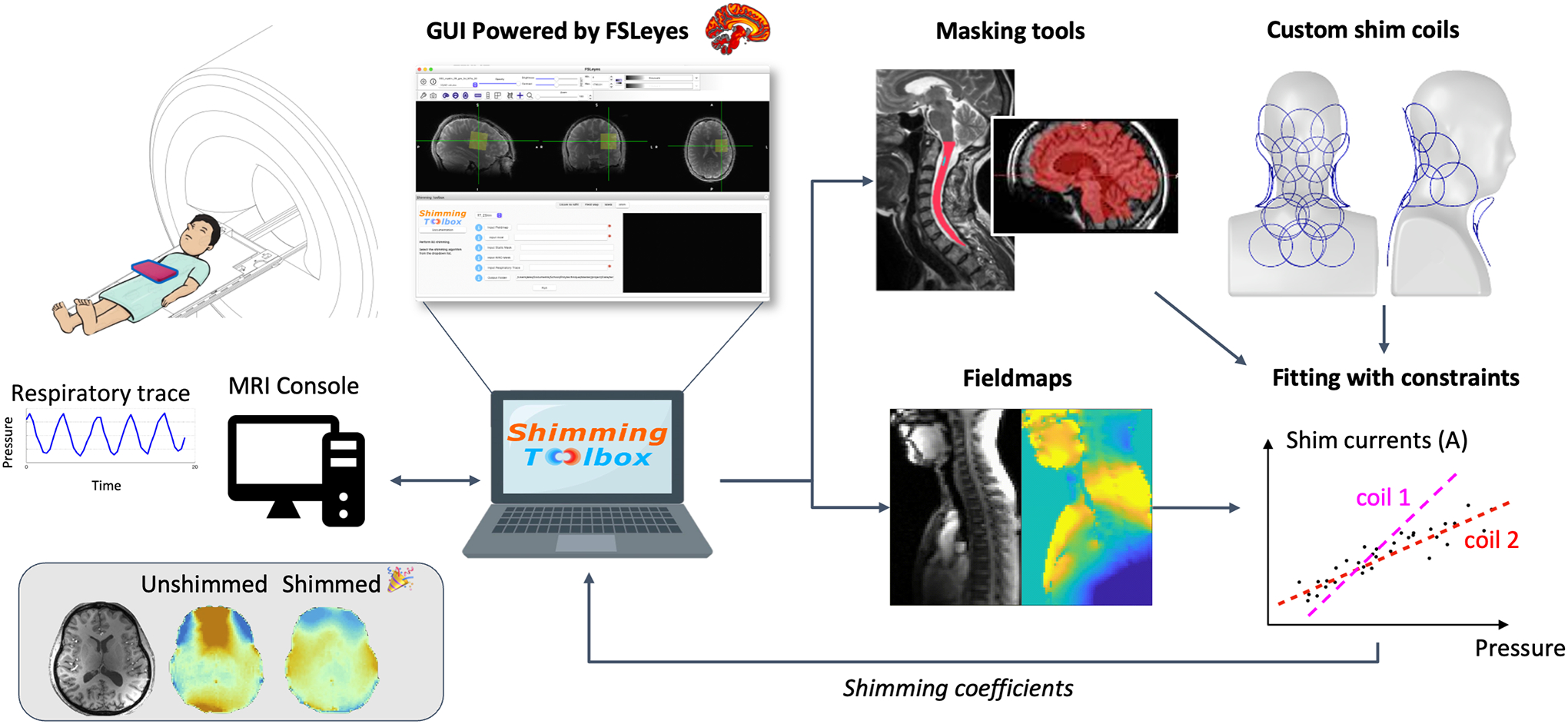
Purpose: Introduce Shimming Toolbox ( https://shimming-toolbox.org), an open-source software package for prototyping new methods and performing static, dynamic, and real-time B0 shimming as well as B1 shimming experiments.
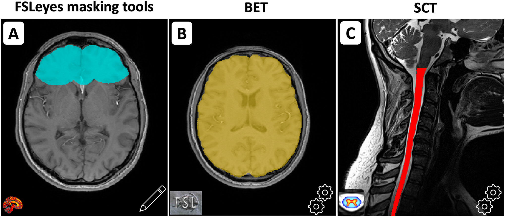
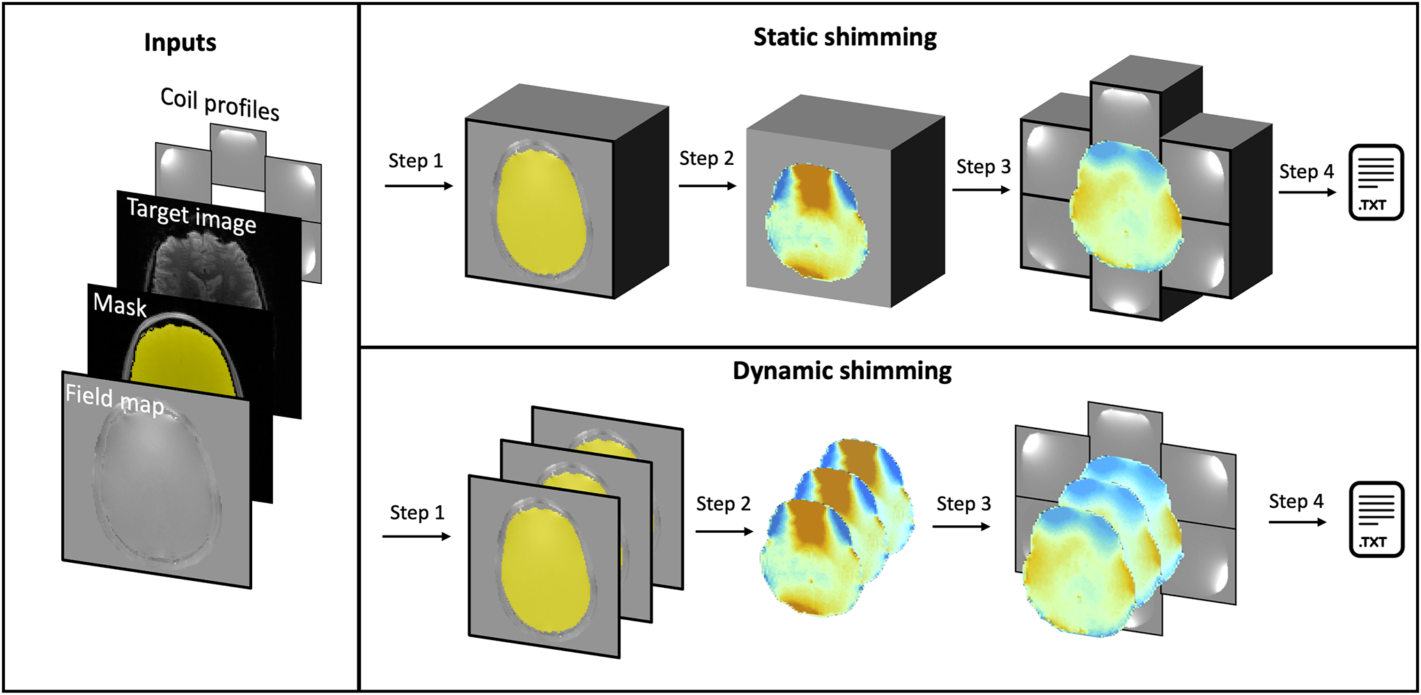
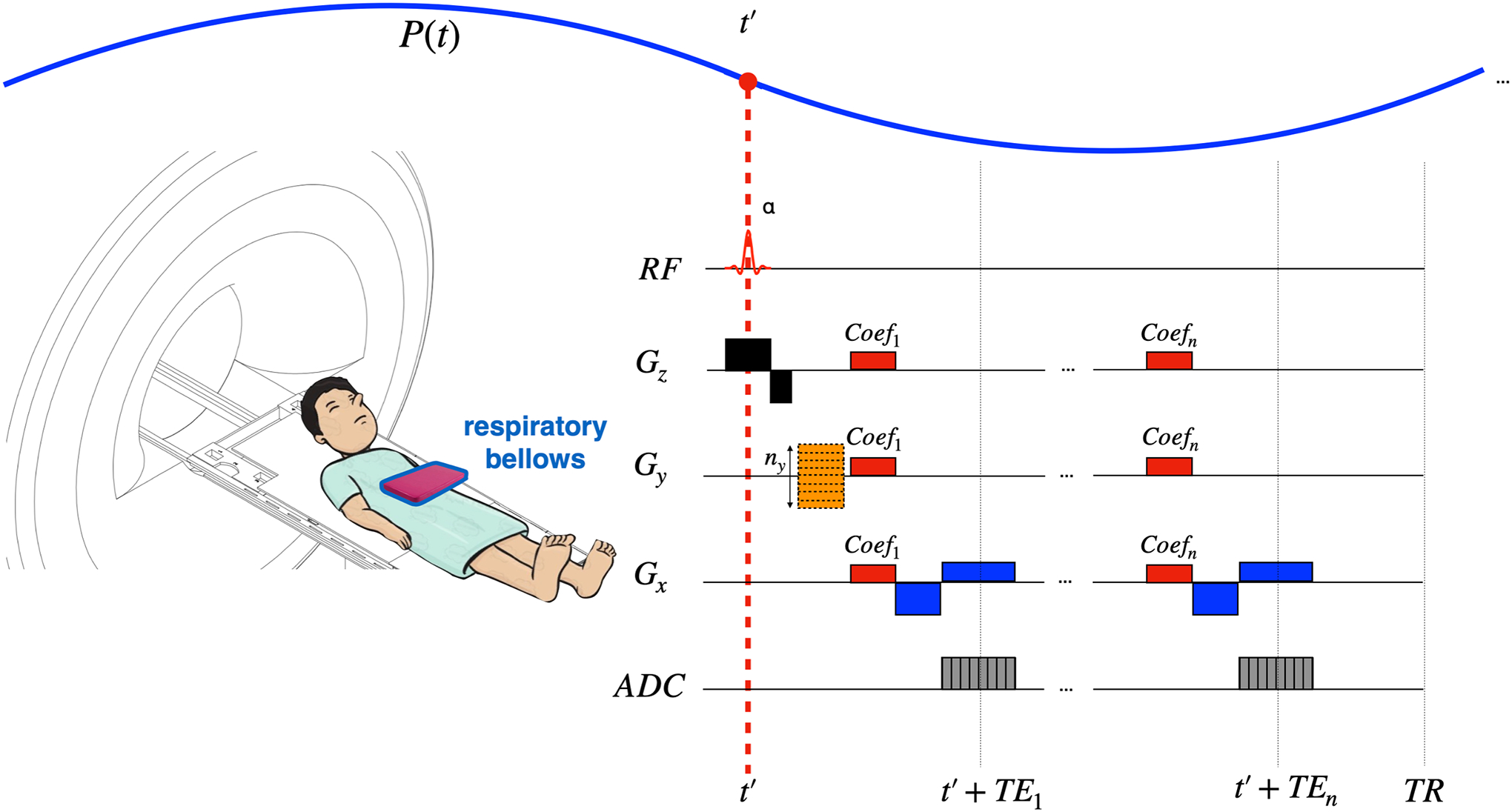
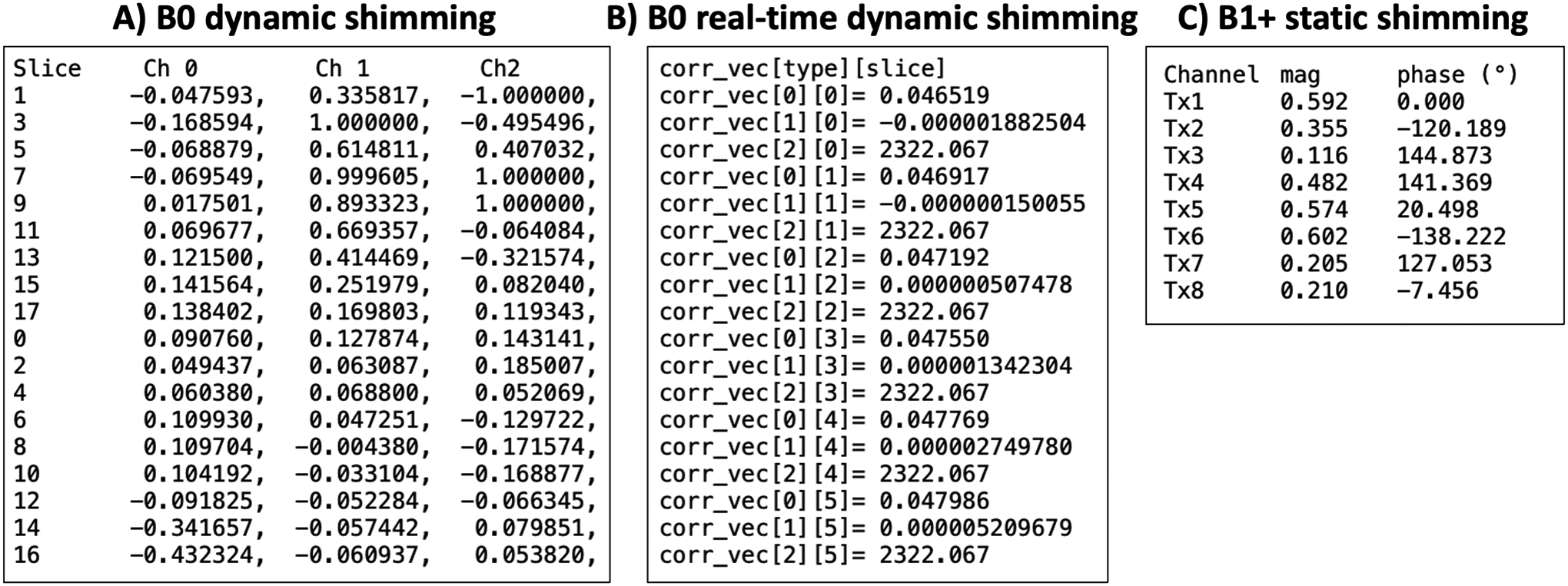
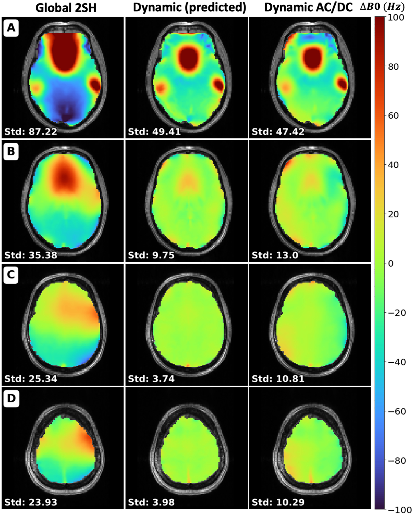
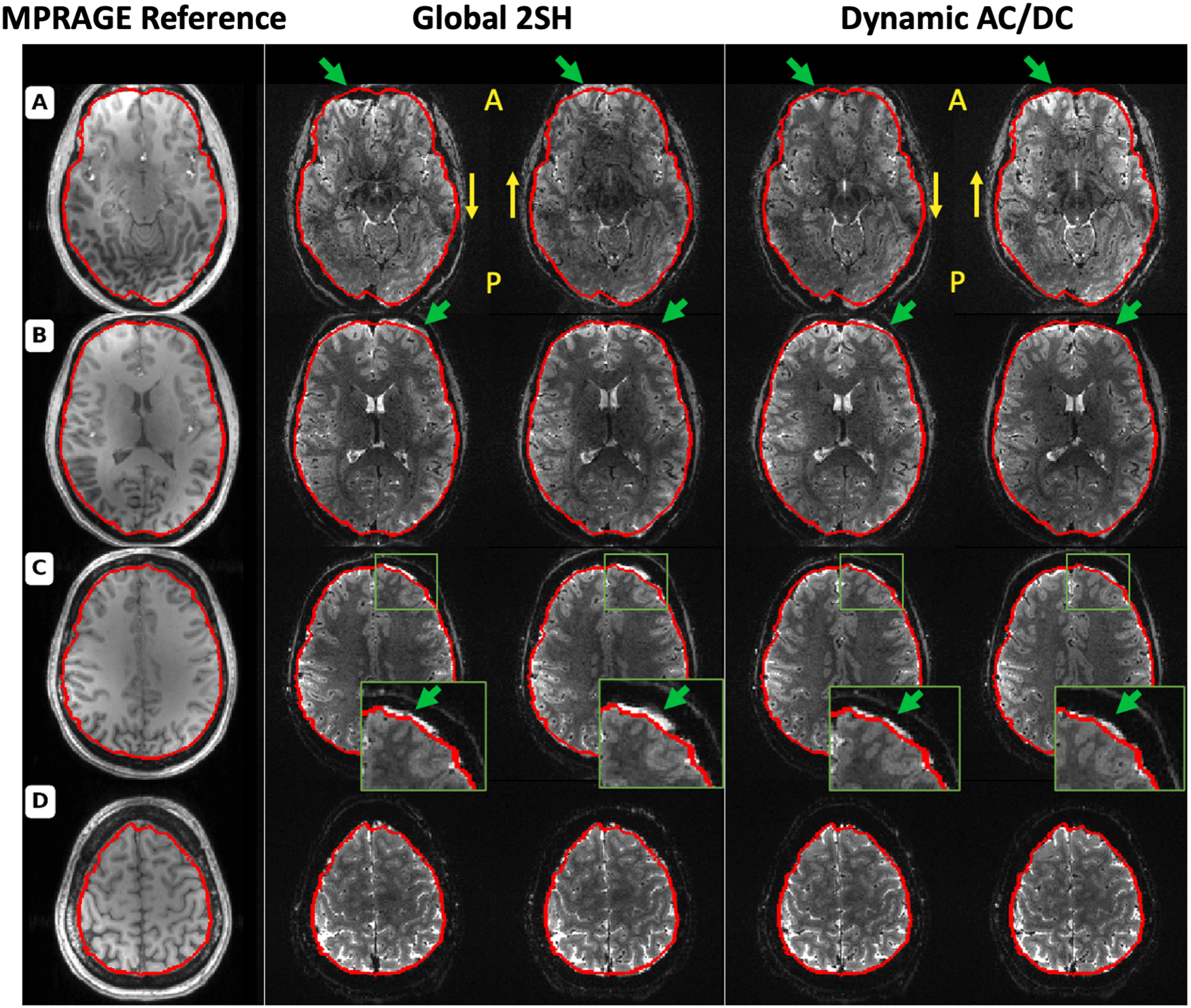
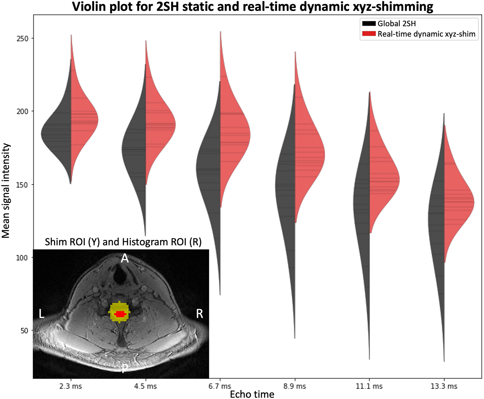
Methods: Shimming Toolbox features various field mapping techniques, manual and automatic masking for the brain and spinal cord, B0 and B1 shimming capabilities accessible through a user-friendly graphical user interface. Validation of Shimming Toolbox was demonstrated in three scenarios: (i) B0 dynamic shimming in the brain at 7T using custom AC/DC coils, (ii) B0 real-time shimming in the spinal cord at 3T, and (iii) B1 static shimming in the spinal cord at 7T.
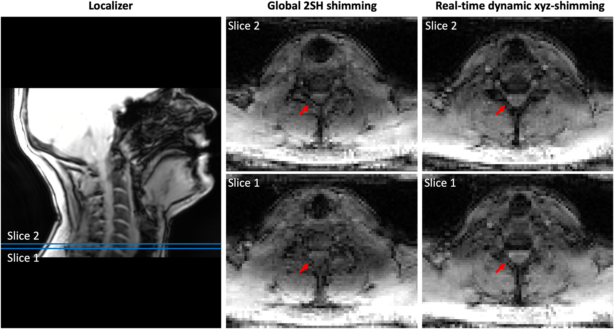
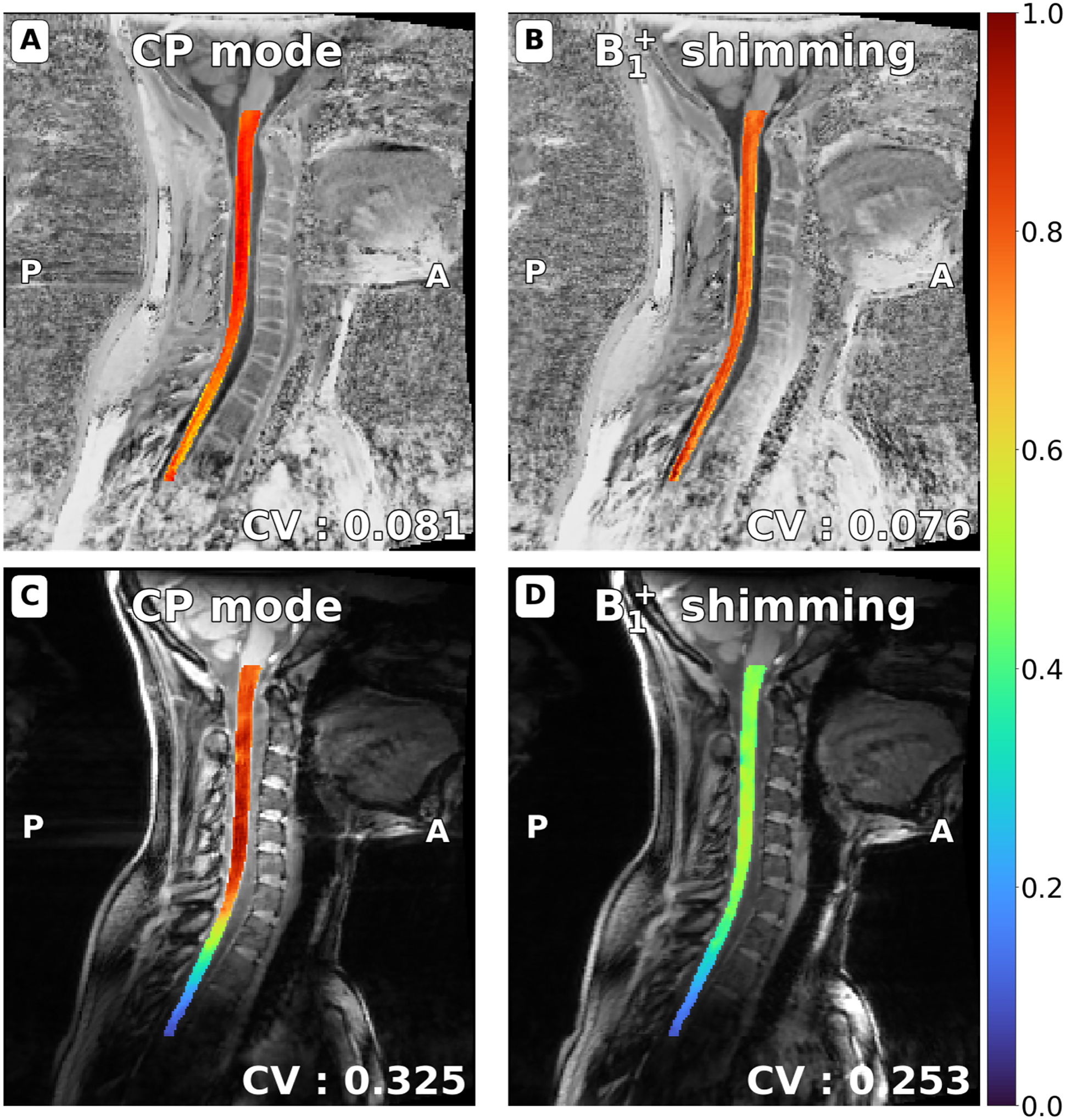
Results: The B0 dynamic shimming of the brain at 7T took about 10 min to perform. It showed a 47% reduction in the standard deviation of the B0 field, associated with noticeable improvements in geometric distortions in EPI images. Real-time dynamic xyz-shimming in the spinal cord took about 5 min and showed a 30% reduction in the standard deviation of the signal distribution. B1 static shimming experiments in the spinal cord took about 10 min to perform and showed a 40% reduction in the coefficient of variation of the B1 field.
Conclusion: Shimming Toolbox provides an open-source platform where researchers can collaborate, prototype and conveniently test B0 and B1 shimming experiments. Future versions will include additional field map preprocessing techniques, optimization algorithms, and compatibility across multiple MRI manufacturers.
Keywords: B0; B1; MRI; Python; inhomogeneities; open-source software; parallel transmit; shimming.
© 2022 International Society for Magnetic Resonance in Medicine.
Figures
Similar articles
-
Radiofrequency-transparent local B0 shimming coils using float traps.Magn Reson Med. 2025 Apr;93(4):1833-1841. doi: 10.1002/mrm.30361. Epub 2024 Nov 4. Magn Reson Med. 2025. PMID: 39497505 Free PMC article.
-
RF shimming in the cervical spinal cord at 7 T.Magn Reson Med. 2024 Dec;92(6):2392-2403. doi: 10.1002/mrm.30225. Epub 2024 Aug 13. Magn Reson Med. 2024. PMID: 39136249
-
Improving brain B0 shimming using an easy and accessible multi-coil shim array at ultra-high field.MAGMA. 2022 Dec;35(6):943-951. doi: 10.1007/s10334-022-01014-6. Epub 2022 May 5. MAGMA. 2022. PMID: 35511312 Free PMC article.
-
In vivo B0 field shimming methods for MRI at 7T.Neuroimage. 2018 Mar;168:71-87. doi: 10.1016/j.neuroimage.2017.06.013. Epub 2017 Jun 7. Neuroimage. 2018. PMID: 28602943 Free PMC article. Review.
-
Spinal cord MRI at 7T.Neuroimage. 2018 Mar;168:437-451. doi: 10.1016/j.neuroimage.2017.07.003. Epub 2017 Jul 3. Neuroimage. 2018. PMID: 28684332 Free PMC article. Review.
Cited by
-
Single-shot echo planar time-resolved imaging for multi-echo functional MRI and distortion-free diffusion imaging.Magn Reson Med. 2025 Mar;93(3):993-1013. doi: 10.1002/mrm.30327. Epub 2024 Oct 20. Magn Reson Med. 2025. PMID: 39428674 Free PMC article.
-
Impact of through-slice gradient optimization for dynamic slice-wise shimming in the cervico-thoracic spinal cord.Magn Reson Med. 2025 Sep;94(3):1090-1102. doi: 10.1002/mrm.30543. Epub 2025 May 1. Magn Reson Med. 2025. PMID: 40312894 Free PMC article.
-
High-Resolution Magnetic Resonance Neurography at 7T: A Pilot Study of Hand Innervation.Diagnostics (Basel). 2024 Jun 12;14(12):1230. doi: 10.3390/diagnostics14121230. Diagnostics (Basel). 2024. PMID: 38928648 Free PMC article.
-
Multi-center benchmarking of cervical spinal cord RF coils for 7 T MRI: A traveling spines study.Magn Reson Med. 2025 Sep;94(3):1339-1355. doi: 10.1002/mrm.30551. Epub 2025 May 20. Magn Reson Med. 2025. PMID: 40391630 Free PMC article.
-
Multi-center benchmarking of cervical spinal cord RF coils for 7 T MRI: A traveling spines study.bioRxiv [Preprint]. 2025 Jan 17:2025.01.13.632825. doi: 10.1101/2025.01.13.632825. bioRxiv. 2025. Update in: Magn Reson Med. 2025 Sep;94(3):1339-1355. doi: 10.1002/mrm.30551. PMID: 39868333 Free PMC article. Updated. Preprint.
References
-
- Karamat MI, Darvish-Molla S, Santos-Diaz A. Opportunities and Challenges of 7 Tesla Magnetic Resonance Imaging: A Review. Crit. Rev. Biomed. Eng 2016;44:73–89. - PubMed
-
- Jezzard P, Balaban RS. Correction for geometric distortion in echo planar images from B0 field variations. Magn. Reson. Med 1995;34:65–73. - PubMed
-
- van Gelderen P, de Zwart JA, Starewicz P, Hinks RS, Duyn JH. Real-time shimming to compensate for respiration-induced B0 fluctuations. Magn. Reson. Med 2007;57:362–368. - PubMed
Publication types
MeSH terms
Grants and funding
LinkOut - more resources
Full Text Sources
Medical
Miscellaneous
