Editorial
. 2023 Apr 14;29(8):1515-1527.
doi: 10.1158/1078-0432.CCR-22-1602.

[18F]FluorThanatrace ([18F]FTT) PET Imaging of PARP-Inhibitor Drug-Target Engagement as a Biomarker of Response in Ovarian Cancer, a Pilot Study

Affiliations
Editorial

[18F]FluorThanatrace ([18F]FTT) PET Imaging of PARP-Inhibitor Drug-Target Engagement as a Biomarker of Response in Ovarian Cancer, a Pilot Study

Austin R Pantel et al. Clin Cancer Res. .

Abstract

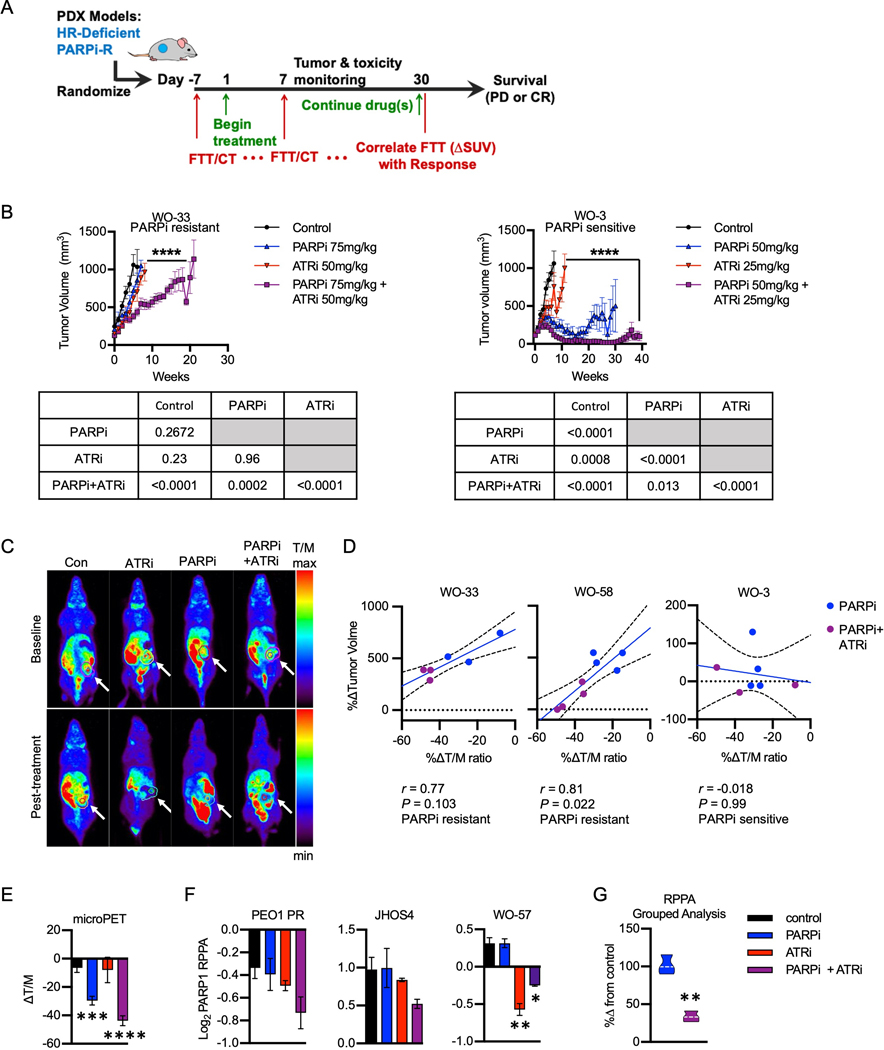
Purpose: PARP inhibitors have become the standard-of-care treatment for homologous recombination deficient (HRD) high-grade serous ovarian cancer (HGSOC). However, not all HRD tumors respond to PARPi. Biomarkers to predict response are needed. [18F]FluorThanatrace ([18F]FTT) is a PARPi-analog PET radiotracer that noninvasively measures PARP-1 expression. Herein, we evaluate [18F]FTT as a biomarker to predict response to PARPi in patient-derived xenograft (PDX) models and subjects with HRD HGSOC.

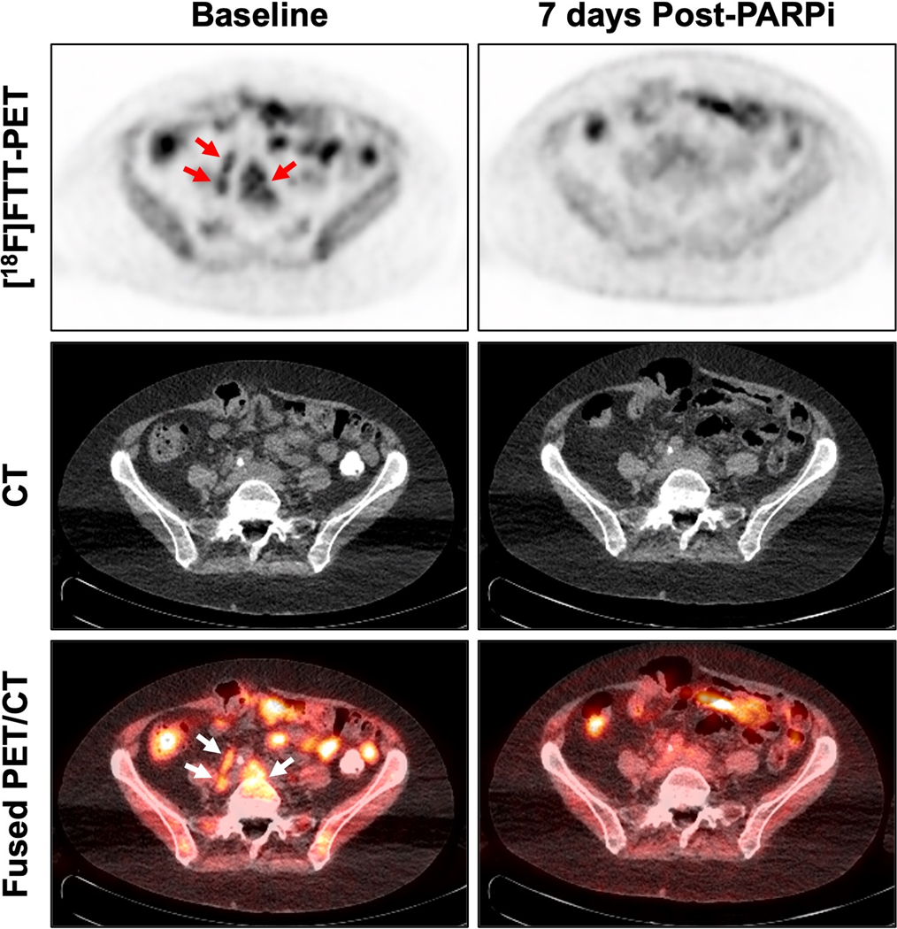
Experimental design: In PDX models, [18F]FTT-PET was performed before and after PARPi (olaparib), ataxia-telangiectasia inhibitor (ATRi), or both (PARPi-ATRi). Changes in [18F]FTT were correlated with tumor volume changes. Subjects were imaged with [18F]FTT-PET at baseline and after ∼1 week of PARPi. Changes in [18F]FTT-PET uptake were compared with changes in tumor size (RECISTv1.1), CA-125, and progression-free survival (PFS).

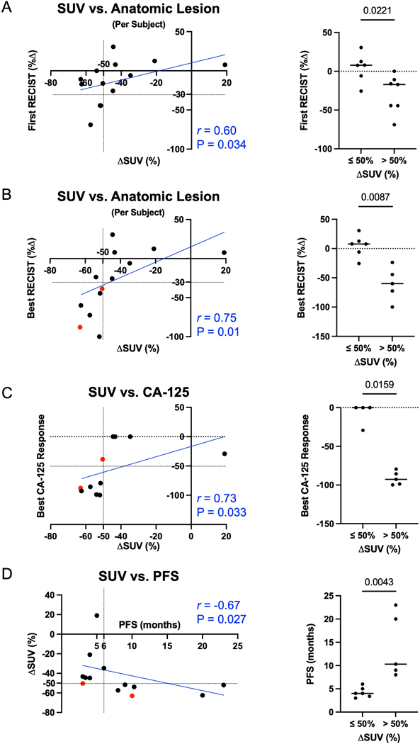
Results: A decrease in [18F]FTT tumor uptake after PARPi correlated with response to PARPi, or PARPi-ATRi treatment in PARPi-resistant PDX models (r = 0.77-0.81). In subjects (n = 11), percent difference in [18F]FTT-PET after ∼7 days of PARPi compared with baseline correlated with best RECIST response (P = 0.01), best CA-125 response (P = 0.033), and PFS (P = 0.027). All subjects with >50% reduction in [18F]FTT uptake had >6-month PFS and >50% reduction in CA-125. Utilizing only baseline [18F]FTT uptake did not predict such responses.

Conclusions: The decline in [18F]FTT uptake shortly after PARPi initiation provides a measure of drug-target engagement and shows promise as a biomarker to guide PARPi therapies in this pilot study. These results support additional preclinical mechanistic and clinical studies in subjects receiving PARPi ± combination therapy. See related commentary by Liu and Zamarin, p. 1384.


Conflict of interest statement

Competing Interests: Drs. Mach and Mankoff are owners in Trevarx Biomedical, a company with a license to [18F]FTT, the radiopharmaceutical investigated in this study. The remaining authors declare no potential conflicts of interest.

Figures

Figure 1:
Figure 1:. Pre-clinical imaging and therapy in PDX ovarian cancer models.
A, Schema of preclinical in vivo efficacy and [18F]FTT-microPET imaging studies. Pre-clinical HRD or PARPi-resistant PDX models were evaluated for PARPi+ATRi efficacy and the change in [18F]FTT uptake before and after therapy. For the imaging cohort, mice were assessed for uptake of [18F]FTT before drug treatment and 1 week after randomization then normalized by a tissue to mass ratio (△SUV). △SUV was correlated to response measured by tumor volume. CR, Complete response; PD, Progressive Disease. B, In vivo efficacy studies testing PARPi and ATRi monotherapy or combination in PARPi-resistant WO-33 (left) and PARPi-sensitive WO-3 (right) PDX models. Tumor volume data over time are shown and represented as mean ± SEM (n = 7–20 per group), and statistical analysis is shown. C-E, PARPi-1 expression as determined by [18F]FTT PET/CT imaging. C, Representative [18F]FTT-PET imaging pre- and post-PARPi (top and bottom, respectively). D, Spearman correlation of percent change of tumor volume from baseline compared to percent change of Tumor/Muscle (T/M) ratio for mice treated with PARPi or PARPi+ATRi for 1 week across three PDX models: WO-33 and WO-58 (PARPi-resistant) and WO-3. E, Change in T/M ratio across all three models (Control, n = 16; ATRi, n = 6; PARPi, n = 11; PARPi+ATRi, n = 10). F, RPPA analysis for PARP-1 expression in PEO1 PR (BRCA2MUT, PARPi-resistant), JHOS4 (BRCA1MUT, PARPi-sensitive) cells and WO-57 (gBRCA1REV, PARPi-resistant) PDX tumors. Data are represented as mean ± SD and analyzed by one-way ANOVA. Combined analysis of PARP-1 expression post-PARPi versus post-PARPi-ATRi across all cell lines are shown and analyzed by two-tailed T-test (G).
Figure 2:
Figure 2:. Clinical trial design and progression-free survival on treatment.
A, Schematic of the clinical trial design. PD, Progressive Disease. B, Swimmer plot for progression-free survival on treatment. Sixteen subjects were evaluated by RECIST 1.1. All subjects received a PARPi unless indicated for PARPi+ATRi (CAPRI) combination. Somatic and germline mutations of interest and HRD tumor status are indicated. Arrows indicate subjects still on trial and undergoing treatment at the time of analysis.
Figure 3:
Figure 3:. Representative clinical imaging of [18F]FTT-PET/CT in PARPi a responder and non-responder.
[18F]FTT-PET and fused PET/CT images are shown for a PARPi responder (subject 10, BRCA1MUT; PFS = 20 months; A) and non-responder (subject 11, BRCA1/2WT, HRD positive; PFS = 5 months; B). A, Pre-PARPi [18F]FTT-PET/CT demonstrates [18F]FTT uptake in a left external iliac lymph node (SUVMAX of 5.2). Imaging performed 8 days later, and after starting olaparib, demonstrates decreased [18F]FTT uptake (SUVMAX of 2), representing successful receptor targeting of the PARPi. B, Pre-PARPi [18F]FTT-PET/CT demonstrate no uptake above background in a right peritoneal implant (SUVMAX of 1.7) suggestive of absent PARP-1 expression and PARPi drug target, thus no therapeutic effect of PARPi. Imaging performed 6 days later, and after starting olaparib, demonstrates no decrease in [18F]FTT uptake (SUVMAX of 2). As expected, this subject had a relatively short PFS.
Figure 4:
Figure 4:. [18F]FTT imaging to predict PARPi response.
A-B, Spearman correlation [18F]FTT △SUV versus the change of lesion size per subject (1–4 target lesions averaged) from anatomic baseline to first RECIST response (obtained 57–158 days after baseline by CT) (A) or best RECIST response (obtained 57–351 days after baseline by CT) (B). C, Spearman correlation △SUV versus the best CA-125 response. D, Spearman correlation of change in [18F]FTT SUV from baseline to after starting PARPi therapy (△SUV) versus PFS. A-D, Two-tailed T-test of PFS, CA-125 response, and RECIST response at ≤ 50% and > 50% reduction are shown. When appropriate, two subjects (shown in red) were excluded from statistical analyses (Spearman and T-test) as these subjects are currently on trial.
Figure 5:
Figure 5:. [18F]FTT-PET on subject previously treated with PARPi.
Subject 16 previously progressed on a PARPi nearly 6 years prior to enrolling in the CAPRI trial. [18F]FTT-PET uptake is seen in retroperitoneal lymph nodes at baseline (left), with abrogation of signal on [18F]FTT-PET obtained 7 days after initiating Olaparib (right). Findings indicate PARPi drug target expression with subsequent therapeutic effect. This subject remains on trial with an encouraging early response.

Comment in

Comment on

References

    1. Moore K, Colombo N, Scambia G, Kim BG, Oaknin A, Friedlander M, et al. Maintenance Olaparib in Patients with Newly Diagnosed Advanced Ovarian Cancer. N Engl J Med 2018;379:2495–505 - PubMed
    1. Poveda A, Floquet A, Ledermann JA, Asher R, Penson RT, Oza AM, et al. Olaparib tablets as maintenance therapy in patients with platinum-sensitive relapsed ovarian cancer and a BRCA1/2 mutation (SOLO2/ENGOT-Ov21): a final analysis of a double-blind, randomised, placebo-controlled, phase 3 trial. Lancet Oncol 2021;22:620–31 - PubMed
    1. Penson RT, Valencia RV, Cibula D, Colombo N, Leath CA 3rd, Bidzinski M, et al. Olaparib Versus Nonplatinum Chemotherapy in Patients With Platinum-Sensitive Relapsed Ovarian Cancer and a Germline BRCA1/2 Mutation (SOLO3): A Randomized Phase III Trial. J Clin Oncol 2020;38:1164–74 - PMC - PubMed
    1. Oza AM, Lorusso D, Aghajanian C, Oaknin A, Dean A, Colombo N, et al. Patient-Centered Outcomes in ARIEL3, a Phase III, Randomized, Placebo-Controlled Trial of Rucaparib Maintenance Treatment in Patients With Recurrent Ovarian Carcinoma. J Clin Oncol 2020;38:3494–505 - PMC - PubMed
    1. Swisher EM, Lin KK, Oza AM, Scott CL, Giordano H, Sun J, et al. Rucaparib in relapsed, platinum-sensitive high-grade ovarian carcinoma (ARIEL2 Part 1): an international, multicentre, open-label, phase 2 trial. Lancet Oncol 2017;18:75–87 - PubMed

Publication types

MeSH terms

Substances