. 2022 Dec 6;119(49):e2214278119.
doi: 10.1073/pnas.2214278119. Epub 2022 Nov 29.

Tumor-targeted delivery of a STING agonist improvescancer immunotherapy

Affiliations

Tumor-targeted delivery of a STING agonist improvescancer immunotherapy

You-Tong Wu et al. Proc Natl Acad Sci U S A. .

Abstract

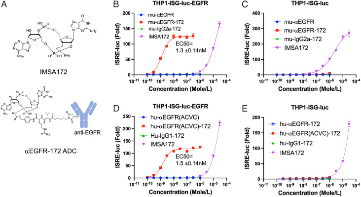
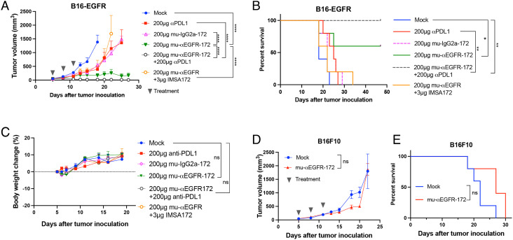
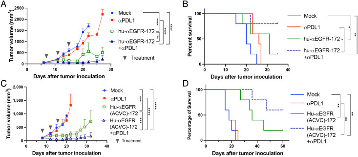
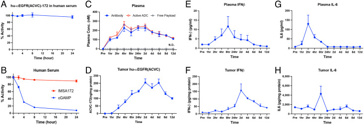
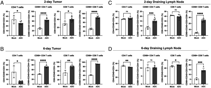
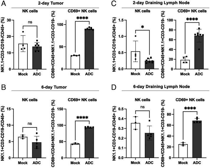
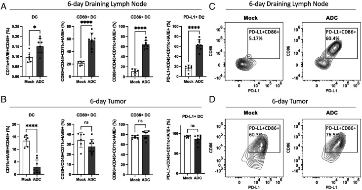
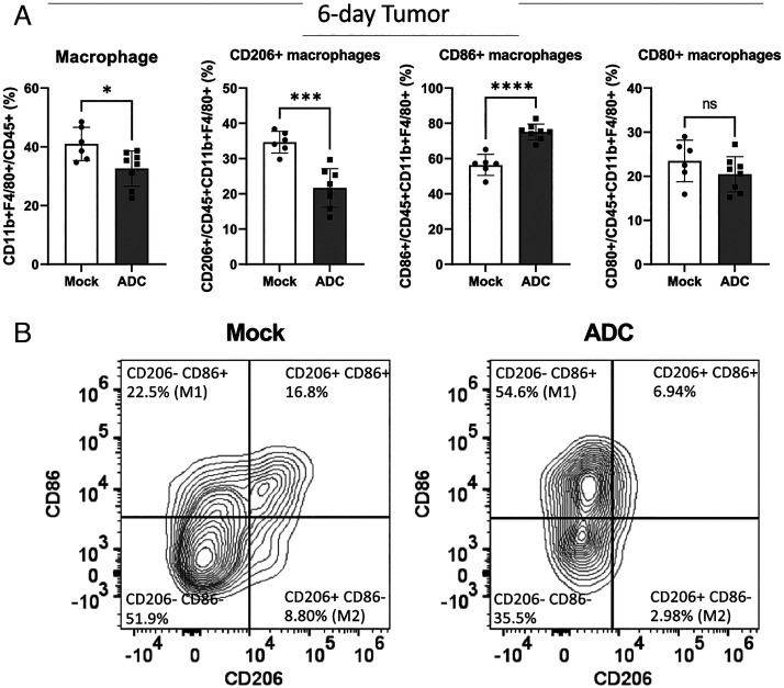
The cGAS-STING pathway is essential for immune defense against microbial pathogens and malignant cells; as such, STING is an attractive target for cancer immunotherapy. However, systemic administration of STING agonists poses safety issues while intratumoral injection is limited by tumor accessibility. Here, we generated antibody-drug conjugates (ADCs) by conjugating a STING agonist through a cleavable linker to antibodies targeting tumor cells. Systemic administration of these ADCs was well tolerated and exhibited potent antitumor efficacy in syngeneic mouse tumor models. The STING ADC further synergized with an anti-PD-L1 antibody to achieve superior antitumor efficacy. The STING ADC promoted multiple aspects of innate and adaptive antitumor immune responses, including activation of dendritic cells, T cells, natural killer cells and natural killer T cells, as well as promotion of M2 to M1 polarization of tumor-associated macrophages. These results provided the proof of concept for clinical development of the STING ADCs.

Keywords: ADC; STING; cGAS; cancer; tumor immunity.


Conflict of interest statement

The authors declare a competing interest. The authors have organizational affiliations to disclose, Z.J.C. is a scientific collaborator with Pfizer and ImmuneSensor Therapeutics, and a scientific advisor for Brii Biosciences, Genor Biopharma and Drug Farm. C.C. is a scientific advisor for ImmuneSensor Therapeutics., Z.J.C. has stock ownership in Brii Biosciences. H.S., L.S., C.C., and Z.J.C. are inventors of US patent 10,336,786. Q.W., H.S., L.S, C.C., and Z.J.C. are inventors of US patent 10,519,188. Y.-t.W., Q.W., H.S., J.Q, L.S., C.C., and Z.J.C. are inventors of US patent 11,033,635. Z.J.C. received research support from Pfizer and ImmuneSensor Therapeutics.

Figures

Fig. 1.
Fig. 1.
Chemical structures and immunostimulatory activities of IMSA172 and its ADCs in cells. (A) Diagrams of chemical structures of IMSA172 and anti-EGFR-172 ADC used in this study. (BE) Serial dilutions of IMSA172 or its conjugates with the indicated antibodies (ADCs) were incubated with THP1-ISG-Luc (C and E) or this cell line stably expressing human EGFR (THP1-ISG-luc-EGFR; B and D) for 16 h, and the interferon response was measured by luciferase assay (see Materials and Methods). mu-αEGFR-172: mouse anti-human EGFR conjugated to IMSA172; mu-IgG2a-172: mouse IgG2a conjugated to IMSA172; mu-αEGFR: unconjugated mouse anti-human EGFR; Hu-αEGFR: human anti-human EGFR (same as cetuximab); hu-αEGFR-172: human anti-human EGFR antibody conjugated to IMSA172; hu-IgG-172: human IgG1 conjugated to IMSA172; hu-αEGFR(ACVC): hu-αEGFR-172 with A114C and V205C mutations for site-specific conjugation; hu-αEGFR(ACVC)-172: hu-αEGFR(ACVC) conjugated to IMSA172 via four specific cysteines. EC50 values were derived using the curve fitting function in Graphpad. Data are representative of at least three independent experiments.
Fig. 2.
Fig. 2.
Antitumor efficacy of mu-αEGFR-172. (A and B) Groups of C57BL/6 mice (n = 5) with established B16F10 tumors expressing human EGFR were treated with the indicated ADCs or PD-L1 antibody or both via intraperitoneal injection. In one group, mice were treated with PBS as mock control. In another group, mu-αEGFR was mixed with IMSA-172 without conjugation. Tumor growth (A) and mouse survival (B) were monitored. (C) Body weight change (%) of mice after treatments. (D and E) Groups of C57BL/6 mice (n = 5) with B16F10 (not expressing EGFR) tumors were treated with mu-αEGFR-172 via intraperitoneal injection. Tumor growth (D) and mouse survival (E) were monitored. Tumor growth and body weights are represented as mean ± SEM, ns: not significant, *P < 0.05, **P < 0.01, ***P < 0.001, ****P < 0.0001 (two-way ANOVA). Log rank (Mantel-Cox) test was used for survival data, ns: not significant, *P < 0.0332, **P < 0.0021, ***P < 0.0002, ****P < 0.0001.
Fig. 3.
Fig. 3.
Antitumor efficacy of human anti-EGFR-172 ADCs. (A and B) Groups of C57BL/6 mice (n = 5) with established B16F10-EGFR tumors were treated intraperitoneally with hu-αEGFR-172 or anti-PD-L1 antibody or both as indicated; tumor growth (A) and mouse survival (B) were monitored. (C and D) Similar to (A) and (B) except that the hu-αEGFR(ACVC)-172 ADC was used. Tumor growth data are presented as mean ± SEM, ns: not significant, *P < 0.05, **P < 0.01, ***< 0.001, ****P < 0.0001 (two-way ANOVA). Log rank (Mantel-Cox) test was used for survival data, ns: not significant, *P < 0.0332, **P < 0.0021, ***P < 0.0002, ****P < 0.0001.
Fig. 4.
Fig. 4.
PK and PD of hu-αEGFR(ACVC)-172. (A) The stability of hu-αEGFR(ACVC)-172 in human sera. hu-αEGFR(ACVC)-172 was incubated with human sera over a time course as indicated. The remaining activity was measured in THP1-ISG-luc-EGFR reporter cells and presented as percentage of untreated samples. (B) Similar to (A) except that the payload IMSA172 and cGAMP were tested in the human sera. (CH) PK and PD of hu-αEGFR(ACVC)-172 in mice. Mice-bearing B16-EGFR tumors were injected with 100 μg of hu-αEGFR(ACVC)-172 intraperitoneally and three mice were sacrificed at each timepoint to collect blood and tumors. ADC and cytokine levels were measured from plasma and tumor homogenates using ELISA. The activity of the ADC and free payload (IMSA172) was measured in THP1-ISG-luc-EGFR cells. Data are presented as mean ± SEM. N.D.: non-detectable.
Fig. 5.
Fig. 5.
ADC treatment activated T cells in tumors and draining lymph nodes and increased CD8/CD4 ratio in tumors. B16F10-EGFR cells were inoculated into C57BL/6 mice subcutaneously. (A and C) On day 7, 200 μg of hu-αEGFR(ACVC)-172 (ADC) or PBS (mock) was administrated intraperitoneally. Two days later, cells from tumors (A) and draining lymph nodes (C) were analyzed by flow cytometry using antibodies against the indicated T cell markers. Mock: n = 4, ADC: n = 7. (B and D) The tumor-bearing mice were treated with ADC or mock on day 7 and day 10. Six days after the first treatment, cells from tumors (B) and draining lymph nodes (D) were analyzed by flow cytometry. Mock: n = 3, ADC: n = 5. Data are shown as mean ± SD and individual values. ns: not significant, *P < 0.05, **P < 0.01, ***P < 0.001, ****P < 0.0001 (student’s t  test).
Fig. 6.
Fig. 6.
ADC treatment activated NKcells in both tumors and draining lymph nodes. Experiments were performed as described in Fig. 5, except that the flow cytometry analysis was focused on NK cells (NK1.1+CD3−CD19−) 2 days (A) or 6 days (B) in tumor and 2 days (C) and 6 days (D) in draining lymph nodes after ADC treatments. ns: not significant, *P < 0.05, ****P < 0.0001 (student’s test).
Fig. 7.
Fig. 7.
ADC treatment activated DCs and enhanced their migration from tumors to draining lymph nodes. Experiments were performed similarly to Fig. 5 B and D. Six days after the first treatment, cells from draining lymph nodes (A) and tumors (B) were analyzed by flow cytometry focusing on DCs (CD11c+IA/IE+). Data are shown as the mean ± SD and individual values. Mock: n = 6, ADC: n = 8. ns: not significant, *< 0.05, ****< 0.0001 (student’s test). (C and D) Representative FACS plots of CD86 and PD-L1 expression levels on DCs from draining lymph nodes (C) and tumors (D).
Fig. 8.
Fig. 8.
ADC treatment polarized M2-like macrophages to M1-like macrophages in tumors. (A) Experiments were performed as described in Fig. 7 except that the flow cytometry analysis was focused on tumor-associated macrophages (CD11b+F4/80+). ns: not significant, *P < 0.05, ***P < 0.001, ****P < 0.0001 (student’s test). (B) Representative FACS plots of CD86 and CD206 expression levels in tumor-associated macrophages.

References

    1. O’Donnell J. S., Teng M. W. L., Smyth M. J., Cancer immunoediting and resistance to T cell-based immunotherapy. Nat. Rev. Clin. Oncol. 16, 151–167 (2019). - PubMed
    1. Bagchi S., Yuan R., Engleman E. G., Immune checkpoint inhibitors for the treatment of cancer: Clinical impact and mechanisms of response and resistance. Annu. Rev. Pathol. 16, 223–249 (2021). - PubMed
    1. Li T., Chen Z. J., The cGAS-cGAMP-STING pathway connects DNA damage to inflammation, senescence, and cancer. J. Exp. Med. 215, 1287–1299 (2018). - PMC - PubMed
    1. Ablasser A., Chen Z. J., cGAS in action: Expanding roles in immunity and inflammation. Science 363, eaat8657 (2019). - PubMed
    1. Sun L., Wu J., Du F., Chen X., Chen Z. J., Cyclic GMP-AMP synthase is a cytosolic DNA sensor that activates the type I interferon pathway. Science 339, 786–791 (2013). - PMC - PubMed

Publication types