. 2022 Dec 6;119(49):e2214150119.
doi: 10.1073/pnas.2214150119. Epub 2022 Nov 28.

The mud-dwelling clam Meretrix petechialis secretes endogenously synthesized erythromycin

Affiliations

The mud-dwelling clam Meretrix petechialis secretes endogenously synthesized erythromycin

Xin Yue et al. Proc Natl Acad Sci U S A. .

Abstract

Although lacking an adaptive immune system and often living in habitats with dense and diverse bacterial populations, marine invertebrates thrive in the presence of potentially challenging microbial pathogens. However, the mechanisms underlying this resistance remain largely unexplored and promise to reveal novel strategies of microbial resistance. Here, we provide evidence that a mud-dwelling clam, Meretrix petechialis, synthesizes, stores, and secretes the antibiotic erythromycin. Liquid chromatography coupled with mass spectrometry, immunocytochemistry, fluorescence in situ hybridization, RNA interference, and enzyme-linked immunosorbent assay revealed that this potent macrolide antimicrobial, thought to be synthesized only by microorganisms, is produced by specific mucus-rich cells beneath the clam's mantle epithelium, which interfaces directly with the bacteria-rich environment. The antibacterial activity was confirmed by bacteriostatic assay. Genetic, ontogenetic, phylogenetic and genomic evidence, including genotypic segregation ratios in a family of full siblings, gene expression in clam larvae, phylogenetic tree, and synteny conservation in the related genome region further revealed that the genes responsible for erythromycin production are of animal origin. The detection of this antibiotic in another clam species showed that the production of this macrolide is not exclusive to M. petechialis and may be a common strategy among marine invertebrates. The finding of erythromycin production by a marine invertebrate offers a striking example of convergent evolution in secondary metabolite synthesis between the animal and bacterial domains. These findings open the possibility of engineering-animal tissues for the localized production of an antibacterial secondary metabolite.

Keywords: antibacterial strategy; clam; macrolide antibiotics; mantle mucus.


Conflict of interest statement

The authors declare no competing interest.

Figures

Fig. 1.
Fig. 1.
The coastal, mud-dwelling clam M. petechialis carries Mp-erythromycin in its mantle. (A) The living environment and anatomical structure of the clam. Left: a diagram of the subsurface habitat of a live clam, with siphons extruded to draw water over the tissues. The mantle margins, which are inside of the shell’s edge, is one of the principal epithelial tissues exposed to the environment. Right: a diagram showing the bipartite mantle (left and right), which protect the other soft body parts of the clam. (B) HPLC/QQQ MS of an erythromycin standard (in red) and the clam mantle extracts (in blue). RT is the retention time of the target peak during HPLC. SRM refers to selected reaction monitoring MS mode. For MS analysis, the parent ion m/z = 734 Da, production m/z = 576 Da, and collision energy were optimized to 18 V. (C) The Left diagram shows ICC using an anti-erythromycin antibody (green fluorescence) on the transversal section of the mantle margin of M. petechialis. The nontargeted tissues were stained using Evans blue dye (purple fluorescence). The Upper Right illustration shows a higher magnification image (the white box in the Left diagram) that revealed Mp-erythromycin was secreted outside the mantle epithelia (the white circle). In the Lower Right diagram, hematoxylin and eosin staining revealed the location of Mp-erythromycin to be confined to a cluster of ovoid basophilia structures along the epithelia of the mantle margin (arrows).
Fig. 2.
Fig. 2.
Mp-erythromycin with antimicrobial activity is stored in the mantle mucus cells and can be secreted outside. (A) AB-PAS staining in the mantle margin section with Mp-erythromycin rich structures (black arrows). The anatomical diagram of the mantle is shown at the Top Left corner. Ep refers to epithelia. (B) Upper illustration shows the ultrastructure (by TEM) of a similar zone to that in the dashed red square of panel A. This figure shows that Mp-erythromycin-rich structures contain tightly packed vesicles (white circle); occasional nuclei (N) are seen as well. The Lower Left figure shows by TEM the ultrastructure of a similar zone to that in the dashed blue square of panel A. It is seen that the vesicles can be secreted through the epithelia (Ep) to the outside. The Lower Right figure presents a schematic illustration of Mp-erythromycin-rich structures. (C) Antibacterial effects of mantle mucus to E. gallinarum and V. parahaemolyticus. 1, the filter paper was soaked with mantle mucus. 2, the filter paper was soaked with fluid from inside the clam mantle cavity (negative control). The bottom filter paper was soaked with 15 μg erythromycin (positive control).
Fig. 3.
Fig. 3.
MpES expression analyses and involvement of MpES in erythromycin biosynthesis. (A) Left: Methyl green-Pyronin staining of the mantle margin confirms that numerous RNA molecules (black arrow) are present in the MpES-mRNA positive position. The green box indicates the mantle margin. Right: FISH targeted to MpES mRNA on the mantle margin section. MpES antisense: FISH using the probe with the reverse complementary sequence of MpES mRNA. MpES sense: FISH using the probe with the same sequence of MpES mRNA. White arrows: MpES mRNA. (B) Changes in the relative mRNA-expression of MpES (Left diagram) and erythromycin level (Right diagram) in the mantle during an RNAi experiment. Control: the clams without injection served as the blank control. EGFP: the clams injected with double-stranded RNA of enhanced green fluorescent protein served as the negative control. ES: the clams injected with dsES; LN refers to the natural logarithm. Horizontal bars represent mean values. Different letters mean that there is a significant difference between values using a nonparametric Mann–Whitney U test (P < 0.05). (C) MpES DNA or cDNA appearance during the larval development of the clam. Upper: stages examined; Lower: presence in the genome (Left) and transcriptome (Right). hpf, hours post fertilization; dpf, days post fertilization.
Fig. 4.
Fig. 4.
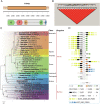
MpES structure and its origin as determined by genetic, phylogenetic, and gene homolog analyses. (A) The domains in MpES as analyzed using the antiSMASH database. T1PKS, type I PKS; KS, ketoacyl synthase; AT, acyltransferase; DH, dehydratase; KR, ketoreductase; ACP, acyl carrier protein. (B) LD structure among the 17 MpES SNPs in a clam family, analyzed by Haploview 4.2 software. The deeper the box color is, the stronger the correlation between SNPs. Red boxes represent the value of correlation coefficient D′ is 1. (C) Maximum likelihood phylogenetic tree. NCBI ID no. for each homolog follows the species name. MpES (in red box) is placed within the animal clade not the bacteria clade. (D) Flanking genes of the homologs according to the genomic graphic. Arrows represent individual genes and their transcriptional orientations. X represents the gene encoding the target homolog in the left tree. EIF, eukaryotic translation initiation factor; CYGB2, cytoglobin 2; ADH, alcohol dehydrogenase; PTCHD3, patched domain containing protein 3; TLR13, Toll-like receptor 13; Pol, DNA polymerase; NEK7, serine/threonine-protein kinase Nek7; YME1L1, ATP-dependent zinc metalloprotease YME1L1; ELOF1, transcription elongation factor 1 homolog; RPL37, 60S ribosomal protein L37; CEP83, centrosomal protein of 83 kDa; RDH12, retinol dehydrogenase 12; ASCC3, activating signal cointegrator 1 complex subunit 3; SIM1, SIM bHLH transcription factor 1; CYB5R4, cytochrome b5 reductase 4; MKX, mohawk homeobox; MASTL, microtubule-associated serine/threonine-protein kinase-like; ACBD5, acyl-CoA-binding domain-containing protein 5; ABI1, abI interactor 1; PDSS1, prenyl (decaprenyl) diphosophate synthase subunit 1; T represents no more flanking genes due to genome assembly gaps.

References

    1. Whitman W. B., Coleman D. C., Wiebe W. J., Prokaryotes: The unseen majority. Proc. Natl. Acad. Sci. U.S.A. 95, 6578–6583 (1998). - PMC - PubMed
    1. Philipp E. E., Abele D., Masters of longevity: Lessons from long-lived bivalves–a mini-review. Gerontology 56, 55–65 (2010). - PubMed
    1. Probandt D., Eickhorst T., Ellrott A., Amann R., Knittel K., Microbial life on a sand grain: From bulk sediment to single grains. ISME J. 12, 623–633 (2018). - PMC - PubMed
    1. Manahan D. T., Amino acid fluxes to and from seawater in axenic veliger larvae of a bivalve (Crassostrea gigas). Mar. Ecol. Prog. Ser. 53, 247–255 (1989).
    1. Stephens G. C., Epidermal amino acid transport in marine invertebrates. Biochim. Biophys. Acta 947, 113–138 (1988). - PubMed

Publication types

LinkOut - more resources