Review
. 2023 Jan;18(1):103-113.
doi: 10.1016/j.cpet.2022.09.006.

Brain PET Imaging: Approach to Cognitive Impairment and Dementia

Affiliations
Review

Brain PET Imaging: Approach to Cognitive Impairment and Dementia

Matthew Spano et al. PET Clin. 2023 Jan.

Erratum in

  • Erratum.
    [No authors listed] [No authors listed] PET Clin. 2023 Apr;18(2):xi. doi: 10.1016/j.cpet.2023.01.002. PET Clin. 2023. PMID: 36858749 No abstract available.

Abstract

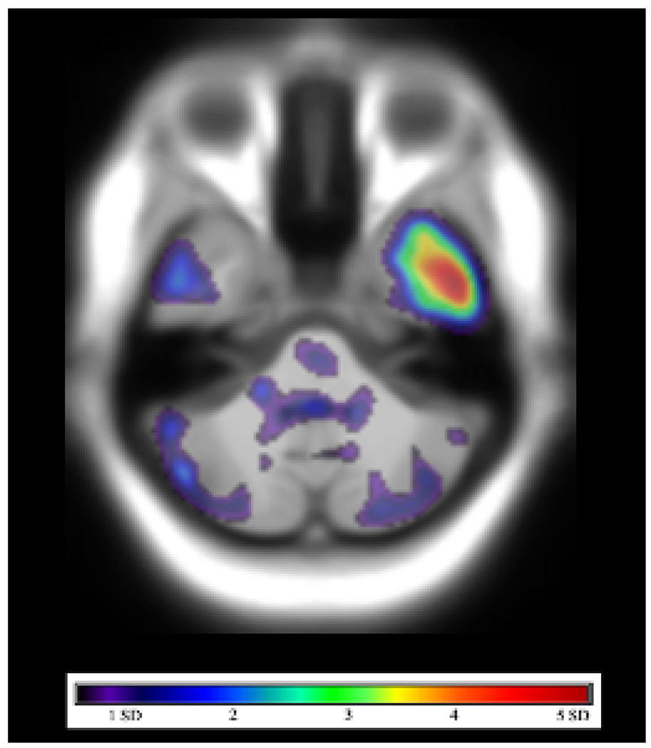
Alzheimer disease (AD) is the most common cause of dementia, accounting for 50% to 60% of cases and affecting nearly 6 million people in the United States. Definitive diagnosis requires either antemortem brain biopsy or postmortem autopsy. However, clinical neuroimaging has been playing a greater role in the diagnosis and management of AD, and several PET tracers approach the sensitivity of tissue diagnosis in identifying AD pathologic condition. This review will focus on the utility of PET imaging in the setting of cognitive impairment, with an emphasis on its role in the diagnosis of AD.

Keywords: Alzheimer disease; Amyloid; Dementia; Neurodegeneration; Positron emission tomography; Tau.


Figures

Fig. 1.
Fig. 1.
Coronal T2 FLAIR image in a patient with normal pressure hydrocephalus. The lateral ventricles are dilated out of proportion to sulcal prominence, there is an acute callosal angle (*), and there is widening of the Sylvian fissures (arrow).
Fig. 2.
Fig. 2.
Typical workflow when comparing a patient’s FDG-PET scan with a normative database. Steps (A) include fusion of the patient’s PET scan with a structural image (B), fusion of the patient’s structural image to a template image in the same space as the normative images (C), and comparison of FDG avidity between the patient and the database across all voxels and regions-of-interest (D). Three-dimensional stereotactic surface projection images (E) with colors representing the patient’s FDG avidity in standard deviations from the normative database.
Fig. 3.
Fig. 3.
Three-dimensional stereotactic surface projection images in a patient with Alzheimer’s disease. The blue color denotes brain regions in which the FDG avidity is at least three standard deviations lower than similarly aged healthy controls from a normative database. The distribution of decreased FDG avidity in the temporal and parietal lobes, particularly the precuneus (arrows), is typical of Alzheimer’s disease.
Fig. 4.
Fig. 4.
Axial FDG-PET image (A) in a patient with amnestic mild cognitive impairment demonstrates decreased FDG avidity in the parietal lobes bilaterally (arrows). Three-dimensional stereotactic surface projection images (B) demonstrate decreased FDG avidity in the precuneus, posterior cingulate gyrus, and temporal and parietal lobes bilaterally, suggestive of underlying AD pathology.
Fig. 5.
Fig. 5.
Axial T1-weighted MR image (A) demonstrates marked bilateral frontal lobe atrophy in a patient with behavioral variant frontotemporal dementia (bvFTD). Three-dimensional stereotactic surface projection images (B) demonstrate decreased FDG avidity in frontal and temporal lobes bilaterally, a distribution typical of bvFTD.
Fig. 6.
Fig. 6.
Three-dimensional stereotactic surface projection images demonstrate decreased FDG avidity in parietal and temporal lobes bilaterally, more pronounced on the left. In addition, there is decreased FDG avidity in the left occipital lobe. The temporoparietal distribution and asymmetric involvement of the left occipital lobe is compatible with posterior cortical atrophy, the visual variant of Alzheimer’s.
Fig. 7.
Fig. 7.
Axial FDG-PET image (A) in a patient with Lewy body dementia showing decreased FDG avidity in the temporal and occipital lobes bilaterally, although with preserved FDG avidity in the medial occipital lobes (arrows). Three-dimensional stereotactic surface projection images (B) demonstrate decreased FDG avidity in the bilateral temporal and occipital lobes and left parietal lobe.
Fig. 8.
Fig. 8.
Axial susceptibility-weighted image (A) shows foci of microhemorrhage, suggestive of amyloid angiopathy. Axial FDG-PET images (B,C) demonstrate decreased FDG avidity in the left occipital lobe in the region of the microhemorrhages.
Fig. 9.
Fig. 9.
Axial FDG-PET image fused to the CT of the head (A) demonstrating asymmetrically decreased FDG avidity in the left temporal lobe (arrow). Three-dimensional stereotactic surface projection images (B) demonstrate asymmetrically decreased FDG avidity in the left parietal and temporal lobes, suggestive of logopenic variant primary progressive aphasia, the language variant of Alzheimer’s.
Fig. 10.
Fig. 10.
Coronal T1-weighted MRI (A), coronal FDG-PET image fused to the MRI (B), and three-dimensional stereotactic surface projection images demonstrating marked bilateral temporal lobe atrophy with associated decreased FDG avidity, compatible with semantic variant primary progressive aphasia.
Fig. 11.
Fig. 11.
Axial image from an amyloid PET scan using the tracer, Pittsburgh Compound B, fused to the MRI of the head (A) demonstrating beta-amyloid deposition in the bilateral frontal lobes and right greater than left parietal lobes. Axial image from a tau PET scan using the tracer, MK6240, fused to the MRI of the head (B) demonstrating tau deposition in the right uncus.
Fig. 12.
Fig. 12.
Axial image from an amyloid PET scan using the tracer, 18F-florbetaben, demonstrating diffuse beta-amyloid deposition (A). Axial T2 FLAIR image (B) fused to the amyloid PET image (C) confirms that the amyloid tracer uptake is in both cortical gray matter and white matter.
Fig. 13.
Fig. 13.
[18F]-flortaucipir PET-MRI in a patient with mild cognitive impairment. SUVR in a combined region-of-interest, including the entorhinal cortex, amygdala, and inferior temporal cortex, was greater than 1.22 relative to cerebellar cortex, compatible with increased tau deposition.

References

    1. Alzheimer’s Association. 2019 Alzheimer’s disease facts and figures. Accessed March 20, 2022. https://www.alz.org/media/documents/alzheimers-facts-and-figures-2019-r.pdf.
    1. Jack CR Jr., Bennett DA, Blennow K, et al. NIA-AA Research Framework: Toward a biological definition of Alzheimer’s disease. Alzheimers Dement 2018;14:535–562. - PMC - PubMed
    1. Thal DR, Rub U, Orantes M, Braak H. Phases of A beta-deposition in the human brain and its relevance for the development of AD. Neurology 2002;58:1791–1800. - PubMed
    1. Braak H, Braak E. Evolution of the neuropathology of Alzheimer’s disease. Acta Neurol Scand Suppl 1996;165:3–12. - PubMed
    1. Kumar ADS. Neuropathology and therapeutic management of Alzheimer’s disease - An update. Drugs Future 2008;33:433.

MeSH terms