. 2023 Jun;17(3):343-349.
doi: 10.14444/8412. Epub 2022 Nov 28.

Endoscopic Techniques for Spinal Oncology: A Systematic Literature Review

Affiliations

Endoscopic Techniques for Spinal Oncology: A Systematic Literature Review

Rohaid Ali et al. Int J Spine Surg. 2023 Jun.

Abstract

Background: Due to its ultraminimally invasive nature, endoscopic spinal surgery is an attractive tool in spinal oncologic care. To date, there has been no comprehensive review of this topic. The authors therefore present a thorough search of the medical literature on endoscopic techniques for spinal oncology.

Methods: A systematic review using PubMed was conducted using the following keywords: endoscopic spine surgery, spinal oncology, and spinal tumors.

Results: Collectively, 19 cases described endoscopic spine surgery for spinal oncologic care. Endoscopic spine surgery has been employed for the care of patients with spinal tumors under the following 4 circumstances: (1) to obtain a reliable tissue diagnosis; (2) to serve as an adjunct during traditional open surgery; (3) to achieve targeted debulking; or (4) to perform definitive resection. These cases employing endoscopic techniques highlight the versatility of this approach and its utility when applied to the right patient and with an experienced surgeon.

Conclusions: Our systematic review suggests that, given the right patient and an experienced surgeon, endoscopic spine surgery should be considered in the armamentarium for spinal oncologic care for both staging and definitive resection.

Clinical relevance: This systematic literature review showed that endoscopic techniques have been successfully applied across the spectrum of care in spinal oncology, from diagnosis to definitive treatment.

Keywords: endoscopic decompression; metastatic lesion; oncologic spine care.


Conflict of interest statement

Declaration of Conflicting Interests : The authors report no conflicts of interest in this work.

Figures
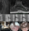
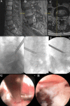
Figure 1
Figure 1
Images from a pediatric case of a ventral epidural thoracic Ewing-like tumor. Panels A and B show sagittal and axial T1 postcontrast magnetic resonance imaging of the thoracic spine with evidence of tumor recurrence in the ventral epidural region. Panels C to F show intraoperative fluoroscopic images. Panel G is an image from the operating room setup. Panels H and I are intraoperative images of the tumor debulking. Source: Figure reprinted with permission from Telfeian AE, Choi DB, Aghion DM. Transforaminal endoscopic surgery under local analgesia for ventral epidural thoracic spinal tumor: case report. Clin Neurol Neurosurg. 2015;134:1–3. doi:10.1016/j.clineuro.2015.03.022.
Figure 2
Figure 2
Images from the case of a 75-year-old patient with metastatic nonsmall cell lung cancer. Panels A and B show a magnetic resonance imaging demonstrating a metastatic focus at the left L5-S1 foramen compressing the exiting left L5 nerve. Panels C and D show intraoperative fluoroscopic imaging localizing this region. Source: Figure reprinted with permission fromTelfeian AE, Oyelese A, Fridley J, Doberstein C, Gokaslan ZL. Endoscopic surgical treatment for symptomatic spinal metastases in long-term cancer survivors. J Spine Surg. 2020;6(2):372-382. doi:10.21037/jss.2019.10.14.
Figure 3
Figure 3
Images from the case of a 76-year-old patient with metastatic prostate cancer status after radiation. Panels A to C demonstrate a magnetic resonance imaging showing a fracture fragment from the L5 superior endplate compressing the traversing L5 nerve root. Panels D to F show intraoperative fluoroscopic localization, and panel G shows intraoperative tumor debulking. Panel H demonstrates the decompressed traversing L5 nerve root. Source: Figure reprinted with permission from Telfeian AE, Oyelese A, Fridley J, Doberstein C, Gokaslan ZL. Endoscopic surgical treatment for symptomatic spinal metastases in long-term cancer survivors. J Spine Surg. 2020;6(2):372-382. doi:10.21037/jss.2019.10.14.
Figure 4
Figure 4
Images from the case of an 86-year-old patient with metastatic prostate cancer. Panel A shows severe left L3-L4 stenosis on magnetic resonance imaging (site marked with arrow), and panels B and C demonstrate intraoperative fluoroscopy localizing to the correct level prior to debulking. Source: Figure reprinted with permission from Telfeian AE, Oyelese A, Fridley J, Doberstein C, Gokaslan ZL. Endoscopic surgical treatment for symptomatic spinal metastases in long-term cancer survivors. J Spine Surg. 2020;6(2):372-382. doi:10.21037/jss.2019.10.14.

References

    1. Wewel JT, O’Toole JE. Epidemiology of spinal cord and column tumors. Neurooncol Pract. 2020;7(Suppl 1):i5–i9. 10.1093/nop/npaa046 - DOI - PMC - PubMed
    1. Rades D, Stalpers LJA, Veninga T, et al. . Evaluation of five radiation schedules and prognostic factors for metastatic spinal cord compression. J Clin Oncol. 2005;23(15):3366–3375. 10.1200/JCO.2005.04.754 - DOI - PubMed
    1. Sutcliffe P, Connock M, Shyangdan D, Court R, Kandala NB, Clarke A. A systematic review of evidence on malignant spinal metastases: natural history and technologies for identifying patients at high risk of vertebral fracture and spinal cord compression. Health Technol Assess. 2013;17(42):1–274. 10.3310/hta17420 - DOI - PMC - PubMed
    1. Prasad D, Schiff D. Malignant spinal-cord compression. Lancet Oncol. 2005;6(1):15–24. 10.1016/S1470-2045(04)01709-7 - DOI - PubMed
    1. Gokaslan ZL, York JE, Walsh GL, et al. . Transthoracic vertebrectomy for metastatic spinal tumors. J Neurosurg. 1998;89(4):599–609. 10.3171/jns.1998.89.4.0599 - DOI - PubMed

LinkOut - more resources