. 2023 Feb 21;100(8):e822-e833.
doi: 10.1212/WNL.0000000000201596. Epub 2022 Nov 28.

Radiomics-Derived Brain Age Predicts Functional Outcome After Acute Ischemic Stroke

Affiliations

Radiomics-Derived Brain Age Predicts Functional Outcome After Acute Ischemic Stroke

Martin Bretzner et al. Neurology. .

Abstract

Background and objectives: While chronological age is one of the most influential determinants of poststroke outcomes, little is known of the impact of neuroimaging-derived biological "brain age." We hypothesized that radiomics analyses of T2-FLAIR images texture would provide brain age estimates and that advanced brain age of patients with stroke will be associated with cardiovascular risk factors and worse functional outcomes.

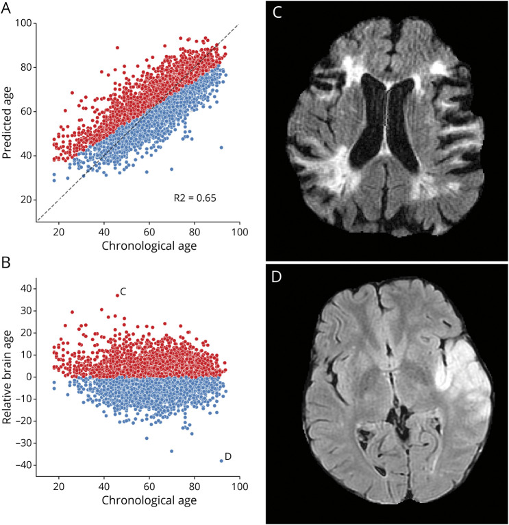
Methods: We extracted radiomics from T2-FLAIR images acquired during acute stroke clinical evaluation. Brain age was determined from brain parenchyma radiomics using an ElasticNet linear regression model. Subsequently, relative brain age (RBA), which expresses brain age in comparison with chronological age-matched peers, was estimated. Finally, we built a linear regression model of RBA using clinical cardiovascular characteristics as inputs and a logistic regression model of favorable functional outcomes taking RBA as input.

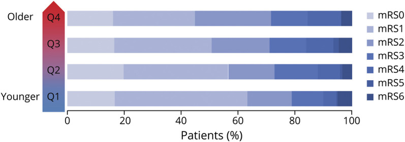
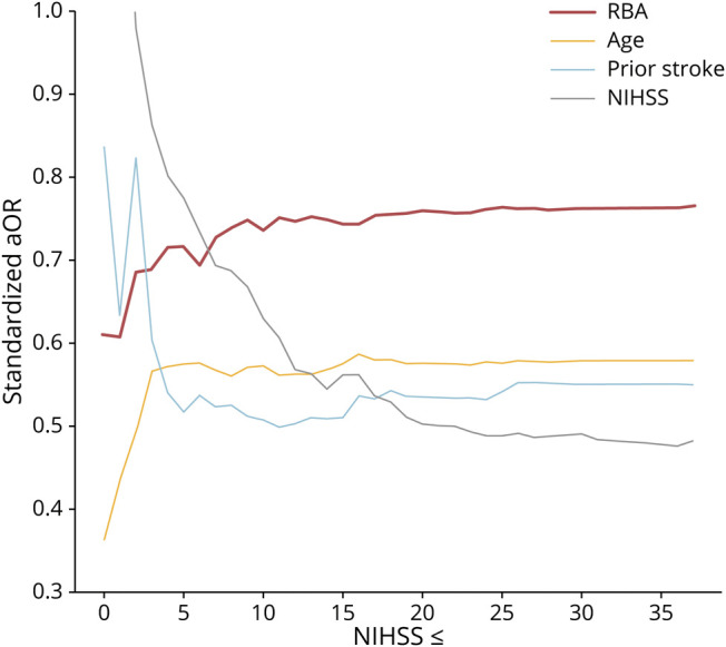
Results: We reviewed 4,163 patients from a large multisite ischemic stroke cohort (mean age = 62.8 years, 42.0% female patients). T2-FLAIR radiomics predicted chronological ages (mean absolute error = 6.9 years, r = 0.81). After adjustment for covariates, RBA was higher and therefore described older-appearing brains in patients with hypertension, diabetes mellitus, a history of smoking, and a history of a prior stroke. In multivariate analyses, age, RBA, NIHSS, and a history of prior stroke were all significantly associated with functional outcome (respective adjusted odds ratios: 0.58, 0.76, 0.48, 0.55; all p-values < 0.001). Moreover, the negative effect of RBA on outcome was especially pronounced in minor strokes.

Discussion: T2-FLAIR radiomics can be used to predict brain age and derive RBA. Older-appearing brains, characterized by a higher RBA, reflect cardiovascular risk factor accumulation and are linked to worse outcomes after stroke.


Figures

Figure 1
Figure 1. Brain Age Prediction Performances and Relative Brain Age
Scatter plots of the T2-FLAIR radiomics (A) predicted brain age and (B) relative brain age (RBA) per chronological age. Patients were colored in red if they had a positive RBA and thus a brain that appeared older to their age-matched peers or in blue if they had a negative RBA and a younger-looking brain. (C) T2-FLAIR axial image of a patient with a positive RBA: predicted brain age = 88, chronological age = 46, RBA = 36.2; this patient's brain exhibits multiple cortical and subcortical sequelae, moderate-to-severe parenchymal atrophy with enlarged ventricles and sulci, and confluent white matter hyperintensities, which extents are unexpectedly large for a 46-year-old patient. (D) T2-FLAIR axial image of a patient with a negative RBA: predicted brain age = 43, chronological age = 92, RBA = −38.6; notwithstanding the left middle cerebral artery lesion, this patient's brain trophicity is maintained; the cortex and the deep gray nuclei are sharply defined, overall describing a healthy brain for this 92-year-old patient.
Figure 2
Figure 2. mRS Shift Plot per Quartile of RBA
Q1-Q4 quartiles of RBA, mRS modified Rankin Scale. Q1-Q2 represents patients with lower RBA and thus younger-looking brains, whereas Q3-Q4 represents older-looking brains. mRS = modified Rankin scale; RBA = relative brain age.
Figure 3
Figure 3. Effect Sizes of the Predictors of Good Functional Outcome by Rank of Baseline NIHSS
RBA had a detrimental impact on achieving a good functional outcome especially in patients with low NIHSS. aOR = adjusted odds ratio; NIHSS = NIH Stroke Scale; RBA = relative brain age.

References

    1. Drozdowska BA, Singh S, Quinn TJ. Thinking about the future: a review of prognostic scales used in acute stroke. Front Neurol. 2019;10:274-274. doi: 10.3389/fneur.2019.00274 - DOI - PMC - PubMed
    1. Franke K, Gaser C. Ten years of BrainAGE as a neuroimaging biomarker of brain aging: what insights have we gained? Front Neurol. 2019;10:789. doi: 10.3389/fneur.2019.00789 - DOI - PMC - PubMed
    1. Hénon H, Pasquier F, Leys D. Poststroke dementia. Cerebrovasc Dis (Basel, Switzerland). 2006;22(1):61-70. doi: 10.1159/000092923 - DOI - PubMed
    1. Ning K, Zhao L, Matloff W, Sun F, Toga AW. Association of relative brain age with tobacco smoking, alcohol consumption, and genetic variants. Sci Rep. 2020;10:10. - PMC - PubMed
    1. Kolbeinsson A, Filippi S, Panagakis Y, et al. . Accelerated MRI-predicted brain ageing and its associations with cardiometabolic and brain disorders. Sci Rep. 2020;10:19940. - PMC - PubMed

Publication types