Review
. 2023 Jul:95:101149.
doi: 10.1016/j.preteyeres.2022.101149. Epub 2022 Nov 25.

Non-canonical Wnt signaling in the eye

Affiliations
Review

Non-canonical Wnt signaling in the eye

Ruchi Shah et al. Prog Retin Eye Res. 2023 Jul.

Abstract

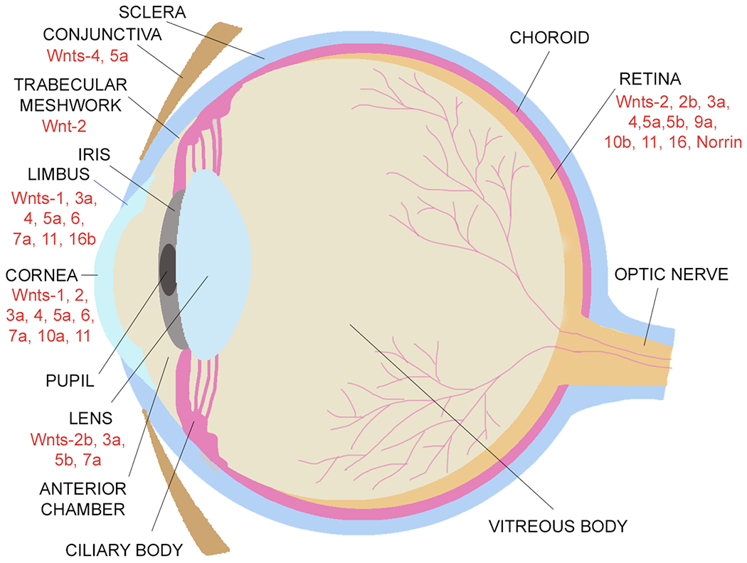
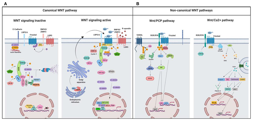
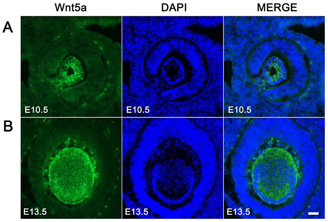
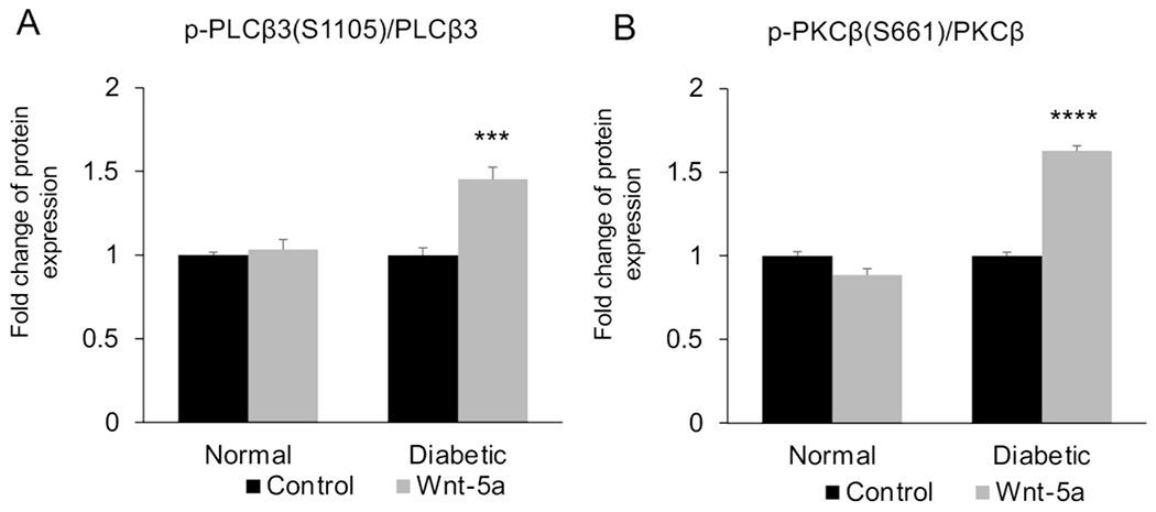
Wnt signaling comprises a group of complex signal transduction pathways that play critical roles in cell proliferation, differentiation, and apoptosis during development, as well as in stem cell maintenance and adult tissue homeostasis. Wnt pathways are classified into two major groups, canonical (β-catenin-dependent) or non-canonical (β-catenin-independent). Most previous studies in the eye have focused on canonical Wnt signaling, and the role of non-canonical signaling remains poorly understood. Additionally, the crosstalk between canonical and non-canonical Wnt signaling in the eye has hardly been explored. In this review, we present an overview of available data on ocular non-canonical Wnt signaling, including developmental and functional aspects in different eye compartments. We also discuss important changes of this signaling in various ocular conditions, such as keratoconus, aniridia-related keratopathy, diabetes, age-related macular degeneration, optic nerve damage, pathological angiogenesis, and abnormalities in the trabecular meshwork and conjunctival cells, and limbal stem cell deficiency.

Keywords: Calcium signaling; Conjunctiva; Cornea; Eye development; Eye diseases; Lens; Planar cell polarity; Retina; Stem cells; Wnt signaling.


Conflict of interest statement

Declaration of competing interest None.

Figures

Figure 1.
Figure 1.
A schematic illustration of the expression of Wnt ligands in the eye.
Figure 2.
Figure 2.
A schematic illustration representing different Wnt signaling pathways. (A) Canonical Wnt signaling. Left panel shows inactive pathway. In the absence of Wnt ligands, β-catenin is phosphorylated by the destruction complex, constituted by the scaffolding proteins APC and AXIN and the kinases GSK3β and CK1α. Then, β-catenin is ubiquitinated and targeted for proteasomal degradation by the complex containing β-TrCP, FBXW7, NEDDL4, and WTX proteins. Thus, β-catenin degradation prevents its presence in the nucleus where a complex formed by TCF/LEF and TLE/Groucho binds HDACs to inhibit transcription of target genes. Right panel shows canonical Wnt signaling active. The binding of Wnt ligands to FZD receptors and LRP co-receptors activates Wnt signaling. LRP receptors are phosphorylated by CK1α and GSK3β. Then, DVL proteins polymerize and are activated at the plasma membrane inhibiting the destruction complex. This results in stabilization and accumulation of β-catenin in the cytosol and its subsequent translocation into the nucleus where it displaces TLE/Groucho repressors forming an active complex with TCF/LEF proteins that bind co-activators such as CBP/p300, BRG1, BCL9, and PYGO. An alternative way of β-catenin signaling includes the disruption of epithelial E-cadherin interactions, which breaks the binding of β-catenin to the cytoplasmic domain of cadherin and leads to the accumulation of β-catenin first in the cytosol, and later in the nucleus. (B) Schematic illustration representing the main non-canonical Wnt pathways. Left panel shows the Wnt/PCP pathway. Wnt ligands bind to the FZD receptor and the co-receptors ROR 1/2 (or RYK). Then, DVL is recruited and activated followed by VANGL activation. Then DVL binds to the small GTPase RHO A with the collaboration of the cytoplasmic protein DAAM1. The small GTPases RAC1 and RHO activate ROCK and JNK. This leads to rearrangements of the cytoskeleton and/or transcriptional responses via for example, ATF2 and/or NFAT. Right panel shows the Wnt/Ca2+ pathway. The signaling is initiated when Wnt ligands bind to the FZD receptor and the co-receptor ROR 1/2 (or RYK). Then, DVL is recruited and activated and binds to the small GTPase which activates phospholipase C leading to intracellular calcium fluxes and downstream calcium dependent cytoskeletal and/or transcriptional responses. APC, adenomatous polyposis coli; BCL9, B-cell CLL/lymphoma 9 protein; β-TrCP, β-Transducin repeat-containing protein; BRG1, Brahma related gene 1; CAMKII, calmodulin-dependent protein kinase II; CBP, CREB-binding protein; CDC42, cell division control protein 42; CELSR, cadherin EGF LAG seven-pass G-type receptor; CK1α,ε,δ, casein kinase 1α,ε,δ; DAAM1, DVL associated activator of morphogenesis; DAG, diacylglycerol; DVL, disheveled; FBXW7, F box/WD repeat-containing protein 7; FZD, Frizzled; GSK3b, glycogen synthase kinase 3β; IP3, inositol 1,4,5 triphosphate; JNK, JUN kinase; LGR5, Leucine-rich repeat-containing G-protein-coupled receptor 5; LRP5/6, low-density lipoprotein receptor-related protein 5/6; NEDD4L, neural precursor cell expressed, developmentally downregulated 4-like; NFAT, nuclear factor of activated T cells; NF-κB, nuclear factor kappa B; PK, Prickle; PKC, protein kinase C; PLC, Phospholipase C; p300, E1A Binding Protein p300; RAC, Ras-related C3 botulinum toxin substrate; RHOA, Ras homolog gene family member A; ROCK, Rho kinase; ROR 1/2, bind tyrosine kinase-like orphan receptor 1 or 2; RYK, receptor-like tyrosine kinase; TBP, TATA-binding protein; PRCN, Porcupine; PYGO, Pygopus; RNF43, Ring finger protein 43; RSPO, R-spondin; TCF/LEF, T-cell factor/lymphoid enhancer factor; TLE, Transducin-Like Enhancer of Split proteins; VANGL, Van Gogh-like; WTX, Wilms tumor suppressor protein complex; YAP/TAZ, Yes-associated protein/Transcriptional co-activator with a PDZ-binding domain; ZNRF3, Zinc and Ring Finger 3. Created with BioRender.com. From: Martin-Orozco E, Sanchez-Fernandez A, Ortiz-Parra I, Ayala-San Nicolas M. WNT signaling in tumors: the way to evade drugs and immunity. Front Immunol. 2019; 10:2854. doi:10.3389/fimmu.2019.02854.
Figure 3.
Figure 3.
Spatiotemporal expression pattern of Wnt-5a during lens development in vivo. Immunofluorescent staining of sagittal sections of mouse embryonic eyes at E10.5 (A) and E13.5 (B) for Wnt-5a (green) and DAPI (blue; nuclei) is shown. Scale bar. 100 μm. From: Han C, Li J, Wang C, Ouyang H, Ding X, Liu Y, Chen S, Luo L. Wnt5a contributes to the differentiation of human embryonic stem cells into lentoid bodies through the noncanonical Wnt/JNK signaling pathway. Invest. Ophthalmol. Vis. Sci. 2018;59:3449-3460. doi:10.1167/iovs.18-23902
Figure 4.
Figure 4.
Wnt phospho array analysis of (A) p-PLCβ3 at Ser 1105 and (B) p-PKCβ at Ser 661 in normal and diabetic human limbal epithelial cells with or without Wnt-5a treatment (200 μg/ml). Values are mean ± SEM. ***, p<0.001; ****, p<0.0001 vs. untreated. From: Shah R, Spektor TM, Punj V, Turjman S, Ghiam S, Kim J, Tolstoff S, Amador; C, Chun ST, Weisenberger DJ, Saghizadeh M, Kramerov AA, Ljubimov AV. Wnt5a promotes diabetic corneal epithelial wound healing and limbal stem cell expression Invest. Ophthalmol. Vis. Sci. 2021;62:847.
Figure 5.
Figure 5.
Single cell RNA sequencing analysis of (A) normal and (B) diabetic human limbal epithelial ex vivo cells showing the gene expression of Wnt receptor FZD6 in various cell clusters. (C) Quantitative RT-PCR analysis also shows increased mRNA levels of FZD6 in human diabetic ex vivo limbal tissue.
Figure 6.
Figure 6.
Working model for the role of non-canonical Wnt ligand Wnt-5a in mechanotransduction. Wnt-5a, through ROR2, activates Cdc42 at adherens junctions, which is necessary for stable binding of vinculin to α-catenin, and efficient mechanocoupling between endothelial cells. Low non-canonical Wnt signaling weakens adherens junctions, impairs force propagation, and disrupts collective cell migration of endothelial cells. From: Carvalho JR, Fortunato IC, Fonseca CG, Pezzarossa A, Barbacena P, Dominguez-Cejudo MA, Vasconcelos FA, Santos NC, Carvalho FA, Franco CA. Non-canonical Wnt signaling regulates junctional mechanocoupling during angiogenic collective cell migration. eLife. 2019;8:e45853. doi: 10.7554/eLife.45853.
Figure 7.
Figure 7.
Wnt-5a treatment induced neurite growth in RGC primary cultures. Representative images of RGC cultures treated with BSA (control), 25 ng, 50 ng and 100 ng recombinant Wnt-5a. Neurites are shown in white, DAPI-stained nuclei are blue. Scale bar: 50 μm. From: Musada GR, Carmy-Bennun T, Hackam AS. Identification of a novel axon regeneration role for non-canonical Wnt signaling in the adult retina after injury. eNeuro. 2022;9:ENEURO.0182-22.2022. doi: 10.1523/ENEURO.0182-22.2022.

References

    1. Ahadome SD, Zhang C, Tannous E, Shen J, Zheng JJ. Small-molecule inhibition of Wnt signaling abrogates dexamethasone-induced phenotype of primary human trabecular meshwork cells. Exp Cell Res. 2017;357:116–123. doi: 10.1016/j.yexcr.2017.05.009. - DOI - PMC - PubMed
    1. Anastas JN, Moon RT. WNT signalling pathways as therapeutic targets in cancer. Nat Rev Cancer. 2013;13:11–26. doi: 10.1038/nrc3419. - DOI - PubMed
    1. Ang SJ, Stump RJ, Lovicu FJ, McAvoy JW. Spatial and temporal expression of Wnt and Dickkopf genes during murine lens development. Gene Expr Patterns. 2004;4:289–295. doi: 10.1016/j.modgep.2003.11.002. - DOI - PubMed
    1. Asimellis G, Kaufman EJ. Keratoconus. [Updated 2022 May 30]. In: StatPearls [Internet]. Treasure Island (FL): StatPearls Publishing; 2022. Jan-. Available from: https://www.ncbi.nlm.nih.gov/books/NBK470435/
    1. Auw-Haedrich C, Agrawal M, Gabbert HE, Meyer P, Arnold N, Reinhard T. Immunohistochemical expression of epithelial cell markers in corneas with congenital aniridia and ocular cicatrizing pemphigoid. Acta Ophthalmol. 2011;89:47–53. doi: 10.1111/j.1755-3768.2009.01603.x. - DOI - PubMed