. 2023 Jan;95(1):e28356.
doi: 10.1002/jmv.28356.

The third inactivated vaccine booster dramatically enhanced SARS-CoV-2 antibody responses and did not influence the profile of prothrombotic antibody

Affiliations

The third inactivated vaccine booster dramatically enhanced SARS-CoV-2 antibody responses and did not influence the profile of prothrombotic antibody

Yunbao Pan et al. J Med Virol. 2023 Jan.

Abstract

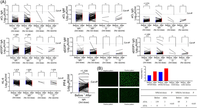
The purpose of this study is to investigate the production of both severe acute respiratory syndrome coronavirus-2 (SARS-CoV-2)-specific antibodies and autoantibodies in serum following the third booster vaccination of the inactivated COVID-19 vaccine, and to study the effect of B cell subsets with CD27 and CD38 phenotypes in peripheral blood on antibody production. Routine blood indexes, SARS-CoV-2 antibodies, platelet factor 4 and seven antiphospholipid antibodies were detected both before and 2 months after vaccination in the medical staff of the Zhongnan Hospital of Wuhan University. Peripheral blood B cell subtypes were detected before vaccination. Following immunization, the positive rate of anti-N-S1 immunoglobulin (IgG) had increased from 24.8% to 91.3% and the average antibody concentration had increased by 11 times. The positive rate of neutralizing antibody had increased from 24.8% to 91.3%, the average antibody concentration had increased by 12 times, and the primary increased anti-S1 IgG subtype was that of IgG1. Peripheral blood CD27 + CD38+ B cells were positively correlated with antibody levels after vaccination and were a predictor of the antibody response. In addition, although some indicators showed slight absolute changes, the blood parameters and antiphospholipid antibodies of most volunteers were normal both before and after COVID-19 inactivated vaccine inoculation, and there was no statistical difference in abnormal rates either before or after inoculation. Antibodies in vivo were increased after vaccination with the inactivated vaccine, and IgG1 was the main subtype involved in response to the vaccine. Vaccination with the inactivated COVID-19 vaccine did not appear to affect thrombus-related autoantibodies.

Keywords: B cell subpopulation; COVID-19; SARS-CoV-2; antiphospholipid antibody; inactivated vaccine.


Conflict of interest statement

The authors declare no conflict of interest.

Figures

Figure 1
Figure 1
Anti‐N‐S1 IgG and NAb levels in donor sera both before and after the 3rd vaccine injection. (A) Vaccination program and blood sample collection of the project participants. (B) Changes in the levels of anti‐N‐S1 IgG and NAb both before (n = 453) and after (n = 183) the 3rd injection. (C) Matched donors were selected and analyzed for changes in serum anti‐N‐S1 IgG and NAb levels, NPI with 3rd dose (n = 149), NPI with no 3rd dose (n = 77), CI with 3rd dose (n = 2) and CI with no vaccine (n = 5). (D) Correlation between anti‐N‐S1 IgG (left), NAb (right) and age before and after the 3rd injection. *p < 0.05, **p < 0.01, ***p < 0.001. CI, convalescent individual of COVID‐19; IgG, immunoglobulin; M, month; NAb, neutralizing antibody; NPI, No previous infection with SARS‐CoV‐2; SARS‐CoV‐2, severe acute respiratory syndrome coronavirus‐2; W, week.
Figure 2
Figure 2
The correlation between anti‐N‐S1 IgG, NAb, anti‐S1 IgG1, anti‐S1 IgG2, anti‐S1 IgG3, and anti‐S1 IgG4 levels. (A) Differences in anti‐S1 IgG1/2/3/4 levels both before (n = 453) and after (n = 183) the 3rd injection. (B) Distribution and association of antibody subtypes rate. (C) Correlation between anti‐N‐S1 IgG and NAb. (D) Correlation between anti‐N‐S1 IgG and anti‐S1 IgG1/2/3/4. (E) Correlation between NAb and anti‐S1 IgG1/2/3/4. IgG, immunoglobulin; NAb, neutralizing antibody.
Figure 3
Figure 3
Hematological parameters after the 3rd dose of vaccination. (A) WBC (white blood cell). (B) RBC (red blood cell). (C) PLT (platelet). (D) YMPH (lymphocyte). (E) NEUT (neutrophils). (F) MONO (mononuclear). (G) EO (eosinophils). (H) BASO (basophils). (I) HGB (hemoglobin).
Figure 4
Figure 4
The correlation between the antibody level after 3rd injection and the lymphocyte subset and blood formed elements before the 3rd injection. (A) CorHeatmap was used to color these correlations: blue indicates negative correlations, red indicates positive correlations, sector size indicates the p Value magnitude, and the color intensity indicates the strength of the correlation. (B−C) Scatter diagrams were used to describe the correlation.
Figure 5
Figure 5
Predictors for antibody response. Logistic models were constructed to identify predictors for antibody response. Routine blood count (A) and B lymphocyte subsets (B) predicted antibody response.
Figure 6
Figure 6
Lymphocyte subsets could serve as predictors of individual responsiveness to the 3rd dose of the vaccine. (A, B) Receiver operator characteristic curves for anti‐N‐S1 IgG and NAb. (C) anti‐N‐S1 IgG and NAb response were correlated with the levels of CD27 + CD19 + CD38+ and CD19 + CD27+ cells. AUC, area under curve; IgG, immunoglobulin; NAb, neutralizing antibody; ROC, receiver operating characteristic.
Figure 7
Figure 7
Expression of autoantibodies both before and after the 3rd dose of the vaccine. (A) Changes in aCL IgA, aCL IgM, aCL IgG, aβ2GP1 IgA, aβ2GP1 IgM, aβ2GP1 IgG, KL‐6 and PF4. (B) Positive rate of ANA antibodies in individuals who had either received the 3rd dose or no 3rd dose (right). Representative immunofluorescence images of the antinuclear antibody (left). ANA, analysis of antinuclear; IgG, immunoglobulin.

References

    1. Wang Q, Zhang Y, Wu L, et al. Structural and functional basis of SARS‐CoV‐2 entry by using human ACE2. Cell. 2020;181(4):894‐904 e9. - PMC - PubMed
    1. Gupta RK, Topol EJ. COVID‐19 vaccine breakthrough infections. Science. 2021;374(6575):1561‐1562. - PubMed
    1. Wang K, Jia Z, Bao L, et al. Memory B cell repertoire from triple vaccinees against diverse SARS‐CoV‐2 variants. Nature. 2022;Mar 603(7903):919‐925. - PMC - PubMed
    1. Figgett WA, Vincent FB, Saulep‐Easton D, Mackay F. Roles of ligands from the TNF superfamily in B cell development, function, and regulation. Sem Immunol. 2014;26(3):191‐202. - PubMed
    1. Vale AM, Schroeder HW, Jr . Clinical consequences of defects in B‐cell development. J Allergy Clin Immunol. 2010;125(4):778‐787. - PMC - PubMed

Publication types