. 2022 Nov 28;13(1):6915.
doi: 10.1038/s41467-022-34480-6.

mTORC1 links pathology in experimental models of Still's disease and macrophage activation syndrome

Affiliations

mTORC1 links pathology in experimental models of Still's disease and macrophage activation syndrome

Zhengping Huang et al. Nat Commun. .

Abstract

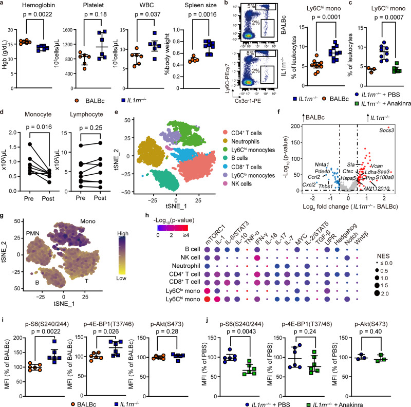
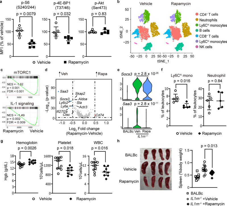
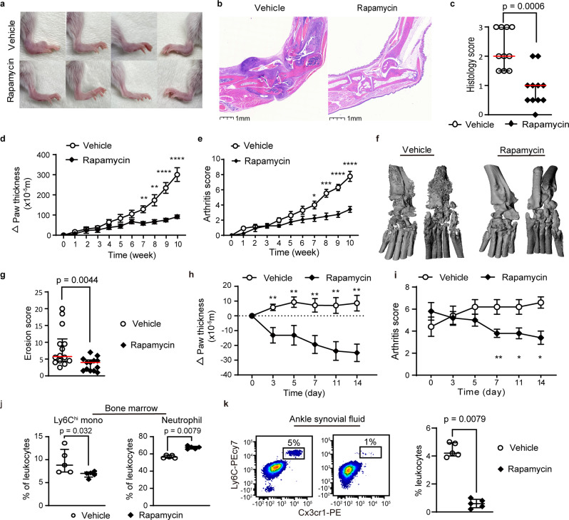
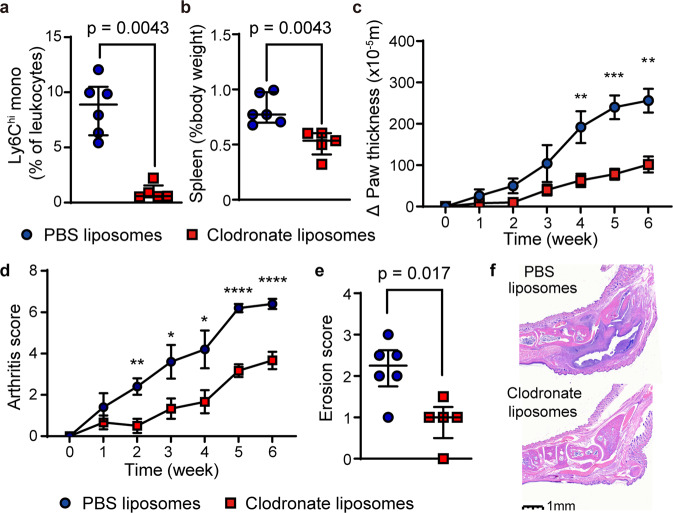
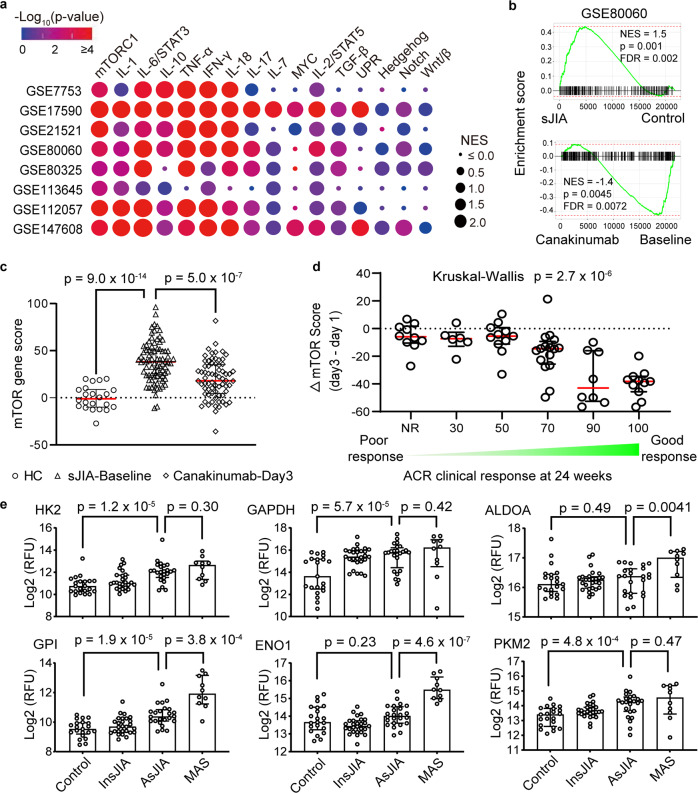
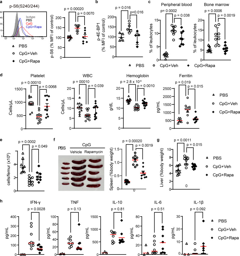
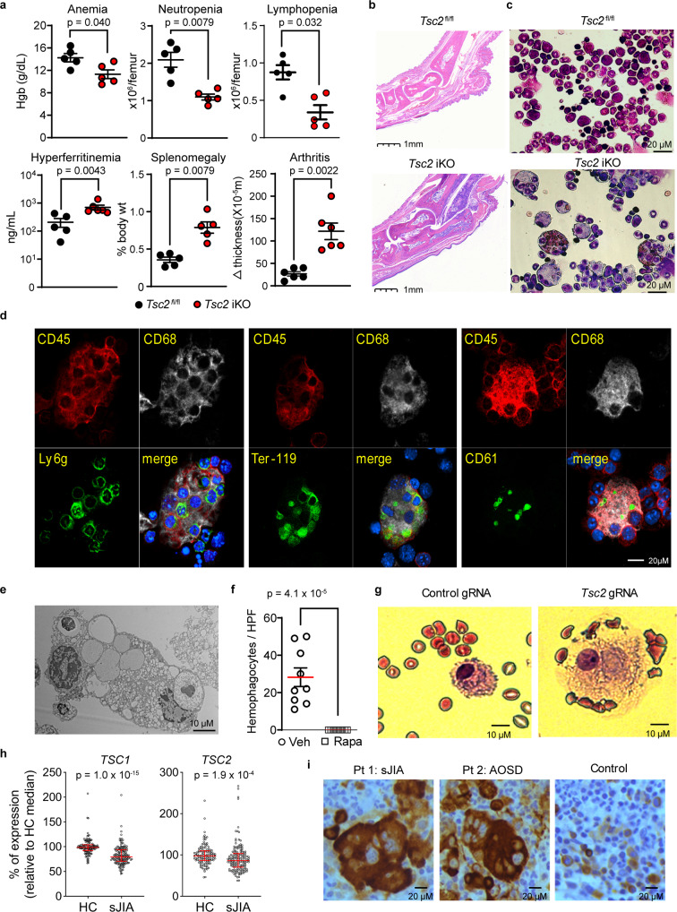
Still's disease is a severe inflammatory syndrome characterized by fever, skin rash and arthritis affecting children and adults. Patients with Still's disease may also develop macrophage activation syndrome, a potentially fatal complication of immune dysregulation resulting in cytokine storm. Here we show that mTORC1 (mechanistic target of rapamycin complex 1) underpins the pathology of Still's disease and macrophage activation syndrome. Single-cell RNA sequencing in a murine model of Still's disease shows preferential activation of mTORC1 in monocytes; both mTOR inhibition and monocyte depletion attenuate disease severity. Transcriptomic data from patients with Still's disease suggest decreased expression of the mTORC1 inhibitors TSC1/TSC2 and an mTORC1 gene signature that strongly correlates with disease activity and treatment response. Unrestricted activation of mTORC1 by Tsc2 deletion in mice is sufficient to trigger a Still's disease-like syndrome, including both inflammatory arthritis and macrophage activation syndrome with hemophagocytosis, a cellular manifestation that is reproduced in human monocytes by CRISPR/Cas-mediated deletion of TSC2. Consistent with this observation, hemophagocytic histiocytes from patients with macrophage activation syndrome display prominent mTORC1 activity. Our study suggests a mechanistic link of mTORC1 to inflammation that connects the pathogenesis of Still's disease and macrophage activation syndrome.


Conflict of interest statement

The authors declare no competing interests.

Figures

Fig. 1
Fig. 1. Monocytosis and enhanced mTORC1 signaling in IL-1-mediated inflammation.
a Complete blood count in BALBc (n = 5) and IL1rn−/− mice (n = 6 per group), and spleen size in BALBc (n = 6) and IL1rn−/− mice (n = 8). b Flow cytometry quantification of circulating Ly6Chi monocytes in BALBc and IL1rn−/− mice at baseline (n = 10 per group) and (c) after anakinra treatment for 2 weeks (WT, n = 3; IL1rn−/− PBS, n = 8; IL1rn−/− + anakinra, n = 6). d Peripheral blood monocyte and lymphocyte count in sJIA patients pre-treatment and 2 weeks after initiation of anakinra (n = 8). e t-SNE display of leukocyte clustering derived from single-cell RNA-seq of peripheral blood cells from BALBc and IL1rn−/− mice (4 mice pooled per group). f Volcano plot of differentially expressed genes in Ly6Chi monocytes of WT and IL1rn−/− mice. g Density plot of Hallmark mTORC1 gene set expression using AddmoduleScore. h Cluster plot of gene set enrichment in peripheral blood leukocyte subsets of BALBc and IL1rn−/− mice based on single-cell RNA-seq (4 mice pooled per group). i Phospho-flow analysis of mTOR substrates in bone marrow Ly6Chi monocytes from WT and IL1rn−/− mice at baseline (n = 6 per group) and (j) after daily anakinra treatment for 2 weeks (n = 6 per group for phospho-S6 and phospho-4EBP1; n = 3 for phosphor-Akt). Data in (a, b, c, i, and j) were pooled from 2 to 3 independent experiments. Mice were 8–9 weeks old for all experiments. Statistical analyses (all two-sided): Mann–Whitney U test (a, b, cd, i, j); Wilcoxon signed-rank test with Bonferroni correction (f). Median and error bars representing interquartile range are displayed in (ab, c, i, j). Source data are provided as a Source Data file.
Fig. 2
Fig. 2. mTORC1 inhibition attenuates features of systemic inflammation in IL1rn−/− mice.
a Phospho-flow quantification of phosphorylated S6, 4EBP1 and Akt expression in bone marrow Ly6Chi monocytes from IL1rn−/− mice treated with rapamycin or vehicle control (n = 5 per group). b t-SNE display of leukocyte clustering derived from single-cell RNA-seq of peripheral blood leukocytes from IL1rn−/− mice treated with vehicle or rapamycin for 4 weeks (4 mice pooled per group). c Gene set enrichment plots of mTORC1 and IL-1 gene sets, (d) volcano plot of differentially expressed genes and (e) violin plots of Socs3 and Saa3 expression in peripheral blood Ly6Chi monocytes from single-cell RNA-seq (n = 4 mice pooled per group). f Quantification of peripheral blood monocytes and neutrophils, (g) complete blood count parameters in IL1rn−/− mice treated with vehicle (n = 11) or rapamycin (n = 8) for 10 weeks. h Spleen size in BALBc mice (n = 9) and IL1rn−/− mice treated with vehicle (n = 11) or rapamycin (n = 9) for 10 weeks. Data in (a, f, g, and h) were pooled from 2 to 3 independent experiments. Mice were 8–9 weeks old for experiments in (ae) and 4 weeks old for experiments in (fh). Statistical analyses (all two-sided): Mann–Whitney U test (a, f, g, and h), permutation test (c), Wilcoxon rank sum test with Bonferroni correction (d, e). Median and error bars representing interquartile range are displayed in (a, f, g, h). Source data are provided as a Source Data file.
Fig. 3
Fig. 3. Rapamycin treatment reduces arthritis severity and bone erosion in IL1rn−/− mice.
a Representative depiction of ankle inflammation, (b) hematoxylin and eosin staining of ankle sections, (c) histologic scores of ankle arthritis (vehicle, n = 10; rapamycin n = 11), (d) ankle and wrist thickness (vehicle, n = 14; rapamycin n = 11), (e) composite arthritis score (vehicle, n = 14; rapamycin n = 11) and (f, g) micro-CT quantification of joint erosion in IL1rn−/− mice treated with rapamycin or vehicle for 10 weeks. h Ankle and wrist joint measurements (n = 8 per group) and (i) composite arthritis score (n = 5 per group) in IL1rn−/− mice with established arthritis treated with vehicle or rapamycin for 2 weeks. j Flow cytometry quantification of Ly6Chi monocytes and neutrophils in the bone marrow and k) synovial fluid of IL1rn−/− mice treated with vehicle control or rapamycin for 10 weeks (n = 5 per group). Data in (ce, gj, k) were pooled from 2 to 3 independent experiments. Mice were 4 weeks old for experiments in (ag, j, k) and 10 weeks old for experiments in (h, i). Statistical analyses (all two-sided): Mann–Whitney U test (c, g, j, k); Student’s t test (d, e, h, i). *p < 0.05, **p < 0.01, ***p < 0.001, ****p < 0.0001. Median and error bars representing interquartile range are displayed in (c, g, j, k). Mean and error bars representing standard errors are displayed in (d, e, h, i). Source data are provided as a Source Data file.
Fig. 4
Fig. 4. Phagocyte depletion reduces arthritis severity and bone erosion in IL1rn−/− mice.
a Peripheral blood monocyte count, (b) spleen size, (c) ankle joint thickness, (d) composite arthritis score, (e) bone erosion score (micro-CT) and (f) representative depiction of joint histology in IL1rn−/− mice treated with PBS-liposomes (n = 6) or clodronate-liposomes (n = 5) for 6 weeks. Data in (a, b, d, e, f) were pooled from 2 independent experiments. Mice were 6 weeks old for experiments in (af). Statistical analyses (all two-sided): Mann–Whitney U test (a, b, e); Student’s t test (c, d). *p < 0.05, **p < 0.01. Median and error bars representing interquartile range are displayed in (a, b, e). Mean and error bars representing standard errors are displayed in (c, d). Source data are provided as a Source Data file.
Fig. 5
Fig. 5. mTORC1 signature in Still’s disease correlates with disease severity and treatment response.
a Cluster plot of gene set enrichment analysis comparing healthy controls and patients with sJIA and AOSD using in publicly available transcriptomic dataset. b Gene set enrichment plot of Hallmark mTORC1 gene set and (c) calculation of mTOR gene set score from healthy controls (n = 22), sJIA patients at baseline (n = 82) and sJIA patients 3 days after canakinumab treatment (n = 69). d Stratification of mTOR gene score before treatment and 3 days after canakinumab based on clinical response after 24 weeks. Clinical response was stratified according to American College of Rheumatology (ACR) Score: ACR 0/Non-responders (NR, n = 10), ACR30 (n = 6), ACR50 (n = 11), ACR70 (n = 16), ACR90 (n = 8), ACR100 (n = 11). e Levels of glycolytic enzymes from serum proteomics analysis of healthy controls (n = 21), sJIA patients with inactive (n = 27) or active disease (n = 24), and sJIA patients with MAS (n = 10). Transcriptomic data for (bd) are derived from GEO data series GSE80060 while proteomics data in (e) were derived from Chen et al.. Statistical analyses (all two-sided): permutation test (a, b), Mann–Whitney U test (c, e), Kruskal–Wallis one-way analysis of variance (d). Median and error bars representing interquartile range are displayed in (ce). Source data are provided as a Source Data file.
Fig. 6
Fig. 6. mTORC1 activation drives CpG-induced macrophage activation syndrome.
a Phospho-S6 staining (n = 8 per group) and (b) phospho-4EBP1 staining (n = 5 per group) in bone marrow Ly6Chi monocytes, (c) flow cytometry quantification of peripheral blood and bone marrow Ly6Chi monocytes (n = 8 per group), (d) hematologic parameters (n = 11 per group) and ferritin levels (PBS, n = 5; CpG + Vehicle, n = 8; CpG + Rapamycin, n = 8), (e) absolute bone marrow cell count (n = 8 per group), (f) representative depiction and quantification of spleen size (n = 8 per group), (g) liver size (n = 8 per group), and (h) plasma cytokine levels (PBS, n = 3; CpG + Vehicle, n = 8; CpG + Rapamycin, n = 8), in PBS-treated or CpG DNA-treated C57BL/6 mice (50 µg every 2 days x 5 doses) given daily rapamycin or vehicle control. Data in (a, b) were normalized to the mean fluorescence intensity (MFI) of the PBS group. Mice were 8 weeks old and data from all panels were pooled from 2 to 3 independent experiments. Statistical analyses (all two-sided): Mann–Whitney U test (all panels). Median and error bars representing interquartile range are displayed in (ah). Source data are provided as a Source Data file.
Fig. 7
Fig. 7. Unrestricted mTORC1 activation drives the development of hemophagocytosis and macrophage activation syndrome.
a Hemoglobin levels (n = 5 per group), bone marrow cell count (n = 5 per group), plasma ferritin levels (Tsc2 fl/fl, n = 5, Tsc2 ikO, n = 6), spleen weight (n = 5 per group), and change in ankle joint thickness (n = 6 per group) and (b) representative ankle pathology (H&E stain) in Tsc2 iKO mice and control mice. c Wright-Giemsa staining of bone marrow leukocytes from Tsc2 iKO and control mice. d Confocal microscopy and e electron microscopy of hemophagocytes from the bone marrow of Tsc2 iKO mice. f Quantification of bone marrow hemophagocytes in Tsc2 iKO mice treated with rapamycin or vehicle control (3 slides analyzed for each mouse; n = 3 per group). Mice were 6–8 weeks of age and analyses in (af) were performed 3 weeks after poly I:C treatment to induce Tsc2 deletion. g Wright-Giemsa staining of human monocytes with targeted disruption of Tsc2 by CRISPR/Cas9 cultured with M-CSF for 14 days. h Pooled transcriptomic analysis of TSC1 and TSC2 expression in peripheral blood cells from patients with sJIA (n = 154) and healthy controls (n = 120). Data are compiled from GEO deposits GSE80325, GSE80060, GSE7753, GSE21521, GSE17590 and GSE112057. i Immunohistochemistry of phospho-S6 (S240/244) on bone marrow section from patients with Still’s disease and controls. Data in (a, f) were pooled from 2 to 3 independent experiments. Images in (be, g) are representative of 2 independent experiments. Images in (i) are derived from one experiment. Statistical analyses (all two-sided): Mann–Whitney U test (a, f, h). Median and error bars representing interquartile range are displayed in (a, f, h). Source data are provided as a Source Data file.

Comment in

References

    1. Still GF. On a form of chronic joint disease in children. Med. Chir. Trans. 1897;80:47–60 49. - PMC - PubMed
    1. Bywaters EG. Still’s disease in the adult. Ann. Rheum. Dis. 1971;30:121–133. - PMC - PubMed
    1. Davies R, et al. Mortality rates are increased in patients with systemic juvenile idiopathic arthritis. Arch. Dis. Child. 2017;102:206–207. - PubMed
    1. Minoia F, et al. Clinical features, treatment, and outcome of macrophage activation syndrome complicating systemic juvenile idiopathic arthritis: a multinational, multicenter study of 362 patients. Arthritis Rheumatol. 2014;66:3160–3169. - PubMed
    1. Crayne CB, Albeituni S, Nichols KE, Cron RQ. The immunology of macrophage activation syndrome. Front. Immunol. 2019;10:119. - PMC - PubMed

Publication types

Substances