. 2022 Nov 28;13(1):7198.
doi: 10.1038/s41467-022-34864-8.

Inactivation of LATS1/2 drives luminal-basal plasticity to initiate basal-like mammary carcinomas

Affiliations

Inactivation of LATS1/2 drives luminal-basal plasticity to initiate basal-like mammary carcinomas

Joseph G Kern et al. Nat Commun. .

Abstract

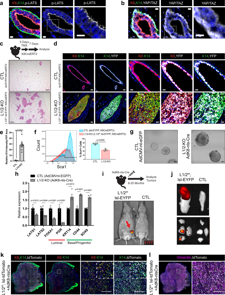
Basal-like breast cancers, an aggressive breast cancer subtype that has poor treatment options, are thought to arise from luminal mammary epithelial cells that undergo basal plasticity through poorly understood mechanisms. Using genetic mouse models and ex vivo primary organoid cultures, we show that conditional co-deletion of the LATS1 and LATS2 kinases, key effectors of Hippo pathway signaling, in mature mammary luminal epithelial cells promotes the development of Krt14 and Sox9-expressing basal-like carcinomas that metastasize over time. Genetic co-deletion experiments revealed that phenotypes resulting from the loss of LATS1/2 activity are dependent on the transcriptional regulators YAP/TAZ. Gene expression analyses of LATS1/2-deleted mammary epithelial cells notably revealed a transcriptional program that associates with human basal-like breast cancers. Our study demonstrates in vivo roles for the LATS1/2 kinases in mammary epithelial homeostasis and luminal-basal fate control and implicates signaling networks induced upon the loss of LATS1/2 activity in the development of basal-like breast cancer.


Conflict of interest statement

The authors declare no competing interests.

Figures

Fig. 1
Fig. 1. Loss of LATS1/2 in luminal mammary cells induces mammary carcinomas exhibiting luminal-basal plasticity.
a, b Immunofluorescence (IF) staining of a phospho-LATS1/2 (Thr1079/1041) or b YAP/TAZ together with K8 and K14 in control mouse mammary ducts (Scale bar, 10 µM). c Diagram of the mouse model used to conditionally delete LATS1/2 in K8+ luminal mammary epithelial cells along with H&E staining of control and Lats1/2f/f;lsl-EYFP;K8CreERT2 mammary glands (Scale bar, 200 µM). d IF staining of K8, K14, and EYFP in control and Lats1/2f/f;lsl-EYFP;K8CreERT2 mammary glands following tamoxifen treatment (Scale bar 50 µM in the left two columns, and 20 µM in the right three columns). e Quantification of K14 intensity/YFP area in mammary ducts of lsl-EYFP;K8CreERT2 (CTL) and Lats1/2f/f;lsl-EYFP;K8CreERT2 (L1/2-KO) mice (n = 5 images from 2 CTL mice, n = 9 images from 2 L1/2-KO mice. Unpaired two-tailed t-test, Data are shown as mean ± SEM). f Flow cytometric profiling for Sca1 expression in CTL and L1/2-KO mammary epithelial cells (n = 4 CTL, n = 3 L1/2-KO. Unpaired two-tailed t-test, Data are shown as mean ± SEM). g Morphology of organoids cultured from control and LATS1/2f/f mice infected with AdK8-nls-Cre (Scale bar, 50 µM). h RNA expression of selected luminal and basal/stem-cell markers in control and LATS1/2f/f organoids infected with AdK8-nls-Cre (n = 4. Unpaired two-tailed t-tests. Data are shown as mean ± SEM). i IVIS live imaging of a primary tumor (red arrow) formed in the mammary gland of a LATS1/2f/f; lsl-EYFP mouse approximately 14 months after intraductal injection with AdK8-nls-Cre compared to an uninjected control mouse. j IVIS imaging of EYFP signal from a representative primary tumor (top) and lung metastases (bottom) formed in a LATS1/2f/f; lsl-EYFP mouse approximately 11 months after intraductal injection with AdK8-nls-Cre compared to organs from an uninjected control mouse. k, l IF staining of k K8, K14, and tdTomato or l Vimentin and tdTomato in a primary tumor formed in a LATS1/2f/f; lsl-tdTomato mouse approximately 13 months after intraductal injection with AdK8-nls-Cre (n = 2 tumors) (Scale bars, 500 µM on low magnification images on left, and 200 µM on high magnification images of regions outlined by white dotted lines on the right). Source data are provided as a source data file.
Fig. 2
Fig. 2. Sox9 expressing luminal cells serve as cells of origin for LATS1/2-null carcinomas.
a IF staining of K8, K14, and Sox9 in control and Lats1/2f/f;K8CreERT2 tamoxifen-treated mammary ducts (Scale bar, 20 µM). b Diagram of Lats1/2f/f;Sox9CreERT2 model used to target Sox9+ mammary epithelial cells and H&E staining of control and Lats1/2-deleted mammary glands (n = 2 for CTL) (Scale bar, 200 µM). c IF of K8, K14, and YFP in control and Lats1/2f/f;lsl-EYFP;Sox9CreERT2 tamoxifen-treated mammary ducts. (Scale bar left two columns, 50 µM, Scale bar right two columns, 20 µM) (n = 2 for CTL). d Quantification of K14 intensity/YFP area in lsl-EYFP;Sox9CreERT2 (CTL) and Lats1/2f/f;lsl-EYFP;Sox9CreERT2 (L1/2-KO) mice (n = 7 images from 2 CTL mice, n = 34 images from 9 L1/2-KO mice. Unpaired two-tailed t-test, Data are shown as mean ± SEM). e IF of K8, K14, and Sox9 in control and Lats1/2f/f;lsl-EYFP;Sox9CreERT2 tamoxifen-treated mammary ducts (Scale bar, 20 µM) (n = 2 for CTL). f Morphology of organoids isolated from control and Lats1/2f/f; Sox9CreERT2 mice and treated with 4OHT (Scale bar, 50 µM). g Expression of selected luminal, basal, and YAP/TAZ-induced markers in control and Lats1/2f/f;lsl-EYFP;Sox9CreERT2 organoids treated with 4OHT (n = 3. Unpaired two-tailed t-tests. Data are shown as mean ± SEM). h Diagram of conditions used to co-delete LATS1/2 and Sox9 in Krt8+ luminal mammary epithelial cells along with H&E staining of control, Lats1/2f/f;K8CreERT2, and Lats1/2f/f;Sox9f/f;K8CreERT2 mammary glands (Scale bar, 200 µM). i IF of K8, K14, and YFP in control, Lats1/2f/f;lsl-EYFP;K8CreERT2, and Lats1/2f/f;Sox9 f/f;lsl-EYFP;K8CreERT2 mammary ducts treated with tamoxifen (Scale bar, 50 µM). j Quantification of K14 intensity/YFP area in mammary ducts of lsl-EYFP;K8CreERT2 (CTL), Lats1/2f/f;lsl-EYFP;K8CreERT2 (L1/2-KO), and Lats1/2f/f;Sox9f/f;lsl-EYFP;K8CreERT2 (L1/2-SOX9-KO) mice (n = 45 images from 5 CTL mice, n = 40 images from 3 L1/2-KO mice, and n = 61 images from 4 L1/2-SOX9-KO mice. One-way ANOVA with Fisher’s least significant difference multiple comparisons test. Data are shown as mean ± SEM). Source data are provided as a source data file.
Fig. 3
Fig. 3. Mammary carcinomas driven by LATS1/2 loss are dependent on YAP and TAZ.
a IF of K8, K14, and YAP/TAZ in control and Lats1/2f/f;lsl-EYFP;K8CreERT2 mammary ducts (Scale bar, 10 µM). b Diagram of conditions used to co-delete LATS1/2, YAP, and TAZ in K8+ luminal mammary epithelial cells and H&E staining of control, Lats1/2f/f;K8CreERT2, and Lats1/2f/f;Yapf/f;Tazf/f;lsl-EYFP;K8CreERT2 mammary glands treated with tamoxifen (Scale bar, 200 µM). c IF of K8, K14, and YFP in control, Lats1/2f/f;lsl-EYFP;K8CreERT2, and Lats1/2f/f;Yapf/f;Tazf/f;lsl-EYFP;K8CreERT2 mammary ducts (Scale bar, 20 µM). d Quantification of K14 intensity/YFP area in mammary ducts of lsl-EYFP;K8CreERT2 (CTL), Lats1/2f/f;lsl-EYFP;K8CreERT2 (L1/2-KO), and Lats1/2f/f;Yapf/f;Tazf/f;lsl-EYFP;K8CreERT2 (L1/2-YAP/TAZ-KO) mice (n = 20 images from 3 CTL mice, n = 14 images from 3 L1/2-KO mice, n = 37 images from 6 L1/2-YAP/TAZ-KO mice. One-way ANOVA with Fisher’s least significant difference multiple comparisons test. Data are shown as mean ± SEM). e Morphology of organoids cultured from control, Lats1/2f/f;K8CreERT2, and Lats1/2f/f;Yapf/f;Tazf/f;K8CreERT2 mice treated with 4OHT (Scale bar, 65 µM). f Expression of selected luminal, basal/stem-cell, and YAP/TAZ-induced markers in organoids cultured from control, Lats1/2f/f;K8CreERT2, and Lats1/2f/f;Yapf/f;Tazf/f;K8CreERT2 mice treated with 4OHT (n = 3. One-way ANOVA with Fisher’s least significant difference multiple comparisons test. Data are shown as mean ± SEM). Source data are provided as a source data file.
Fig. 4
Fig. 4. Luminal LATS1/2 loss promotes a basal-like transcriptional program phenocopying human basal-like breast cancer.
a Heatmap of gene expression changes in EYFP+ mammary cells sorted from Lats1/2f/f;lsl-EYFP;K8CreERT2 mice compared to lsl-EYFP;K8CreERT2 control mice as analyzed by RNA-sequencing (n = 3 per condition, 2-fold change and FDR < 0.05 cutoff). b Selected GSEA-generated genesets enriched in the upregulated (red) and downregulated (blue) signatures of LATS1/2-null cells compared to control. c GSVA analysis comparing the upregulated and downregulated signatures of LATS1/2-null cells to those of normal mammary luminal mature, luminal progenitor, and basal cells (GSE63310). d Analysis of copy number alterations of Hippo pathway-related factors in human breast cancers. Heatmap representing one or two allele deletions or amplifications are shown on top and the frequency of these alterations in the different breast cancer subtypes are shown at the bottom. e Activity levels of the LATS-up/downregulated genes in TCGA-BRCA samples across the Basal, HER2+, Luminal A, Luminal B, and Normal-like (NL) subtypes (n = 189(Basal), 564(LumA), 215(LumB), 82(Her2), 40(NL). Two-sided t-test. p-values represent comparisons to the Basal group. No adjustment for multiple comparisons. Each box plot draws the center line at the median value, the upper and lower box boundaries at the first and third quartiles (25th and 75th percentiles), and the whiskers at ±1.5 × the interquartile range). f Breast cancer patients with survival information (n = 1102) were divided into two groups based on high and low gene expression following LATS1/2 deletion based on the median LATS1/2 gene expression scores (n = 551 per group), and these gene expression signatures were compared for their overall survival outcomes. The statistical significance of survival difference between groups was determined using a log-rank test.

References

    1. Greaves M, Maley CC. Clonal evolution in cancer. Nature. 2012;481:306–313. doi: 10.1038/nature10762. - DOI - PMC - PubMed
    1. Vermeulen L, Sprick MR, Kemper K, Stassi G, Medema JP. Cancer stem cells—old concepts, new insights. Cell Death Differ. 2008;15:947–958. doi: 10.1038/cdd.2008.20. - DOI - PubMed
    1. Batlle E, Clevers H. Cancer stem cells revisited. Nat. Med. 2017;23:1124–1134. doi: 10.1038/nm.4409. - DOI - PubMed
    1. Ge Y, Fuchs E. Stretching the limits: from homeostasis to stem cell plasticity in wound healing and cancer. Nat. Rev. Genet. 2018;19:311–325. doi: 10.1038/nrg.2018.9. - DOI - PMC - PubMed
    1. Gupta PB, Pastushenko I, Skibinski A, Blanpain C, Kuperwasser C. Phenotypic plasticity: driver of cancer initiation, progression, and therapy resistance. Cell Stem Cell. 2019;24:65–78. doi: 10.1016/j.stem.2018.11.011. - DOI - PMC - PubMed

Publication types