

The pathogenicity comparison of Lagovirus europaeus GI.1 and GI.2 strains in China by using relative quantitative assay
- PMID: 36443356
- PMCID: PMC9705280
- DOI: 10.1038/s41598-022-25118-0
The pathogenicity comparison of Lagovirus europaeus GI.1 and GI.2 strains in China by using relative quantitative assay
Abstract
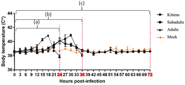
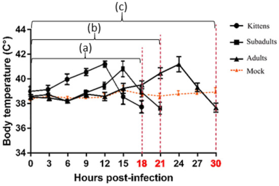
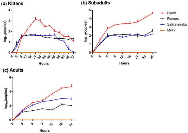
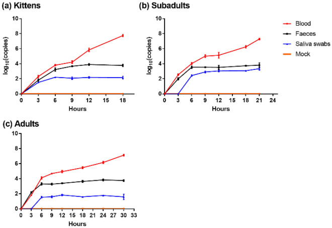
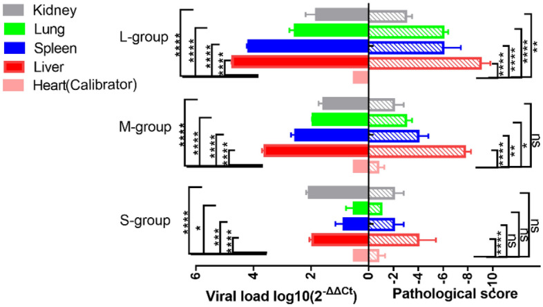
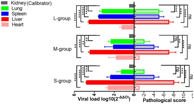
Lagovirus europaeus GI.1 belongs to Lagovirus in the Caliciviridae family. GI.1 causes an acute, septic, and highly lethal disease in rabbits. Lagovirus europaeus GI.2, a new variant of GI.1, has caused explosive mortality in rabbits of all ages in Sichuan Province, China. To explore the differences in pathogenicity of rabbits infected with GI.1/GI.2, we investigated the virulence and disease progression of a naturally occurring GI.1/GI.2 in 4-week-old, 13-week-old, and 25-week-old New Zealand White laboratory rabbits after GI.1/GI.2 infection. Objective measures of disease progression were recorded using continuous body-temperature monitoring. We observed the kittens were infected with GI.2 during the most urgent course of the disease, and GI.1 was not lethal to kittens. We found that the target organ of both GI.1 and GI.2 was the liver, but the disease course of the two viruses was differed. Our study enriches the research on the pathogenicity of GI.1 and GI.2 under the same conditions.
© 2022. The Author(s).
Conflict of interest statement
The authors declare no competing interests.
Figures
Similar articles
-
Immune response in the recombinant strain of Lagovirus europaeus GI.1a.Infect Genet Evol. 2023 Jun;110:105427. doi: 10.1016/j.meegid.2023.105427. Epub 2023 Mar 21. Infect Genet Evol. 2023. PMID: 36958414
-
Occurrence of Lagovirus europaeus (Rabbit Hemorrhagic Disease Virus) in Domestic Rabbits in Southwestern Poland in 2019: Case Report.Microbiol Spectr. 2022 Dec 21;10(6):e0229822. doi: 10.1128/spectrum.02298-22. Epub 2022 Nov 29. Microbiol Spectr. 2022. PMID: 36445093 Free PMC article.
-
GI.1b/GI.1b/GI.2 recombinant rabbit hemorrhagic disease virus 2 (Lagovirus europaeus/GI.2) in Morocco, Africa.Arch Virol. 2019 Jan;164(1):279-283. doi: 10.1007/s00705-018-4052-y. Epub 2018 Oct 4. Arch Virol. 2019. PMID: 30284631
-
Lack of evidence for differences in the spread of classic (Lagovirus europaeus/GI.1) and novel (Lagovirus europaeus/GI.2) rabbit haemorrhagic disease viruses in Europe and North Africa.Vet Rec. 2022 Feb;190(3):e1067. doi: 10.1002/vetr.1067. Epub 2021 Oct 28. Vet Rec. 2022. PMID: 34713453 Review.
-
Immunity against Lagovirus europaeus and the Impact of the Immunological Studies on Vaccination.Vaccines (Basel). 2021 Mar 13;9(3):255. doi: 10.3390/vaccines9030255. Vaccines (Basel). 2021. PMID: 33805607 Free PMC article. Review.
Cited by
-
Rabbit hemorrhagic disease virus 2, 2010-2023: a review of global detections and affected species.J Vet Diagn Invest. 2024 Sep;36(5):617-637. doi: 10.1177/10406387241260281. Epub 2024 Jun 19. J Vet Diagn Invest. 2024. PMID: 39344909 Free PMC article. Review.
-
An RT-qPCR Assay from Rectal Swabs for the Detection of Rabbit Hemorrhagic Disease Virus 2 in Natural Cases.Transbound Emerg Dis. 2023 Mar 24;2023:1869692. doi: 10.1155/2023/1869692. eCollection 2023. Transbound Emerg Dis. 2023. PMID: 40303805 Free PMC article.
-
Characterisation of Lagovirus europaeus GI-RHDVs (Rabbit Haemorrhagic Disease Viruses) in Terms of Their Pathogenicity and Immunogenicity.Int J Mol Sci. 2024 May 14;25(10):5342. doi: 10.3390/ijms25105342. Int J Mol Sci. 2024. PMID: 38791380 Free PMC article. Review.
-
A Standardised Method to Quantify the Infectious Titre of Rabbit Haemorrhagic Disease Virus.Viruses. 2025 Apr 24;17(5):609. doi: 10.3390/v17050609. Viruses. 2025. PMID: 40431621 Free PMC article.
References
-
- Liu SJ, Xue HP, Pu BQ, Qian NH. A new viral disease in rabbits. J. Vet. Diagn. Investig. 1984;16:253–255.
Publication types
MeSH terms
LinkOut - more resources
Full Text Sources
Research Materials
