

Multi-modal retinal scanning to measure retinal thickness and peripheral blood vessels in multiple sclerosis
- PMID: 36443364
- PMCID: PMC9705292
- DOI: 10.1038/s41598-022-24312-4
Multi-modal retinal scanning to measure retinal thickness and peripheral blood vessels in multiple sclerosis
Abstract
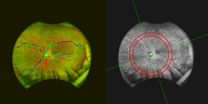
Our purpose was to investigate changes to the retina in multiple sclerosis (MS) using established and novel modes of retinal image acquisition and analysis. 72 participants with MS and 80 healthy volunteers underwent retinal scanning with optical coherence tomography (OCT) and ultra-widefield (UWF) scanning laser ophthalmoscopy (SLO), over a two-year period. Changes in retinal nerve fibre layer (RNFL) thickness, macular volume and retinal blood vessel diameter were measured and parameters were then tested for associations with MS. Measurements from OCT showed that individuals with MS had a thinner RNFL and reduced macular volume when compared to healthy volunteers. On UWF images, participants with MS had reduced arterial widths in the inferior nasal quadrant of both eyes and reduced venous widths in the inferior nasal quadrant of right eyes. Longitudinal analysis showed that participants with MS had an accelerated annual rate of RNFL thinning in several regions of the retina. In conclusion, the assessment of OCT showed thinning of the RNFL and macula in concordance with previous reports on MS, while analysis of blood vessels in the retinal periphery from UWF-SLO images revealed novel changes.
© 2022. The Author(s).
Conflict of interest statement
This work was partially funded by Optos Plc and The Medical Research Council. Dr Jano van Hemert is the imaging research manager and academic liaison at Optos Plc. The authors declare no other competing interests.
Figures
Similar articles
-
Peripapillary retinal nerve fiber layer thickness measured by optical coherence tomography in different clinical subtypes of multiple sclerosis.Mult Scler Relat Disord. 2019 Jan;27:260-268. doi: 10.1016/j.msard.2018.11.003. Epub 2018 Nov 5. Mult Scler Relat Disord. 2019. PMID: 30423530
-
[Quantification of retinal nerve fiber thickness. A comparison of laser scanning ophthalmoscopy, polarimetry and optical coherence tomography in healthy and glaucomatous eyes].Ophthalmologe. 2001 Sep;98(9):832-43. doi: 10.1007/s003470170058. Ophthalmologe. 2001. PMID: 11594222 German.
-
Baseline retinal nerve fiber layer thickness and macular volume quantified by OCT in the North American phase 3 fingolimod trial for relapsing-remitting multiple sclerosis.J Neuroophthalmol. 2013 Dec;33(4):322-9. doi: 10.1097/WNO.0b013e31829c51f7. J Neuroophthalmol. 2013. PMID: 24051419 Free PMC article. Clinical Trial.
-
Optical coherence tomography in mild cognitive impairment - Systematic review and meta-analysis.Clin Neurol Neurosurg. 2020 Sep;196:106036. doi: 10.1016/j.clineuro.2020.106036. Epub 2020 Jun 22. Clin Neurol Neurosurg. 2020. PMID: 32623211
-
[New examination methods for macular disorders--application of diagnosis and treatment].Nippon Ganka Gakkai Zasshi. 2000 Dec;104(12):899-942. Nippon Ganka Gakkai Zasshi. 2000. PMID: 11193944 Review. Japanese.
Cited by
-
An Open-Source Deep Learning Algorithm for Efficient and Fully Automatic Analysis of the Choroid in Optical Coherence Tomography.Transl Vis Sci Technol. 2023 Nov 1;12(11):27. doi: 10.1167/tvst.12.11.27. Transl Vis Sci Technol. 2023. PMID: 37988073 Free PMC article.
-
De-escalation of Disease-Modifying Therapy for People with Multiple Sclerosis Due to Safety Considerations: Characterizing 1-Year Outcomes in 25 People Who Switched from Ocrelizumab to Diroximel Fumarate.Adv Ther. 2024 Aug;41(8):3059-3075. doi: 10.1007/s12325-024-02902-0. Epub 2024 Jun 11. Adv Ther. 2024. PMID: 38861218 Free PMC article.
-
Non-invasive in vivo imaging of brain and retinal microglia in neurodegenerative diseases.Front Cell Neurosci. 2024 Jan 29;18:1355557. doi: 10.3389/fncel.2024.1355557. eCollection 2024. Front Cell Neurosci. 2024. PMID: 38348116 Free PMC article. Review.
-
Regional retinal vulnerability in multiple sclerosis: integrating OCT, MRI, and clinical data for enhanced diagnosis and automated monitoring.Rom J Morphol Embryol. 2025 Jan-Mar;66(1):119-130. doi: 10.47162/RJME.66.1.11. Rom J Morphol Embryol. 2025. PMID: 40384198 Free PMC article.
-
Evaluating Fundoscopy as a Screening Tool for Optic Nerve Atrophy in Multiple Sclerosis: An Optical Coherence Tomography (OCT) Comparative Study.J Clin Med. 2025 Mar 22;14(7):2166. doi: 10.3390/jcm14072166. J Clin Med. 2025. PMID: 40217617 Free PMC article.
References
-
- McAlpine D, Compston A. McAlpine’s Multiple Sclerosis. Elsevier Health Sciences; 2005.
Publication types
MeSH terms
Grants and funding
LinkOut - more resources
Full Text Sources
Medical
