. 2022 Nov 28;12(1):20443.
doi: 10.1038/s41598-022-24982-0.

Terlipressin combined with conservative fluid management attenuates hemorrhagic shock-induced acute kidney injury in rats

Affiliations

Terlipressin combined with conservative fluid management attenuates hemorrhagic shock-induced acute kidney injury in rats

Leticia Urbano Cardoso Castro et al. Sci Rep. .

Abstract

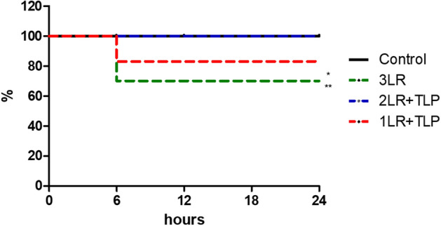
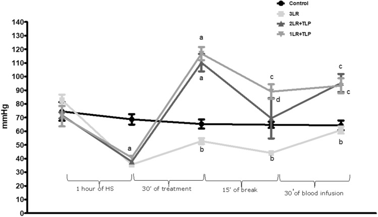
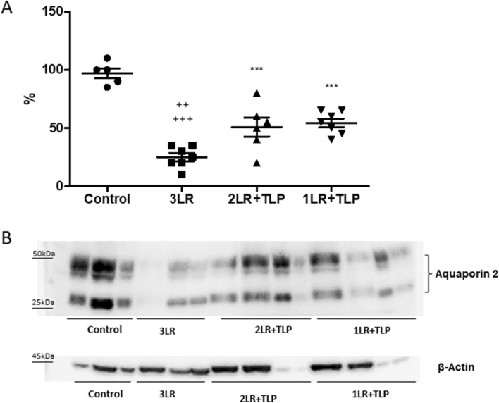
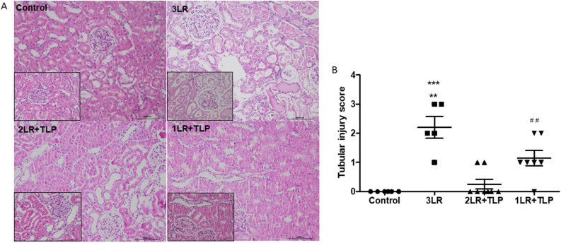
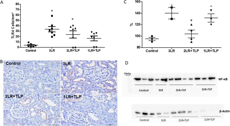
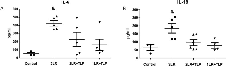
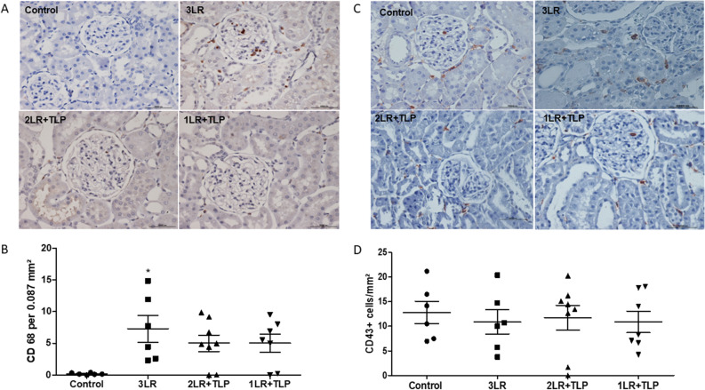
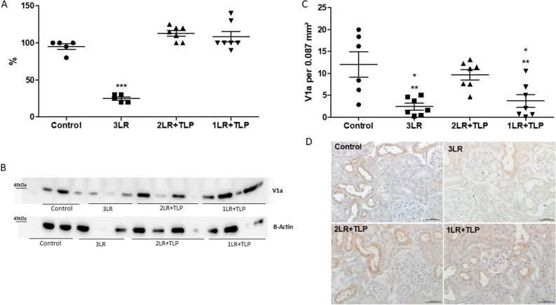
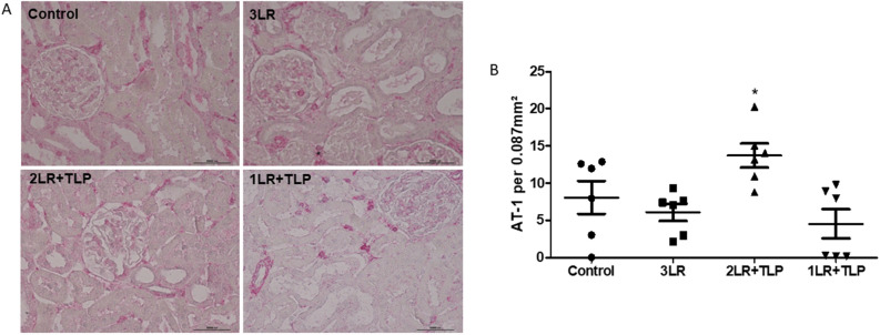
Hemorrhagic shock (HS), a major cause of trauma-related mortality, is mainly treated by crystalloid fluid administration, typically with lactated Ringer's (LR). Despite beneficial hemodynamic effects, such as the restoration of mean arterial pressure (MAP), LR administration has major side effects, including organ damage due to edema. One strategy to avoid such effects is pre-hospitalization intravenous administration of the potent vasoconstrictor terlipressin, which can restore hemodynamic stability/homeostasis and has anti-inflammatory effects. Wistar rats were subjected to HS for 60 min, at a target MAP of 30-40 mmHg, thereafter being allocated to receive LR infusion at 3 times the volume of the blood withdrawn (liberal fluid management); at 2 times the volume (conservative fluid management), plus terlipressin (10 µg/100 g body weight); and at an equal volume (conservative fluid management), plus terlipressin (10 µg/100 g body weight). A control group comprised rats not subjected to HS and receiving no fluid resuscitation or treatment. At 15 min after fluid resuscitation/treatment, the blood previously withdrawn was reinfused. At 24 h after HS, MAP was higher among the terlipressin-treated animals. Terlipressin also improved post-HS survival and provided significant improvements in glomerular/tubular function (creatinine clearance), neutrophil gelatinase-associated lipocalin expression, fractional excretion of sodium, aquaporin 2 expression, tubular injury, macrophage infiltration, interleukin 6 levels, interleukin 18 levels, and nuclear factor kappa B expression. In terlipressin-treated animals, there was also significantly higher angiotensin II type 1 receptor expression and normalization of arginine vasopressin 1a receptor expression. Terlipressin associated with conservative fluid management could be a viable therapy for HS-induced acute kidney injury, likely attenuating such injury by modulating the inflammatory response via the arginine vasopressin 1a receptor.


Conflict of interest statement

The authors declare no competing interests.

Figures

Figure 1
Figure 1
Curve of survival after hemorrhagic shock. *P < 0.05 vs. 2LR + TLP; **P < 0.01 vs. control; Control, no intervention; 3LR, induction of hemorrhagic shock, followed by infusion of lactated Ringer’s at 3 times the volume of the blood withdrawn; 2LR + TLP, induction of hemorrhagic shock, followed by infusion of lactated Ringer’s at 2 times the volume of the blood withdrawn, together with intravenous injection of terlipressin at 10 µg/100 g body weight; 1LR + TLP, induction of hemorrhagic shock, followed by infusion of lactated Ringer’s at a volume equal to that of the blood withdrawn, together with intravenous injection of terlipressin at 10 µg/100 g body weight.
Figure 2
Figure 2
Mean arterial pressure during the experiment. aP < 0.001 vs. control; bP < 0.001 vs. 2LR + TLP and 1LR + TLP; cP < 0.01 vs. control; dP < 0.05 vs. control; HS, hemorrhagic shock; Control, no intervention; 3LR, induction of HS, followed by infusion of lactated Ringer’s at 3 times the volume of the blood withdrawn; 2LR + TLP, induction of HS, followed by infusion of lactated Ringer’s at 2 times the volume of the blood withdrawn, together with intravenous injection of terlipressin at 10 µg/100 g body weight; 1LR + TLP, induction of HS, followed by infusion of lactated Ringer’s at a volume equal to that of the blood withdrawn, together with intravenous injection of terlipressin at 10 µg/100 g body weight.
Figure 3
Figure 3
Aquaporin 2 expression in renal tissue at 24 h after hemorrhagic shock induction. Densitometric analysis (A) and immunoblotting (B). Immunoblots reacted with anti-AQP2 revealed 29- and 35 to 50-kD AQP2 bands, representing nonglycosylated and glycosylated forms of AQP2, respectively (Supplementary Fig. 1). ***P < 0.001 vs. control; ++P < 0.01 vs. 2LR + TLP; +++P < 0.001 vs. 1LR + TLP; Control, no intervention; 3LR, induction of hemorrhagic shock, followed by infusion of lactated Ringer’s at 3 times the volume of the blood withdrawn; 2LR + TLP, induction of hemorrhagic shock, followed by infusion of lactated Ringer’s at 2 times the volume of the blood withdrawn, together with intravenous injection of terlipressin at 10 µg/100 g body weight; 1LR + TLP, induction of hemorrhagic shock, followed by infusion of lactated Ringer’s at a volume equal to that of the blood withdrawn, together with intravenous injection of terlipressin at 10 µg/100 g body weight.
Figure 4
Figure 4
Acute tubular injury at 24 h after hemorrhagic shock induction. Kidney tissue sections stained with periodic acid-Schiff (A). Magnification, × 200 and × 400. Tubular damage score measured in the cortex (B). ***P < 0.001 vs. control and 2LR + TLP; **P < 0.01 vs. 1LR + TLP; ##P < 0.01 vs. control and 2LR + TLP; Control, no intervention; 3LR, induction of hemorrhagic shock, followed by infusion of lactated Ringer’s at 3 times the volume of the blood withdrawn; 2LR + TLP, induction of hemorrhagic shock, followed by infusion of lactated Ringer’s at 2 times the volume of the blood withdrawn, together with intravenous injection of terlipressin at 10 µg/100 g body weight; 1LR + TLP, induction of hemorrhagic shock, followed by infusion of lactated Ringer’s at a volume equal to that of the blood withdrawn, together with intravenous injection of terlipressin at 10 µg/100 g body weight.
Figure 5
Figure 5
Immunohistochemical analysis of Toll-like receptor 4 (TLR4) expression in rat kidney tissue. (A) Bar graph of TLR4 expression. Immunostaining (brown, in B) for TLR4+ cells in kidney cortex samples from control, 3LR, 2LR + TLP, and 1LR + TLP group rats. Magnification, × 400. Densitometric analysis (C) and immunoblotting (D) of nuclear factor kappa B (NF-κB) expression in the renal tissue at 24 h after hemorrhagic shock induction (Supplementary Fig. 2). *P < 0.05 vs. control; +P < 0.05 vs. 3LR and 1LR + TLP. Control, no intervention; 3LR, induction of hemorrhagic shock, followed by infusion of lactated Ringer’s at 3 times the volume of the blood withdrawn; 2LR + TLP, induction of hemorrhagic shock, followed by infusion of lactated Ringer’s at 2 times the volume of the blood withdrawn, together with intravenous injection of terlipressin at 10 µg/100 g body weight; 1LR + TLP, induction of hemorrhagic shock, followed by infusion of lactated Ringer’s at a volume equal to that of the blood withdrawn, together with intravenous injection of terlipressin at 10 µg/100 g body weight.
Figure 6
Figure 6
Cytokine levels in renal tissue at 24 h after hemorrhagic shock induction. Interleukin (IL)-6 (A) and IL-18 (B). &P < 0.05 vs. the other groups. Control, no intervention; 3LR, induction of hemorrhagic shock, followed by infusion of lactated Ringer’s at 3 times the volume of the blood withdrawn; 2LR + TLP, induction of hemorrhagic shock, followed by infusion of lactated Ringer’s at 2 times the volume of the blood withdrawn, together with intravenous injection of terlipressin at 10 µg/100 g body weight; 1LR + TLP, induction of hemorrhagic shock, followed by infusion of lactated Ringer’s at a volume equal to that of the blood withdrawn, together with intravenous injection of terlipressin at 10 µg/100 g body weight.
Figure 7
Figure 7
Immunohistochemical analysis of CD68 expression in rat kidney tissue. Immunostaining [brown, in (A)] for CD68 in kidney cortex samples from control, 3LR, 2LR + TLP, and 1LR + TLP group rats. Magnification, × 400. (B) Bar graph of CD68 expression. Immunohistochemical analysis of CD43 expression in rat kidney tissue. Immunostaining [brown, in (C)] for CD43 in kidney cortex samples from control, 3LR, 2LR + TLP, and 1LR + TLP group rats. Magnification, × 400. (D) Bar graph of CD43 expression. *P < 0.05 vs. control; Control, no intervention; 3LR, induction of hemorrhagic shock, followed by infusion of lactated Ringer’s at 3 times the volume of the blood withdrawn; 2LR + TLP, induction of hemorrhagic shock, followed by infusion of lactated Ringer’s at 2 times the volume of the blood withdrawn, together with intravenous injection of terlipressin at 10 µg/100 g body weight; 1LR + TLP, induction of hemorrhagic shock, followed by infusion of lactated Ringer’s at a volume equal to that of the blood withdrawn, together with intravenous injection of terlipressin at 10 µg/100 g body weight.
Figure 8
Figure 8
Immunohistochemical analysis of uromodulin expression in rat kidney tissue. Immunostaining [brown, in (A)] for uromodulin+ cells in kidney cortex samples from control, 3LR, 2LR + TLP, and 1LR + TLP group rats. Magnification, × 400. (B) Bar graph of uromodulin expression. Control, no intervention; 3LR, induction of hemorrhagic shock, followed by infusion of lactated Ringer’s at 3 times the volume of the blood withdrawn; 2LR + TLP, induction of hemorrhagic shock, followed by infusion of lactated Ringer’s at 2 times the volume of the blood withdrawn, together with intravenous injection of terlipressin at 10 µg/100 g body weight; 1LR + TLP, induction of hemorrhagic shock, followed by infusion of lactated Ringer’s at a volume equal to that of the blood withdrawn, together with intravenous injection of terlipressin at 10 µg/100 g body weight.
Figure 9
Figure 9
Arginine vasopressin 1a receptor (V1aR) expression in the renal tissue at 24 h after hemorrhagic shock induction. Immunohistochemical analysis of V1aR expression in rat kidney tissue at 24 h after hemorrhagic shock induction, by densitometric analysis (A) and immunoblotting (B) (Supplementary Fig. 3). Bar graph of V1aR expression (C) and immunostaining [brown, in (D)] for V1aR in kidney cortex samples from control, 3LR, 2LR + TLP, and 1LR + TLP group rats. Magnification, × 40. ***P < 0.001 vs. control, 2LR + TLP, and 1LR + TLP. *P < 0.05 vs. 2LR + TLP; **P < 0.01 vs. control; Control, no intervention; 3LR, induction of hemorrhagic shock, followed by infusion of lactated Ringer’s at 3 times the volume of the blood withdrawn; 2LR + TLP, induction of hemorrhagic shock, followed by infusion of lactated Ringer’s at 2 times the volume of the blood withdrawn, together with intravenous injection of terlipressin at 10 µg/100 g body weight; 1LR + TLP, induction of hemorrhagic shock, followed by infusion of lactated Ringer’s at a volume equal to that of the blood withdrawn, together with intravenous injection of terlipressin at 10 µg/100 g body weight.
Figure 10
Figure 10
Immunohistochemical analysis of angiotensin II type 1 receptor (AT-1R) expression in rat kidney tissue. Immunostaining [violet, in (A)] for AT-1R in kidney cortex samples from control, 3LR, 2LR + TLP, and 1LR + TLP group rats. Magnification, × 400. (B) Bar graph of AT-1R expression. *P < 0.05 vs. control, 3LR and 1LR + TLP; Control, no intervention; 3LR, induction of hemorrhagic shock, followed by infusion of lactated Ringer’s at 3 times the volume of the blood withdrawn; 2LR + TLP, induction of hemorrhagic shock, followed by infusion of lactated Ringer’s at 2 times the volume of the blood withdrawn, together with intravenous injection of terlipressin at 10 µg/100 g body weight; 1LR + TLP, induction of hemorrhagic shock, followed by infusion of lactated Ringer’s at a volume equal to that of the blood withdrawn, together with intravenous injection of terlipressin at 10 µg/100 g body weight.

Similar articles

Cited by

  • Obesity aggravates acute kidney injury resulting from ischemia and reperfusion in mice.
    da Silva IO, de Menezes NK, Jacobina HD, Parra AC, Souza FL, Castro LC, Roelofs JJTH, Tammaro A, Gomes SA, Sanches TR, Andrade L. da Silva IO, et al. Sci Rep. 2024 Apr 29;14(1):9820. doi: 10.1038/s41598-024-60365-3. Sci Rep. 2024. PMID: 38684767 Free PMC article.
  • Novel model of cardiac hypertrophy with cardiorenal dysfunction.
    de Almeida Silva A, Jensen L, Marques JR, Nascimento-Carvalho B, de Souza LE, da Silva MB, Moreira NJD, Veras MM, Waked D, Souza FL, Santinho MAR, Lunardon G, Caldini EG, da Conceição Andrade L, Diniz GP, Irigoyen MC. de Almeida Silva A, et al. Sci Rep. 2025 Jul 19;15(1):26242. doi: 10.1038/s41598-025-90435-z. Sci Rep. 2025. PMID: 40683937 Free PMC article.

References

    1. Jacob M, Kumar P. The challenge in management of hemorrhagic shock in trauma. Med. J. Armed Forces India. 2014;70:163–169. doi: 10.1016/j.mjafi.2014.03.001. - DOI - PMC - PubMed
    1. Nandra KK, et al. Pharmacological preconditioning with erythropoietin attenuates the organ injury and dysfunction induced in a rat model of hemorrhagic shock. Dis. Model Mech. 2013;6(3):701–709. - PMC - PubMed
    1. Gutierrez G, Reines HD, Wulf-Gutierrez ME. Clinical review: hemorrhagic shock. Crit. Care. 2004;8(5):373–381. doi: 10.1186/cc2851. - DOI - PMC - PubMed
    1. Hostmann A, et al. Biphasic onset of splenic apoptosis following hemorrhagic shock: Critical implications for Bax, Bcl-2, and Mcl-1 proteins. Crit. Care. 2008;12(1):R8. doi: 10.1186/cc6772. - DOI - PMC - PubMed
    1. Lee CC, et al. A comparison of vasopressin, terlipressin and lactated ringers for resuscitation of uncontrolled hemorrhagic shock in an animal model. PLoS ONE. 2014;9(4):e95821. doi: 10.1371/journal.pone.0095821. - DOI - PMC - PubMed

Publication types