. 2023 Jan;34(1):1-42.
doi: 10.1007/s00192-022-05397-x. Epub 2022 Nov 28.

An International Continence Society (ICS)/ International Urogynecological Association (IUGA) joint report on the terminology for the assessment and management of obstetric pelvic floor disorders

Affiliations

An International Continence Society (ICS)/ International Urogynecological Association (IUGA) joint report on the terminology for the assessment and management of obstetric pelvic floor disorders

Stergios K Doumouchtsis et al. Int Urogynecol J. 2023 Jan.

Erratum in

Abstract

Aims: The terminology of obstetric pelvic floor disorders should be defined and reported as part of a wider clinically oriented consensus.

Methods: This Report combines the input of members of two International Organizations, the International Continence Society (ICS) and the International Urogynecological Association (IUGA). The process was supported by external referees. Appropriate clinical categories and a sub-classification were developed to give coding to definitions. An extensive process of 12 main rounds of internal and 2 rounds of external review was involved to exhaustively examine each definition, with decision-making by consensus.

Results: A terminology report for obstetric pelvic floor disorders, encompassing 357 separate definitions, has been developed. It is clinically-based with the most common diagnoses defined. Clarity and user-friendliness have been key aims to make it usable by different specialty groups and disciplines involved in the study and management of pregnancy, childbirth and female pelvic floor disorders. Clinical assessment, investigations, diagnosis, conservative and surgical treatments are major components. Illustrations have been included to supplement and clarify the text. Emerging concepts, in use in the literature and offering further research potential but requiring further validation, have been included as an Appendix. As with similar reports, interval (5-10 year) review is anticipated to maintain relevance of the document and ensure it remains as widely applicable as possible.

Conclusion: A consensus-based Terminology Report for obstetric pelvic floor disorders has been produced to support clinical practice and research.

Keywords: Childbirth trauma; Obstetric injuries; Obstetric pelvic floor disorders; Perineal trauma; Terminology.


Figures

Fig. 1
Fig. 1
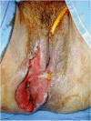
Obstetric vesico-vaginal fistula during repair [37]. Source: Reprinted by permission from Springer Nature
Fig. 2
Fig. 2
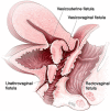
Pelvic floor fistula anatomy © Levent Efe [10]
Fig. 3
Fig. 3
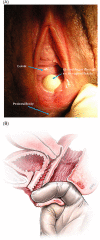
Rectovaginal fistula [10]. Figure 3A: Recto-vaginal fistula, low in the vagina, just proximal to the anus © J Goh; 3B: © Levent Efe
Fig. 4
Fig. 4
Uroflowmetry A schematic representation of urine flow over time and parameters of uroflowmetry [10]. (Republished with permission of John Wiley & Sons—Books, permission conveyed through Copyright Clearance Center, Inc)
Fig. 5
Fig. 5
Endoanal ultrasound images showing normal appearance of the internal (IAS) and external (EAS) anal sphincters [47]. Source: Reprinted by permission from Springer Nature
Fig. 6
Fig. 6
Ultrasound image of the pelvic floor in a woman following forceps vaginal delivery, showing avulsion of the levator plate from the right pubic ramus. Courtesy of Professor HP Dietz [49]. Source: Reproduced with permission of the Licensor through PLSclear
Fig. 7
Fig. 7
Tomographic ultrasound imaging assessment of the levator ani muscles Intact LAM [8, 14]. (Republished with permission of John Wiley & Sons—Books, permission conveyed through Copyright Clearance Center, Inc)
Fig. 8
Fig. 8
Ballooning of the genital hiatus on straining manoeuvre—levator defect [8, 14]. (Republished with permission of John Wiley & Sons—Books, permission conveyed through Copyright Clearance Center, Inc)
Fig. 9
Fig. 9
External appearance of a third degree tear [47] Source: Reprinted by permission from Springer Nature
Fig. 10
Fig. 10
Third-degree perineal tear, with complete division of the internal and external anal sphincters [49]. Source: Reproduced with permission of the Licensor through PLSclear
Fig. 11
Fig. 11
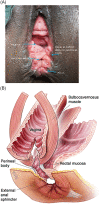
A, B: (A) Fourth degree rectovaginal tear with perineal body disruption. Congenital defects of a similar configuration may also occur. © J Goh. (B) © Levent Efe [10]
Fig. 12
Fig. 12
Classification of anal sphincter injuries [76] Source: Reproduced with kind permission from publishers
Fig. 13
Fig. 13
Necrotizing perineal infection [79]. Source: Reprinted by permission from Springer Nature
Fig. 14
Fig. 14
Debridement of necrotizing perineal infection [79]. Source: Reprinted by permission from Springer Nature
Fig. 15
Fig. 15
Infralevator hematoma [79]. Source: Reprinted by permission from Springer Nature
Fig. 16
Fig. 16
Supralevator hematoma [79]. Source: Reprinted by permission from Springer Nature
Fig. 17
Fig. 17
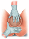
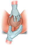
Central palmar support Reprinted/adapted by permission from Springer Nature Customer Service Centre GmbH Springer Nature: Perineal Mapping. in Perineal Trauma at Childbirth (KMK Ismail) [COPYRIGHT] (2017) [105]
Fig. 18
Fig. 18
Viennese method. Reprinted/adapted by permission from Springer Nature Customer Service Centre GmbH Springer Nature: Perineal Mapping. in Perineal Trauma at Childbirth (KMK Ismail) [COPYRIGHT] (2017) [105]
Fig. 19
Fig. 19
Finnish method. (a) Finnish method—application of the accoucheur’s hands on the fetal head and perineum; (b) Finnish method—accoucheur’s dominant hand, view from above; (c) Finnish method—accoucheur’s dominant hand, view from the front Reprinted/adapted by permission from Springer Nature Customer Service Centre GmbH Springer Nature: Perineal Mapping. in Perineal Trauma at Childbirth (KMK Ismail) [COPYRIGHT] (2017) [105]
Fig. 20
Fig. 20
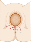
Types of episiotomy [110]. Key: a midline episiotomy; b modified median episiotomy; c J-shaped episiotomy; d mediolateral episiotomy; e lateral episiotomy; f radical lateral (Schuchardt incision). Source: Reprinted by permission from Springer Nature
Fig. 21
Fig. 21
Repair of obstetric anal sphincter injuries [76]. Source: Reproduced with kind permission
Fig. 22
Fig. 22
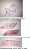
The two different methods of external anal sphincter repair. A Overlapping technique. B End-to-end technique. Extract from: V. Letouzey, E. Mousty, B. Fatton, J.-F. Bourgaux, M. Bertrand, M. Prudhomme, P. Marès, R. de Tayrac. Traumatisme anal chez la parturiente. EMC Gynecologie; Vol 11, n o 3, July 2016: pp 1–12. Copyright © 2016 Elsevier Masson SAS. All rights reserved. http://dx.doi.org/10.1016/S0246-1064(15)65086-1 [129]
Fig. 23
Fig. 23
Episiotomy repair using continuous non-locking suture (all layers) [110]. (a) Episiotomy repair, suturing of vaginal wall. Insert first stitch to the vagina above the apex of episiotomy cut and tie a knot. (b) Episiotomy repair, suturing of perineal muscles. (c) Episiotomy repair, suturing of perineal skin. Starting from the bottom edge of the episiotomy cut, close perineal skin in the opposite direction towards the vaginal orifice using subcuticular continuous suture. (d) Episiotomy repair, knotting of the stitch. Place the subcuticular stitch in the vagina just above the level of the hymen and tie a knot. Source: Reprinted by permission from Springer Nature

References

    1. Smith LA, Price N, Simonite V, Burns EE. Incidence of and risk factors for perineal trauma: a prospective observational study. BMC Pregnancy Childbirth. 2013;13:59. doi: 10.1186/1471-2393-13-59. - DOI - PMC - PubMed
    1. Keighley MRB, Perston Y, Bradshaw E, et al. The social, psychological, emotional morbidity and adjustment techniques for women with anal incontinence following Obstetric Anal Sphincter Injury: use of a word picture to identify a hidden syndrome. BMC Pregnancy Childbirth. 2016;16:275. doi: 10.1186/s12884-016-1065-y. - DOI - PMC - PubMed
    1. Dudding TC, Vaizey CJ, Kamm MA. Obstetric anal sphincter injury. Ann Surg. 2008;247:224–237. doi: 10.1097/SLA.0b013e318142cdf4. - DOI - PubMed
    1. The Management of Third- and Fourth-Degree Perineal Tears. Green-Top Guideline No 29, London; 2015.
    1. Concise Oxford English Dictionary . ninth ed. Clarendon Press Oxford; 1995.