

Hydatid disease of the brain and spine
- PMID: 36443475
- PMCID: PMC9707099
- DOI: 10.1007/s00381-022-05770-7
Hydatid disease of the brain and spine
Abstract
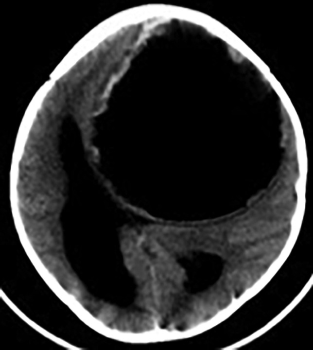
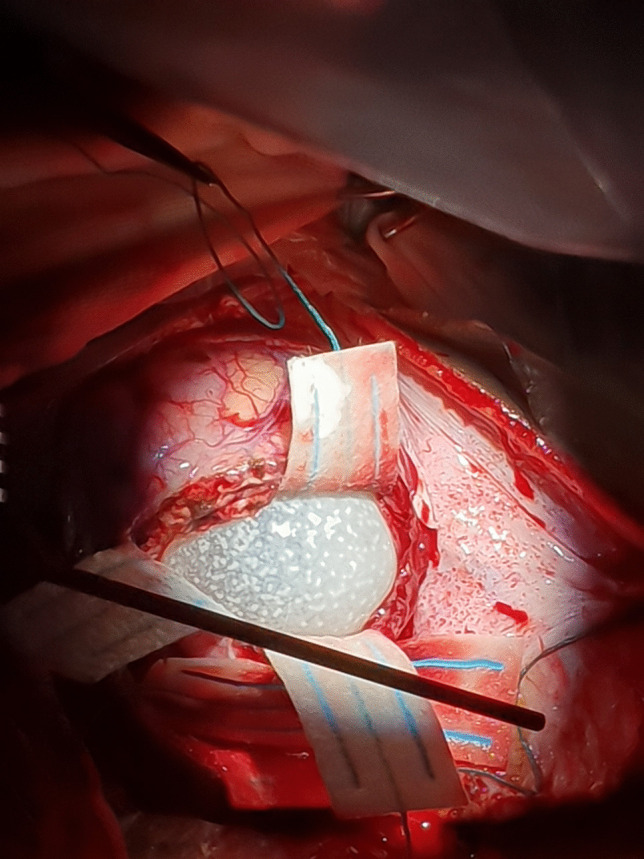
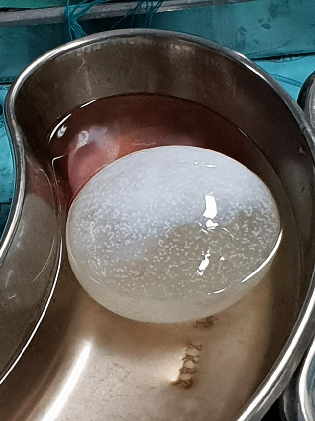
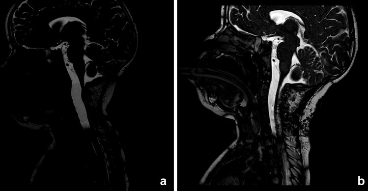
Hydatid disease of the central nervous system is relatively rare and comprises about 2-3% of all the hydatid cyst cases reported in the world. Spinal hydatid disease is an even rarer entity. It is endemic in sheep and cattle-raising regions, seen mainly in Mediterranean countries including Turkey and Syria. Pediatric neurosurgeons in non-endemic countries face a challenge when they encounter children with hydatid cysts of the central nervous system, mostly due to lack of awareness and the ensuing diagnostic dilemmas. It is also a significant socioeconomic problem in developing countries, due to improper hygiene and lack of dedicated veterinary practice. The clinical features are largely nonspecific and very according to location and severity of disease. However, with the advent of advances in MR imaging, the diagnostic accuracy of hydatic disease involving the brain and spine has increased. Intact removal of the cyst/s, without causing any spillage, and appropriate antihelminthic therapy is the goal and key to cure and prevention of recurrence. In this manuscript, the current literature on hydatid cyst of the brain and spine is reviewed to better understand the epidemiology, pathophysiology, diagnostic accuracy, and advances in therapeutic options. A heightened clinical suspicion, awareness of MR imaging features, improved surgical strategies, and options for prevention are discussed.
Keywords: Echinococcus; Hydatid disease; Intracranial infestation; Spinal infection.
© 2022. The Author(s), under exclusive licence to Springer-Verlag GmbH Germany, part of Springer Nature.
Conflict of interest statement
The authors have no conflict of interest or funding to declare for this manuscript.
Figures


References
-
- Fuchs R (1895) Hippokrates. Sämtliche Werke, München: Lüneburg
-
- Hosemann G (1928) Die Echinokokkenkrankheit. Enke
-
- Eckert J, Thompson RC. Historical aspects of echinococcosis. Adv Parasitol. 2017;1(95):1–64. - PubMed
-
- Abbasioun K, Amirjam SA. Diagnosis and management of hydatid cyst of the central nervous system. Neurosurgery. 2001;11:1–9. doi: 10.1097/00013414-200103000-00001. - DOI
Publication types
MeSH terms
LinkOut - more resources
Full Text Sources
Medical
