. 2022 Dec;4(12):1732-1745.
doi: 10.1038/s42255-022-00693-8. Epub 2022 Nov 28.

Maternal diet disrupts the placenta-brain axis in a sex-specific manner

Affiliations

Maternal diet disrupts the placenta-brain axis in a sex-specific manner

Alexis M Ceasrine et al. Nat Metab. 2022 Dec.

Abstract

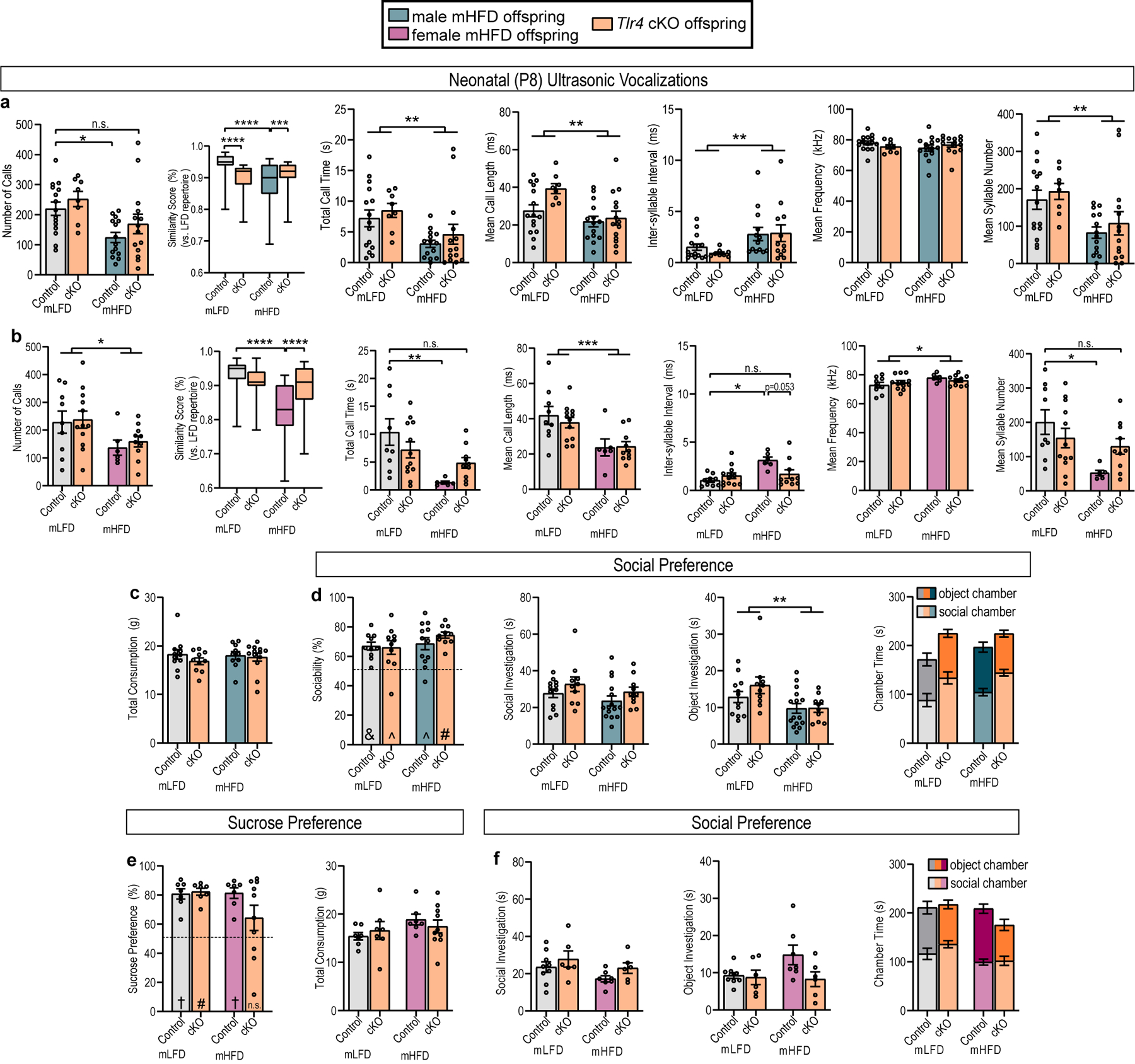
High maternal weight is associated with detrimental outcomes in offspring, including increased susceptibility to neurological disorders such as anxiety, depression and communicative disorders. Despite widespread acknowledgement of sex biases in the development of these disorders, few studies have investigated potential sex-biased mechanisms underlying disorder susceptibility. Here, we show that a maternal high-fat diet causes endotoxin accumulation in fetal tissue, and subsequent perinatal inflammation contributes to sex-specific behavioural outcomes in offspring. In male offspring exposed to a maternal high-fat diet, increased macrophage Toll-like receptor 4 signalling results in excess microglial phagocytosis of serotonin (5-HT) neurons in the developing dorsal raphe nucleus, decreasing 5-HT bioavailability in the fetal and adult brains. Bulk sequencing from a large cohort of matched first-trimester human samples reveals sex-specific transcriptome-wide changes in placental and brain tissue in response to maternal triglyceride accumulation (a proxy for dietary fat content). Further, fetal brain 5-HT levels decrease as placental triglycerides increase in male mice and male human samples. These findings uncover a microglia-dependent mechanism through which maternal diet can impact offspring susceptibility for neuropsychiatric disorder development in a sex-specific manner.


Conflict of interest statement

The authors declare no competing interests.

Figures

Extended Data Fig. 1
Extended Data Fig. 1. Maternal high-fat diet increases maternal and offspring weight, but does not impact litter size, sex ratio, or maternal care
a, mHFD increases weight before mating (n=20 females/diet) b, mHFD dams are heavier during gestation than mLFD dams (n=15 dams/diet). c, Litter size and sex ratio are not impacted by mHFD (n=15 mLFD, 13 mHFD litters). d, Percent of time spent on nest is unchanged by HFD (n=5 dams/diet) e, Percent of time spent nursing is unchanged by HFD (n=5 dams/diet). f, Male and female mHFD offspring weights are increased compared to sex-matched mLFD offspring (*P < 0.05, **P < 0.01, *** P < 0.001; exact P and n values can be found in Source Data). g, Placenta weight is not changed by mHFD (n=6 mLFD and 8 mHFD litters (large solid circles); individual placenta weights are represented in small open circles)). Data are mean ± s.e.m. P values are derived from mixed-effects two-way ANOVA (diet × time; a, b), two-way ANOVA (time of day × diet; d, e), (sex × maternal diet; g), or unpaired two-tailed t-tests (c, f).
Extended Data Fig. 2
Extended Data Fig. 2. Maternal high-fat diet imparts sex-specific offspring behavioral outcomes
a-e, mHFD decreases total USV call time and mean call length, increases mean inter-syllable interval, does not affect mean call frequency, and decreases mean syllable number in male offspring (n=9 mLFD, 8 mHFD male offspring from 4 mLFD and 3 mHFD litters). f, mHFD dependent decrease in USV number is apparent throughout neonatal development in male offspring (P8 data as previously shown, n= 11 P7 and 7 P10 mLFD offspring from 3 P7 litters and 4 P10 litters, 14 P7 and 8 P10 mHFD offspring from 4 P7 and 3 P10 litters). g-k, mHFD decreases total USV call time and mean USV call length, increases mean inter-syllable interval and mean call frequency, and decreases mean syllable number in female offspring (n=18 mLFD, 14 mHFD female offspring from 5 mLFD and 3 mHFD litters). l, mHFD dependent decrease in USV number is apparent throughout neonatal development in female offspring (P8 data as shown previously, n=13 P7 and 14 P10 mLFD offspring from 5 P7 and 5 P10 litters, 5 P7 and 10 P10 mHFD offspring from 3 P7 and 3 P10 litters) m, mHFD does not alter male offspring social or object investigation times or chamber times during a 3-chamber social preference test (n=13 mLFD, 15 mHFD male offspring from 5 mLFD and 6 mHFD litters). n, Male mLFD and mHFD offspring display a strong preference for a novel social stimulus over a familiar one. mHFD does not alter male offspring novel or familiar investigation times or chamber times in a social novelty preference test (n=11 mLFD and 18 mHFD offspring from 4 mLFD and 7 mHFD litters). o, Female mHFD offspring spend less time investigating a social stimulus than female mLFD offspring, but overall chamber time is not different (n=17 mLFD, 16 mHFD female offspring from 6 mLFD and 6 mHFD litters). p, Female mHFD offspring have decreased preference for a novel social stimulus compared to mLFD female offspring. Female mHFD offspring spend less time investigating a novel social stimulus than female mLFD offspring and spend less time in the chamber containing the novel conspecific (n=14 mLFD and 14 mHFD offspring from 5 mLFD and 5 mHFD litters). Data are mean ± s.e.m.; P values are derived from unpaired two-tailed t-tests (a, b, c, e, g, h, i, j, k, o, p), 2-way ANOVA (diet × age; f, l) or one-sample t-tests assessing difference from chance (50%; n, p; †P < 0.001, &P < 0.01, ^P < 0.05).
Extended Data Fig. 3
Extended Data Fig. 3. Maternal high-fat diet imparts sex-specific offspring behavioral outcomes
a, Male offspring total consumption (water + sucrose) is unaffected by maternal diet (n=10 mLFD, 15 mHFD male offspring from 5 mLFD and 5 mHFD litters). b, Schematic of forced swim test (FST) created with Biorender.com. c-e, Male mHFD offspring spend less time immobile than mLFD offspring, move a greater cumulative distance, and have a higher swimming velocity than mLFD offspring during an FST (n= 6 mLFD and 7 mHFD offspring from 3 mLFD and 4 mHFD litters). f, Female offspring total consumption is unaffected by maternal diet (n=14 mLFD, 12 mHFD female offspring from 5 mLFD and 5 mHFD litters). g-i, Female mHFD perform similarly to female mLFD offspring in a forced swim test (n=9 mLFD, 6 mHFD offspring from 4 mLFD and 3 mHFD litters). j, Schematic of an open field test created with biorender.com. k-n, Distance traveled, velocity, number of middle entries, and percent of time in center in an open field test are unchanged in male mHFD offspring (n=10 mLFD, 10 mHFD male offspring from 3 mLFD and 4 mHFD litters). o-r, Distance traveled, velocity, number of middle entries, and percent of time in center in an open field test are unchanged in female mHFD offspring (n=11 mLFD, 11 mHFD female offspring from 3 mLFD and 3 mHFD litters). Data are mean ± s.e.m.; P values are derived from unpaired two-tailed t-tests (c, d, e).
Extended Data Fig. 4
Extended Data Fig. 4. Maternal high-fat diet decreases serotonin in male offspring
a, Maternal plasma 5-HT levels are unaffected by HFD at gd14.5 (n=9 LFD and 8 HFD pregnant dams). b, Tph2 and 5HTT are significantly decreased in male mHFD placenta (n=10 mLFD, 9 mHFD offspring from 3 mLFD and 4 mHFD litters). c, MAOA is significantly decreased in female mHFD placenta (n= 13 mLFD, 16 mHFD offspring from 3 mLFD and 4 mHFD litters). d, 5-HT and 5-HT synthesis (5-HT/tryptophan (Trp)) are significantly decreased while 5-HT turnover (5-HIAA/5-HT) is increased in male mHFD placenta (n= 8 mLFD and 8 mHFD offspring from 8 mLFD and 8 mHFD litters). e, 5-HT and Tryptophan are significantly decreased in male mHFD forebrain tissue (n=8 mLFD and 9 mHFD offspring from 8 mLFD and 9 mHFD dams). f, mHFD does not influence 5-HT, 5-HT synthesis, or 5-HT turnover in female placenta (n=6 mLFD and 7 mHFD from 6 mLFD and 7 mHFD litters). g, mHFD does not influence 5-HT but decreases tryptophan levels in female forebrain (n=8 mLFD and 6 mHFD from 8 mLFD and 6 mHFD litters). h, Quinolinic acid levels are unaffected by mHFD in male placenta and fetal brain (n=5 mLFD and 5mHFD offspring from 5 mLFD and 5 mHFD litters). i, Quinolinic acid levels are unaffected by mHFD in female placenta and fetal brain (n=5 mLFD and 5 mHFD offspring from 5 mLFD and 5 mHFD litters). Data are mean ± s.e.m. P values are derived from unpaired one-sample t-tests (b, c), or unpaired two-tailed t-tests (d, e, g)
Extended Data Fig. 5
Extended Data Fig. 5. Maternal tryptophan supplementation rescues mHFD-induced behavioral changes in male offspring
a, Dietary tryptophan enrichment does not impact maternal weight (n=15 LFD, 16 LFD+Trp, 15 HFD, 17 HFD+Trp females) b, Dietary tryptophan enrichment does not impact litter size or sex ratio (n=10 mLFD, 12 mLFD+Trp, 11 mHFD, 11 mHFD+Trp litters) c-d, Maternal dietary tryptophan enrichment does not impact offspring weight (n values can be found in Source Data) e, Maternal dietary tryptophan enrichment increases serotonin in mLFD, but not mHFD, male placenta (n=11 mLFD, 15 mLFD+Trp, 17 mHFD, 12 mHFD+Trp offspring from 4 mLFD, 5 mLFD+Trp, 4 mHFD, and 4 mHFD+Trp litters). f, Maternal dietary tryptophan enrichment disrupts the positive correlation between brain and placenta serotonin in male offspring (n=15 mLFD +/− Trp offspring from 4 mLFD and 5 mLFD+Trp litters). g, Brain and placenta serotonin levels are not correlated in male mHFD +/− tryptophan offspring (n=14 mHFD, 17 mHFD+Trp offspring from 4 mHFD and 5 mHFD+Trp litters). h, Maternal dietary tryptophan enrichment increases serotonin levels in mHFD adult male midbrain (n=6 mLFD, 6 mLFD+Trp, 8 mHFD, 6 mHFD+Trp offspring from 4 mLFD, 6 mLFD+Trp, 6 mHFD, and 6 mHFD+Trp litters). i-m, Total call time, mean call length, mean inter-syllable interval, mean frequency, and mean syllable number in male mLFD and mHFD +/− tryptophan offspring (n=13 mLFD, 21 mLFD+Trp, 23 mHFD, 19 mHFD+Trp offspring from 5 mLFD, 7 mLFD+Trp, 8 mHFD, and 6 mHFD+Trp litters). n-q, Maternal dietary tryptophan enrichment does not impact male offspring juvenile social behavior (n=8 mLFD, 15 mLFD+Trp, 6 mHFD, and 14 mHFD+Trp male offspring from 3 mLFD, 6 mLFD+Trp, 3 mHFD, and 4 mHFD+Trp litters). r-u, Maternal dietary tryptophan enrichment does not impact open field behavior in adult male offspring (12 mLFD, 9 mLFD+Trp, 13 mHFD, 12 mHFD+Trp male offspring from 4 mLFD, 4 mLFD+Trp, 3 mHFD, and 5 mHFD+Trp litters). Data are mean ± s.e.m.; P values are derived from 2-way ANOVA (Fat × Trp content; b, e, h, i, j, k, m), 3-way ANOVA (Fat Content × Trp content × Days on Diet/Age; a, c, d), two-tailed Pearson’s correlation (f, g), or one-sample t-tests assessing difference from chance (50%; n; #P < 0.0001, †P < 0.001, ^P < 0.05).
Extended Data Fig. 6
Extended Data Fig. 6. Maternal tryptophan supplementation does not rescue behavior in female mHFD offspring
a, Maternal dietary tryptophan enrichment does not impact adult female offspring midbrain serotonin levels (n=11 mLFD, 11 mLFD+Trp, 9 mHFD, 10 mHFD+Trp offspring from 6 mLFD, 9 mLFD+Trp, 7 mHFD, and 8 mHFD+Trp litters). b-h, Maternal dietary tryptophan enrichment does not rescue mHFD-dependent changes to neonatal female ultrasonic vocalizations (n=19 mLFD, 28 mLFD+Trp, 23 mHFD, 22 mHFD+Trp offspring from 6 mLFD, 8 mLFD+Trp, 8 mHFD, and 9 mHFD+Trp litters). i, Maternal tryptophan enrichment increases female offspring sucrose preference regardless of maternal dietary fat intake (n=8 mLFD, 8 mLFD+Trp, 8 mHFD, 11 mHFD+Trp from 3 mLFD, 5 mLFD+Trp, 4 mHFD, and 6 mHFD+Trp litters) j-m, Maternal tryptophan enrichment influence on juvenile social preference (n=8 mLFD, 11 mLFD+Trp, 6 mHFD, and 10 mHFD+Trp offspring from 3 mLFD, 5 mLFD+Trp, 4 mHFD, and 3 mHFD+Trp litters). n-q, Maternal tryptophan enrichment influence on open field behavior (n=13 mLFD, 11 mLFD+Trp, 9 mHFD, and 12 mHFD+Trp offspring from 4 mLFD, 4 mLFD+Trp, 4 mHFD, and 4 mHFD+Trp litters). Data are mean ± s.e.m. except for the box plot (c) where whiskers are min. to max., hinges of boxes are 25th and 75th percentiles, and the middle line is the median; P values are derived from 2-way ANOVA (fat content × tryptophan content; b, c, d, e, f, g, h, i, n, o) or one-sample t-tests assessing difference from chance (50%; j; &P < 0.01). n.s. not significant
Extended Data Fig. 7
Extended Data Fig. 7. mHFD induces Tlr4-dependent embryonic inflammation driving behavior changes in males and females
a, Representative e14.5 placenta delineating decidua (D), junctional zone (JZ), and fetal labyrinth (L). Clear delineation was observed in all samples (n=3 animals/sex/diet) b, Placental macrophages are primarily located in the stroma at e14.5 (inset 2), though some are within the vasculature (inset 1). Similar results were obtained across 6 animals (approximately 10 sections/animal). c, Increased macrophage density in male and female mHFD placenta (scale=50μm; n=3 animals per sex/diet). d, qPCR (Mean fold change values shown normalized to 18S) from male and female e14.5 midbrain microglia (n=4 male and 5 female/diet from 4 mLFD and 5 mHFD litters) e, IMARIS reconstruction quantification from female e14.5 dorsal raphe nucleus microglia. Statistics shown for animal averages (large circles), small circles are individual microglia. (n=3 mLFD, 5, mLFD+Trp, 6 mHFD, 5 mHFD+Trp from 3 litters/diet). f, Representative images of male placenta macrophages. Macrophage density in the placenta remains increased in mHFD+Trp male offspring. Statistics shown for animal averages (large circles), small circles are individual images. (scale=50μm; n=6 mLFD, 5 mLFD+Trp, 5 mHFD, 5 mHFD+Trp). g, qPCR from male and female e14.5 placenta (male: n=11 mLFD, 10 mHFD; female: n=13 mLFD, 15 mHFD from 3 mLFD and 4 mHFD litters). h, Schematic of macrophage-specific Tlr4 knockout created with Biorender.com and confirmation of Tlr4 knockdown in microglia (n=3 control, 5 Tlr4 cKO mice). i-j, Serotonin levels are significantly increased by macrophage-specific loss of Tlr4 in male mHFD offspring fetal forebrain and adult midbrain (n=8 mHFD control, 11 mHFD cKO fetal forebrain from 6 mHFD litters; n=9 mHFD, 9 mHFD cKO adult midbrain from 4 litters). k, Placenta 5-HT in mHFD male offspring with or without macrophage Tlr4-signaling (n=6 mHFD control, 11 mHFD cKO from 6 mHFD litters). l, IMARIS reconstructions from female mLFD and mHFD control and cKO e14.5 DRN. Statistics ran for animal averages (large circles), small circles are individual microglia. (n=7 mLFD control, 5 mLFD cKO, 5 mHFD control, and 4 mHFD cKO from 7 mLFD and 5 mHFD litters) m, Tlr4-reporter activity in response to lipopolysaccharide (LPS) and saturated fatty acids. (n=5 biological replicates (each replicate was run in triplicate, and the average is represented as one open circle)). Data are mean ± s.e.m.; P values are derived from unpaired two-tailed t-tests (g, i, j, k), two-way ANOVA (c, e, f, h), or one-sample t-tests assessing difference from 0 (baseline; m).
Extended Data Fig. 8
Extended Data Fig. 8. mHFD induces Tlr4-dependent inflammation driving offspring behavior changes
a, USV metrics from male and b, female control and cKO mLFD and mHFD offspring (n=15 male mLFD control and 8 cKO from 5 litters and 14 male mHFD control and 14 cKO from 6 litters; n=9 female mLFD control and 12 cKO from 7 litters and 6 female mHFD and 10 cKO from 5 litters). c, Total liquid consumption in mLFD and mHFD control and cKO males (n=11 mLFD, 10 mLFD cKO, 11 mHFD, and 10 mHFD cKO from 5 mLFD and 6 mHFD litters) d, Social preference in male mLFD and mHFD control and Tlr4 cKO offspring (n=10 mLFD, 10 mLFD cKO, 12 mHFD, 10 mHFD cKO from 5 mLFD and 6 mHFD litters) e, Sucrose preference in female mLFD and mHFD control and Tlr4 cKO offspring (n=7 mLFD, 7 mLFD cKO, 7 mHFD, 10 mHFD cKO from 5 mLFD and 4 mHFD litters) f, Social preference metrics in female mLFD and mHFD control and Tlr4 cKO offspring (n=9 mLFD, 6 mLFD cKO, 7 mHFD, and 6 mHFD cKO from 5 mLFD and 5 mHFD litters). Data are mean ± s.e.m. except for the box plots (a, b) where whiskers are min. to max., hinges of boxes are 25th and 75th percentiles, and the middle line is the median; P values are derived from two-way ANOVA (a, b, d, f) or one-sample t-tests assessing difference from chance (50%; d, e; #P < 0.0001, †P < 0.001, &P < 0.01, ^P < 0.05).
Extended Data Fig. 9
Extended Data Fig. 9. Human maternal decidual triglyceride accumulation negatively correlates with fetal brain serotonin in males only
a, Average gestational age was equal in male and female human tissue samples (n=17 male and 20 female). b, Triglyceride levels are increased in mHFD placenta (n=7 mLFD and 8 mHFD male placenta (open circles from 5 mLFD and 3 mHFD dams; statistics shown for litter average (diamonds)). c, Fetal brain serotonin levels are significantly negatively correlated with placental triglyceride accumulation (n=11 individuals from 5 litters). d, Average decidual triglyceride levels were statistically equal but trended higher in female pregnancies versus male. e, Violin plot showing log normalized expression of marker genes for striatum and dorsal thalamus, midbrain and cerebellum, and frontal cortex and hippocampal formation regions from human brain samples in males and females. The color of the dot representing each sample shows the brain 5-HT concentration for that sample. f, Heatmap depicting strength Pearson’s correlations for select genes marking syncytiotrophoblasts, trophoblasts, and immune-related signaling in the placenta. Data are mean ± s.e.m.; P values (* P < 0.05) are derived from unpaired two-tailed t-tests (b) or two-tailed Pearson’s correlations (c, f).
Figure 1.
Figure 1.. Maternal high-fat diet imparts sex-specific offspring behavioral outcomes
a, Schematic of maternal low-fat and high-fat diet paradigm (10% and 45% represent %kcal from fat in diet). b, Schematic of neonatal maternal separation-induced ultrasonic vocalization (USV) recording and analysis. c-f, mHFD decreases neonatal USV number and syllable repertoire similarity in male and female offspring (n=9 mLFD, 8 mHFD male offspring; 18 mLFD, 14 mHFD female offspring from 5 mLFD and 4 mHFD litters). g, Schematic of 3-chambered social preference test. h, Male mLFD and mHFD offspring display a strong social preference (n=13 mLFD, 15 mHFD male offspring from 5 mLFD and 6 mHFD litters). i, Female mHFD offspring have a reduced social preference versus mLFD offspring (n=17 mLFD, 16 mHFD female offspring from 6 mLFD and 6 mHFD litters). j, Schematic of sucrose preference test. k, Male mHFD offspring display a decreased preference for sucrose versus mLFD offspring (n=10 mLFD, 15 mHFD male offspring from 5 mLFD and 5 mHFD litters). l, Female mLFD and mHFD offspring display a strong sucrose preference (n=14 mLFD, 12 mHFD female offspring from 5 mLFD and 5 mHFD litters). Data are mean ± s.e.m. except for the box plots (d, f) where whiskers are min. to max., hinges of boxes are 25th and 75th percentiles, and the middle line is the median; P values are derived from unpaired two-tailed t-tests (c, e, i, k), paired two-tailed t-tests (d, f), or one-sample t-tests assessing difference from chance (50%; h, i, k, l; #P < 0.0001, †P < 0.001, &P < 0.01). Schematics (a, b, g, j) created with Biorender.com.
Figure 2.
Figure 2.. mHFD decreases 5-HT in male offspring and increasing 5-HT levels is sufficient to prevent behavioral phenotypes
a, Male mHFD offspring have decreased fetal forebrain serotonin (n=12 mLFD, 3 mHFD male offspring from 3 mLFD and 3 mHFD litters). b, Male mHFD offspring have decreased placenta serotonin (n=18 mLFD, 20 mHFD offspring from 4 mLFD and 6 mHFD litters). c-d, mHFD does not impact fetal serotonin levels in female offspring (forebrain: n=11 mLFD, 13 mHFD from 3 mLFD and 3 mHFD litters; placenta: n=11 mLFD, 24 mHFD female offspring from 4 mLFD and 6 mHFD litters) e-f, Fetal serotonin levels are significantly correlated between brain and placenta in mLFD males only (n=17 male, 10 female mLFD offspring from 4 mLFD litters; 3 male, 9 female mHFD offspring from 3 mHFD litters). g, Maternal dietary tryptophan enrichment increases serotonin levels in mHFD fetal male forebrain (n=14 mLFD, 15 mLFD+Trp, 12 mHFD, 17 mHFD+Trp offspring from 5 mLFD, 5 mLFD+Trp, 3 mHFD, and 5 mHFD+Trp litters) h-i, Maternal dietary tryptophan enrichment rescues mHFD-induced decrease in USV number and syllable repertoire similarity (n= 13 mLFD, 21 mLFD+Trp, 23 mHFD, 19 mHFD+Trp offspring from 5 mLFD, 7 mLFD+Trp, 8 mHFD, and 7 mHFD+Trp litters). j-k, Maternal dietary tryptophan enrichment rescues sucrose preference in mHFD male offspring and does not affect total consumption (n= 12 mLFD, 9 mLFD+Trp, 11 mHFD, 7 mHFD+Trp male offspring from 6 mLFD, 5 mLFD+Trp, 5 mHFD, and 6 mHFD+Trp litters). Data are mean ± s.e.m. except for the box plot (i) where whiskers are min. to max., hinges of boxes are 25th and 75th percentiles, and the middle line is the median; P values are derived from unpaired two-tailed t-tests (a, b), two-tailed Pearson’s correlations (e, f), two-way ANOVA (fat content × tryptophan content; g, h, i, j), or one-sample t-tests assessing difference from chance (50%; j; #P < 0.0001, ^P < 0.05).
Figure 3.
Figure 3.. mHFD induces Tlr4-dependent inflammation driving offspring behavior changes
a, Increased microglia and phagosome immunoreactivity in male and female mHFD DRN at e14.5 (scale=50μm; n=4 male and 3 female/diet from 3mLFD and 3mHFD litters). b, IMARIS reconstructions of DRN microglia reveals increased microglial phagocytosis of serotonin in males at e14.5. Statistics are displayed for animal average (large circles), small circles are individual microglia reconstructions (n=6 mLFD, 5mLFD+Trp, 5 mHFD, 6 mHFD+Trp mice from 3 mLFD, 2 mLFD+Trp, 3 mHFD, and 3mHFD+Trp litters). c, IMARIS reconstruction of DRN microglia in male control and Tlr4 cKO e14.5 offspring (n=6 mLFD control, 6 mLFD cKO, 5 mHFD control, and 3 mHFD cKO male offspring from 7 mLFD and 5 mHFD litters). d, Decreased sucrose preference is rescued in male mHFD offspring lacking macrophage-Tlr4 signaling. (n=11 mLFD, 10 mLFD cKO, 11 mHFD, and 10 mHFD cKO from 5 mLFD and 6 mHFD litters). e, Decreased social preference is rescued in female mHFD offspring lacking macrophage-Tlr4 signaling. (n=9 mLFD, 6 mLFD cKO, 7 mHFD, and 6 mHFD cKO from 5 mLFD and 5 mHFD litters). f-g, Tlr4-reporter activity in response to e14.5 brain (f) and placenta (g) lysate from mLFD and mHFD offspring (n=6 offspring/sex/diet from 5 mLFD and 5 mHFD litters). h-i, Endotoxin levels in mLFD and mHFD brain (h) and placenta (i) tissue lysate (n=3–5 mLFD male, 4–5 mLFD female, 5 mHFD male, 4–5 mHFD female from 5 mLFD and 5 mHFD litters). Data are mean ± s.e.m.; P values are derived from two-way ANOVA (a-i) or one-sample t-tests assessing difference from chance (50%; d, e; #P < 0.0001, †P < 0.001, &P < 0.01). n.s. not significant.
Figure 4.
Figure 4.. Maternal triglycerides negatively correlate with male fetal brain 5-HT
a, Human fetal development at 63–81 days post conception (d.p.c.) is roughly equivalent to mouse embryonic development at 14–16 d.p.c. (schematic created with Biorender.com) b, Scatter plots showing transcript (TPM) correlation with maternal triglycerides versus −Log10(P value) in male and female placenta and brain bulk sequencing data (n=16 matched male tissue sets (3 male brain excluded for cerebellum/midbrain overrepresentation, 19 female). Red dots indicate a gene that is significantly correlated (P < 0.05; two-tailed Pearson’s correlation). c, GO Enrichment analysis of transcripts significantly correlated with maternal triglyceride accumulation demonstrates robust and distinct upregulation in immune responses in male and female placenta in response to increasing maternal triglyceride accumulation. GO enrichment of transcripts significantly correlated with maternal triglyceride accumulation shows downregulation of neuronal development and function in female brain tissue in response to increasing maternal triglyceride accumulation. d, Multivariate dot plots show two-tailed Spearman correlations between brain and placental serotonin from male and female human fetal tissues. Dot color represents the age of the matched tissue set, and dot size represents the maternal decidual triglyceride level (mg/dL), a proxy for maternal dietary fat intake. Brain and placental serotonin levels are positively correlated in male, but not female, tissues between 72 and 82 d.p.c. (n=17 matched male tissue sets, 20 female). e, Brain serotonin levels are significantly negatively correlated with decidual triglyceride levels in males only. Placental serotonin levels trend towards a negative correlation with decidual triglycerides in males only. Data are mean ± s.e.m.; n represents biologically independent matched human tissue set. P values are derived from two-tailed Spearman correlations.

Comment in

References

    1. Vickers MH, Breier BH, Cutfield WS, Hofman PL & Gluckman PD Fetal origins of hyperphagia, obesity, and hypertension and postnatal amplification by hypercaloric nutrition. Am J Physiol Endocrinol Metab 279, E83–7 (2000). - PubMed
    1. Edlow AG Maternal obesity and neurodevelopmental and psychiatric disorders in offspring. Prenat Diagn 37, 95–110 (2017). - PMC - PubMed
    1. Kott J & Brummelte S Trick or treat? Evaluating contributing factors and sex-differences for developmental effects of maternal depression and its treatment. Horm Behav 111, 31–45 (2019). - PubMed
    1. Krakowiak P et al. Maternal metabolic conditions and risk for autism and other neurodevelopmental disorders. Pediatrics 129, e1121–8 (2012). - PMC - PubMed
    1. Mina TH et al. Prenatal exposure to very severe maternal obesity is associated with adverse neuropsychiatric outcomes in children. Psychological Medicine 47, 353–362 (2017). - PubMed

Publication types