

Life-threatening paradoxical thromboembolism in a patient with patent foramen ovale
- PMID: 36443797
- PMCID: PMC9703718
- DOI: 10.1186/s12947-022-00298-x
Life-threatening paradoxical thromboembolism in a patient with patent foramen ovale
Abstract
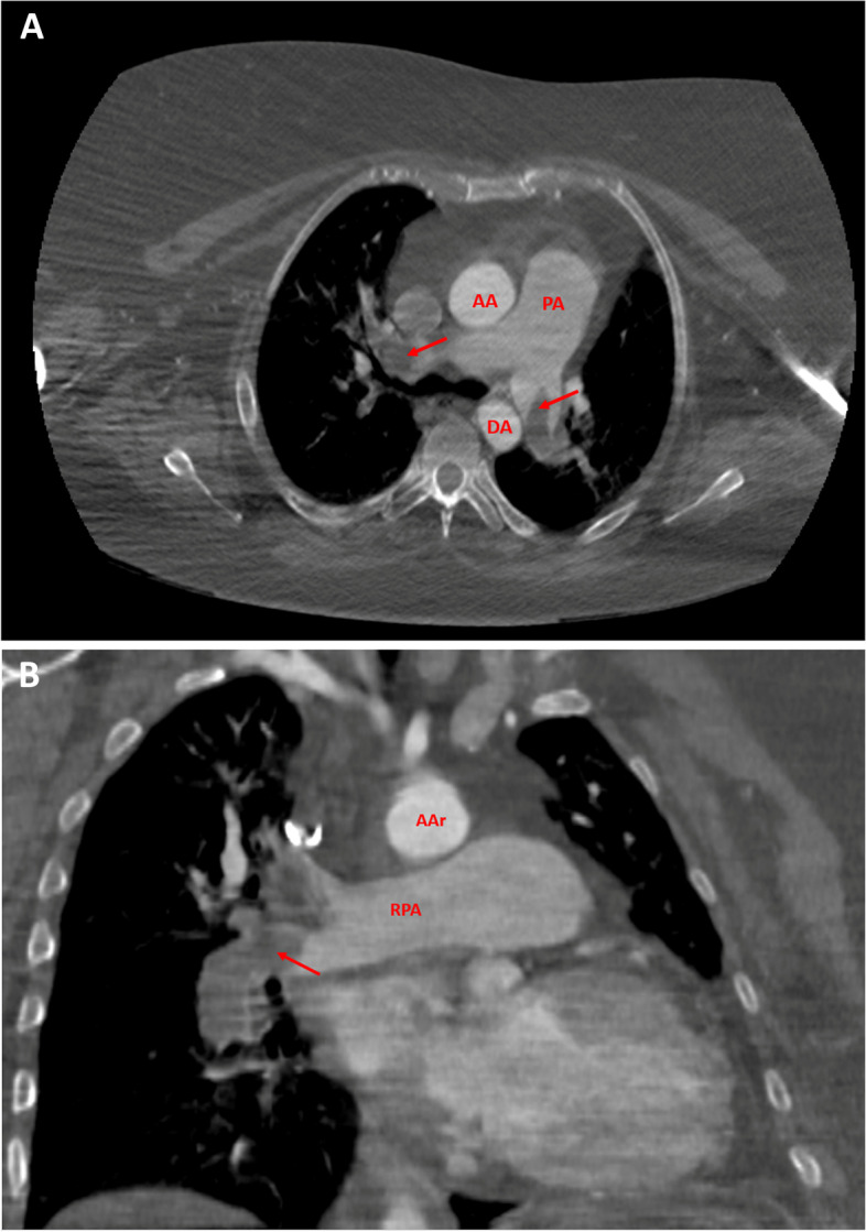
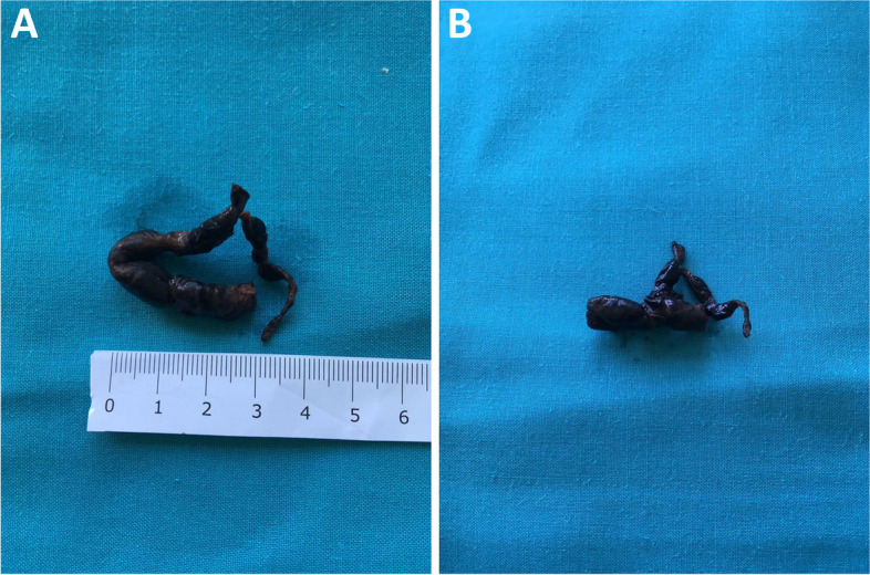
Background: Venous thromboembolism represents the third most frequent acute cardiovascular syndrome worldwide. Its clinical manifestations are deep vein thrombosis and/or pulmonary embolism. Despite a considerable mortality, diagnosis is often missed. CASE PRESENTATION: We report the management of a female patient with high-risk pulmonary thromboembolism treated initially with thromboaspiration, complicated by embolus jailing in a patent foramen ovale. In this situation, left cardiac chambers and systemic circulation were jeopardized by this floating embolus.
Conclusions: High-risk pulmonary embolism requires reperfusion strategy but sometimes mechanical thromboaspiration may be not fully successful; transesophageal echocardiography led to a prompt diagnosis of this unexpected finding; in this very particular case, open surgery represented a bail-out procedure to avoid cerebral and systemic embolism.
Keywords: Deep vein thrombosis; Paradoxical embolization; Patent foramen ovale; Pulmonary embolism.
© 2022. The Author(s).
Conflict of interest statement
The authors declare that they have no competing interests.
Figures

References
-
- Pristipino C, Sievert H, D’Ascenzo F, et al. European position paper on the management of patients with patent foramen ovale. General approach and left circulation thromboembolism. EuroIntervention. 2019;40(38):3182–95. - PubMed
Publication types
MeSH terms
LinkOut - more resources
Full Text Sources
Medical
