

Long-Acting Fluocinolone Acetonide Intravitreal Implant for Recurrent Bilateral Non-Infectious Posterior Uveitis
- PMID: 36444172
- PMCID: PMC9700445
- DOI: 10.2147/IMCRJ.S384356
Long-Acting Fluocinolone Acetonide Intravitreal Implant for Recurrent Bilateral Non-Infectious Posterior Uveitis
Abstract
Purpose: Our case emphasizes the utility of long-acting intravitreal fluocinolone implants (YUTIQ) for managing recalcitrant forms of non-infectious posterior uveitis, NIPU.
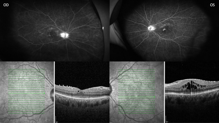
Patient: We present a case of bilateral NIPU refractory to topical corticosteroids and intravitreal triamcinolone and dexamethasone.
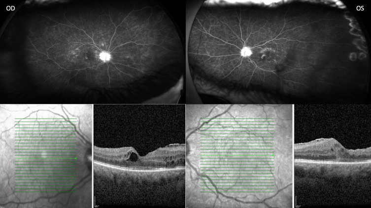
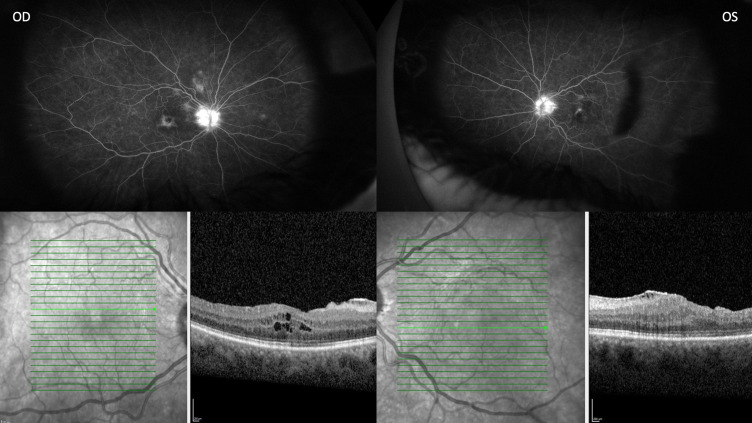
Results: Management with bilateral YUTIQ improved local ocular inflammatory control with improved vision and anatomical outcomes.
Conclusion: NIPU can develop into recurrent forms resistant to short-acting therapeutics. Long-acting efficacy with YUTIQ emphasizes the need to recognize such refractory NIPU cases.
Keywords: NIU; fluocinolone acetonide implant; non-infectious uveitis.
© 2022 Babel et al.
Conflict of interest statement
Dr Eric K Chin was involved in research and clinical trials for Opthea, Genentech, Novartis, Iveric, Chendgu Kanghong Biosciences, Kodiak Sciences, and Bayer, outside the submitted work. The authors report no other conflicts of interest in this work.
Figures
Similar articles
-
Fluocinolone acetonide 0.2 µg/day intravitreal implant in non-infectious uveitis affecting the posterior segment: EU expert user panel consensus-based clinical recommendations.J Ophthalmic Inflamm Infect. 2024 May 30;14(1):22. doi: 10.1186/s12348-024-00402-4. J Ophthalmic Inflamm Infect. 2024. PMID: 38814386 Free PMC article. Review.
-
Efficacy of the Fluocinolone Acetonide (Yutiq) Intravitreal Implant as Monotherapy for Uveitis.Ocul Immunol Inflamm. 2023 Oct;31(8):1603-1607. doi: 10.1080/09273948.2022.2076131. Epub 2022 Jul 6. Ocul Immunol Inflamm. 2023. PMID: 35793136
-
Fluocinolone acetonide 0.18-mg implant for treatment of recurrent inflammation due to non-infectious uveitis: a case series of 15 patients.J Ophthalmic Inflamm Infect. 2024 Sep 19;14(1):44. doi: 10.1186/s12348-024-00427-9. J Ophthalmic Inflamm Infect. 2024. PMID: 39298051 Free PMC article.
-
Preventing relapse in non-infectious uveitis affecting the posterior segment of the eye - evaluating the 0.2 μg/day fluocinolone acetonide intravitreal implant (ILUVIEN®).J Ophthalmic Inflamm Infect. 2020 Nov 30;10(1):32. doi: 10.1186/s12348-020-00225-z. J Ophthalmic Inflamm Infect. 2020. PMID: 33251553 Free PMC article. Review.
-
Use of fluocinolone acetonide intravitreal implant to manage chronic panuveitis for long-term inflammatory control without interfering with systemic immunity.Digit J Ophthalmol. 2022 Dec 28;28(4):119-125. doi: 10.5693/djo.02.2022.10.002. eCollection 2022. Digit J Ophthalmol. 2022. PMID: 36660185 Free PMC article.
Cited by
-
Fluocinolone acetonide 0.2 µg/day intravitreal implant in non-infectious uveitis affecting the posterior segment: EU expert user panel consensus-based clinical recommendations.J Ophthalmic Inflamm Infect. 2024 May 30;14(1):22. doi: 10.1186/s12348-024-00402-4. J Ophthalmic Inflamm Infect. 2024. PMID: 38814386 Free PMC article. Review.
References
-
- Kempen JH, Altaweel MM; Multicenter Uveitis Steroid Treatment (MUST) Trial Research Group. Benefits of systemic anti-inflammatory therapy versus fluocinolone acetonide intraocular implant for intermediate uveitis, posterior uveitis, and panuveitis: fifty-four-Month Results of the Multicenter Uveitis Steroid Treatment (MUST) trial and follow-up study. Ophthalmology. 2015;122(10):1967–1975. doi:10.1016/j.ophtha.2015.06.042 - DOI - PMC - PubMed
Publication types
LinkOut - more resources
Full Text Sources
