

The Athletic ECG: A Line of Defense Against Misinterpretation
- PMID: 36444192
- PMCID: PMC9700076
- DOI: 10.1016/j.jaccas.2022.08.024
The Athletic ECG: A Line of Defense Against Misinterpretation
Abstract
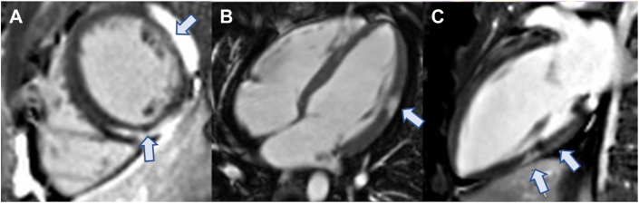
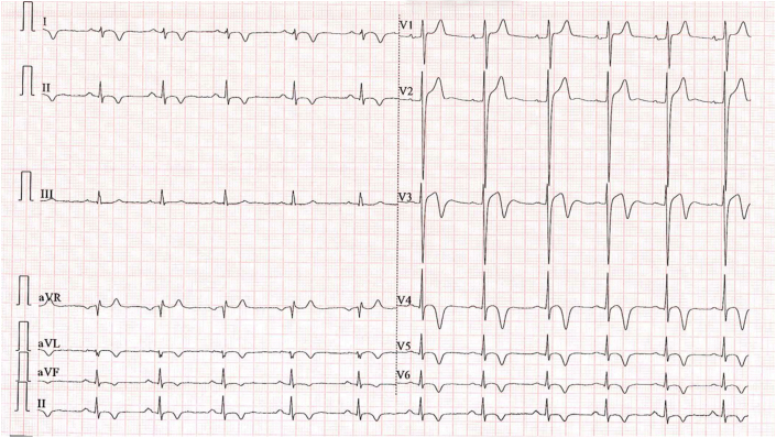
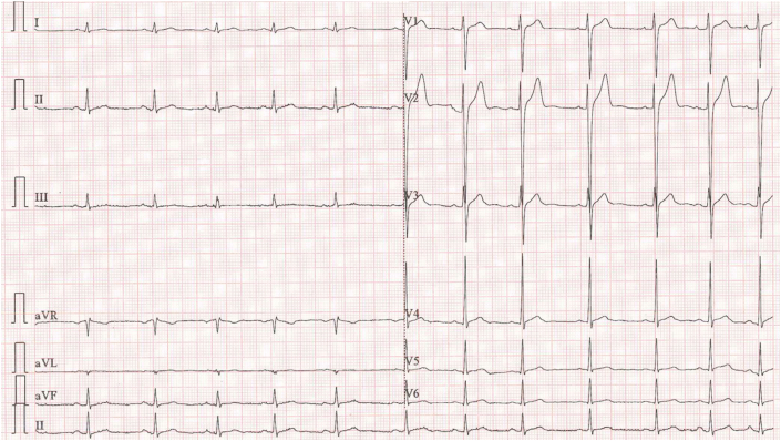
A 17-year-old competitive athlete was found to have a minor electrocardiogram abnormality on routine screening. Cardiac magnetic resonance revealed evidence of marked myocarditis, allowing a subsequent safe abstinence from exercise. The case highlights the importance of careful electrocardiogram interpretation, especially in athletes, where physiologic adaptive changes can pose a diagnostic challenge. (Level of Difficulty: Intermediate.).
Keywords: CMR, cardiac magnetic resonance; ECG, electrocardiogram; EMB, endomyocardial biopsy; cardiac risk; echocardiography; electrocardiogram; exercise; imaging; myocarditis; sports cardiology.
Crown Copyright © 2022 Published by Elsevier on behalf of the American College of Cardiology Foundation.
Conflict of interest statement
The authors have reported that they have no relationships relevant to the contents of this paper to disclose.
Figures


References
-
- Alida L.P., Caforio S.P. Current state of knowledge on aetiology, diagnosis, management, and therapy of myocarditis: a position statement of the European Society of Cardiology Working Group on Myocardial and Pericardial Diseases. Eur Heart J. 2013;34(33):2636–2648. - PubMed
-
- Pelliccia A., Sharma S., Gati S., et al. 2020 ESC guidelines on sports cardiology and exercise in patients with cardiovascular disease: the Task Force on sports cardiology and exercise in patients with cardiovascular disease of the European Society of Cardiology (ESC) Eur Heart J. 2021;42(1):17–96. - PubMed
-
- Barry J., Maron J.E. Eligibility and disqualification recommendations for competitive athletes with cardiovascular abnormalities: task force 3: hypertrophic cardiomyopathy, arrhythmogenic right ventricular cardiomyopathy and other cardiomyopathies, and myocarditis: a scientific statement from the American Heart Association and American College of Cardiology. Circulation. 2015;132(22):273–280. - PubMed
-
- Sharma S., Drezner J., Baggish A., et al. International recommendations for electrocardiographic interpretation in athletes. Eur Heart J. 2018;39(16):1466–1480. - PubMed
-
- O’Leary D. Why bioethics should be concerned with medically unexplained symptoms. Am J Bioeth. 2018;18(5):6–15. - PubMed
Publication types
LinkOut - more resources
Full Text Sources
