. 2022 Nov 10;25(12):105552.
doi: 10.1016/j.isci.2022.105552. eCollection 2022 Dec 22.

Chromodomain helicase DNA-binding domain 2 maintains spermatogonial self-renewal by promoting chromatin accessibility and mRNA stability

Affiliations

Chromodomain helicase DNA-binding domain 2 maintains spermatogonial self-renewal by promoting chromatin accessibility and mRNA stability

Ziqian Min et al. iScience. .

Abstract

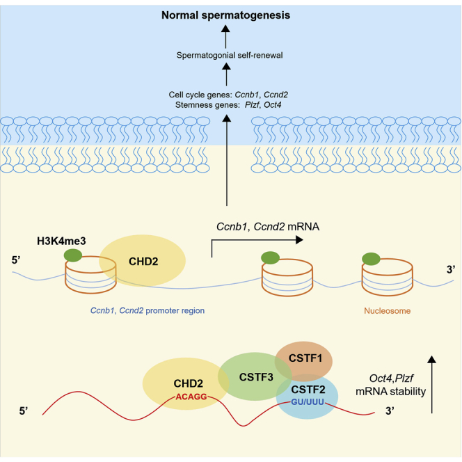
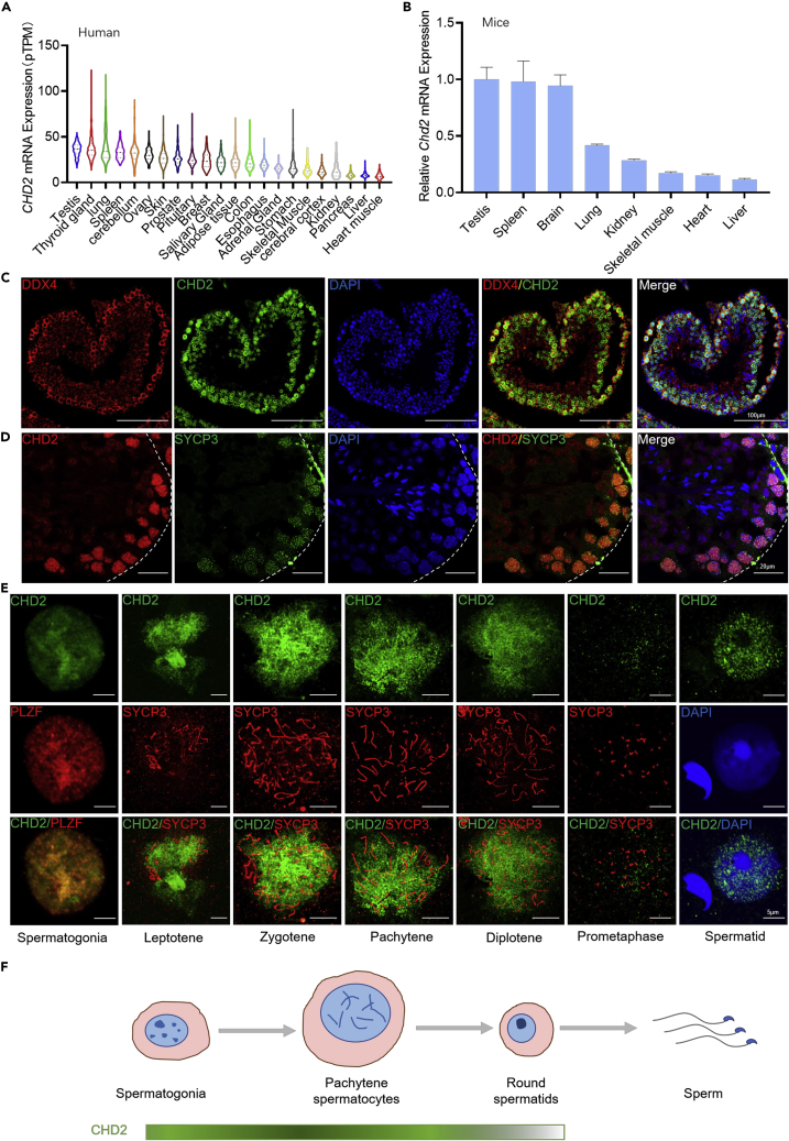
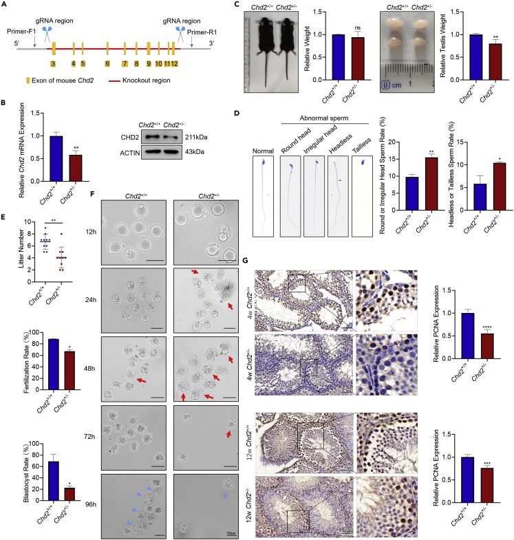
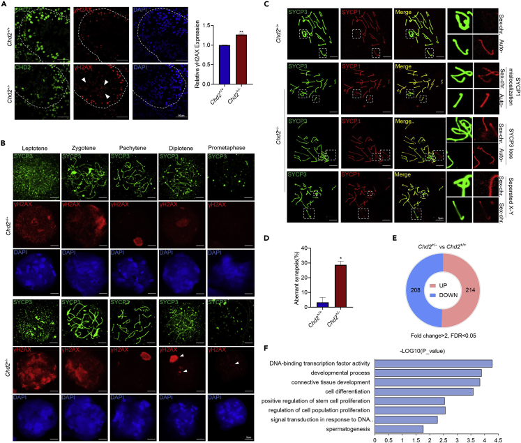
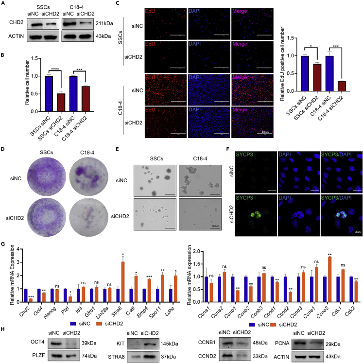
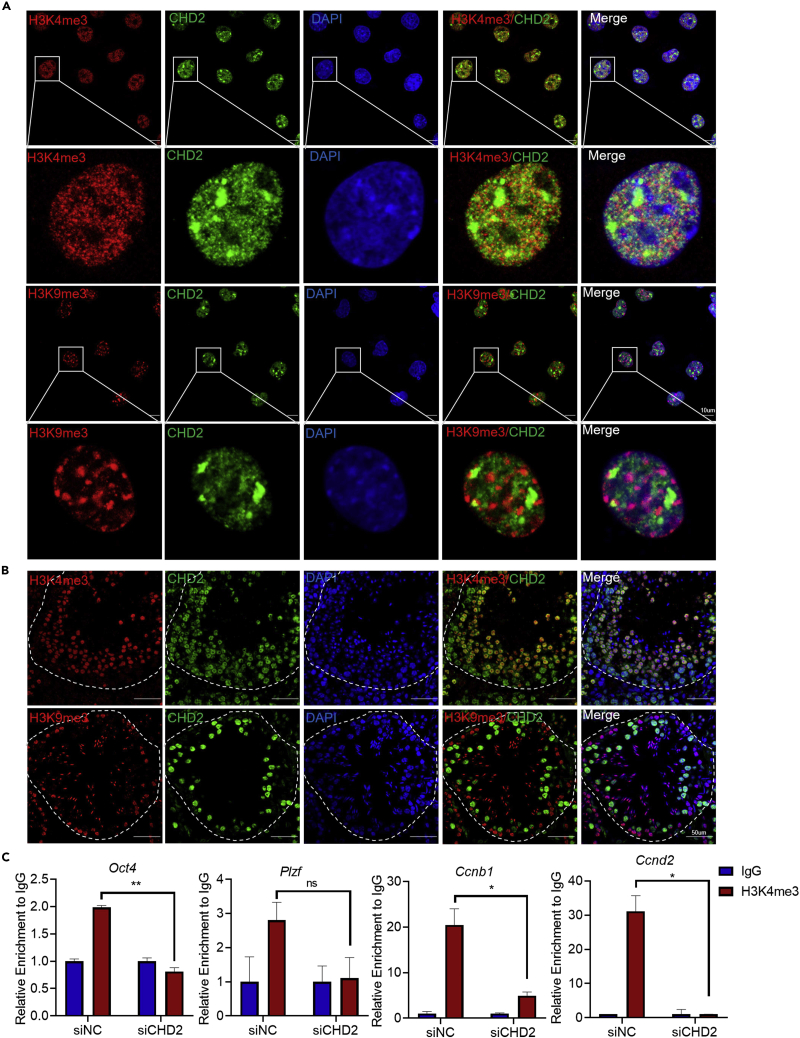
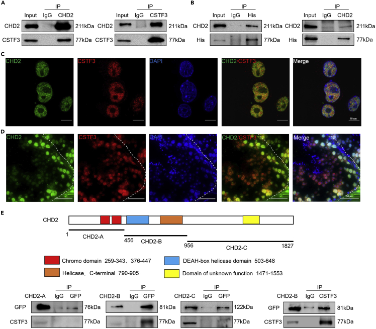
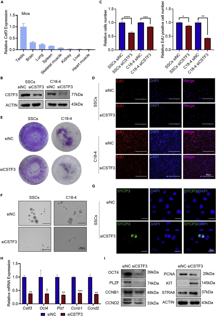
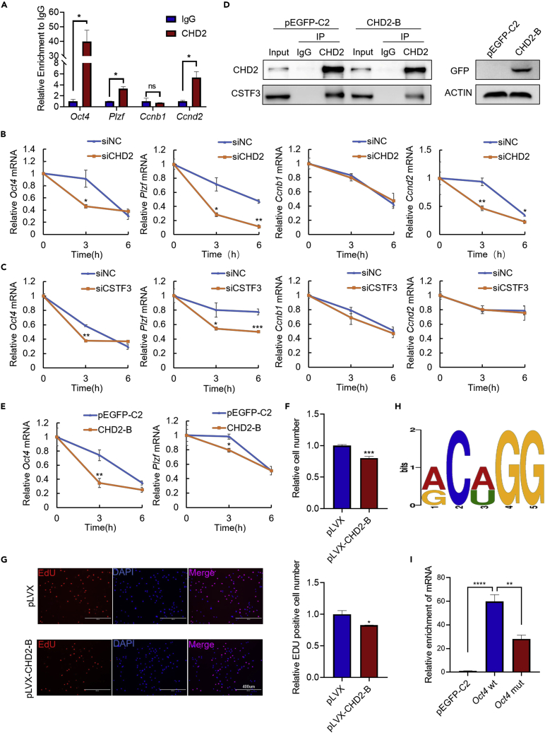
Chromodomain helicase DNA-binding domain 2 (CHD2) is a chromatin remodeling factor involved in many developmental processes. However, its role in male germ cell development has not been elucidated. Here, we confirm that CHD2 expression is enriched in the male germline. In a heterozygous knockout mouse model of Chd2 (Chd2 +/-), we demonstrated that Chd2 haploinsufficiency resulted in testicular developmental delay, an increased rate of abnormal sperm, and impaired fertility in mice. In vitro experiments in mouse spermatogonia showed that CHD2 knockdown inhibits spermatogonial self-renewal. Mechanistically, CHD2 maintains the enrichment of H3K4me3 in the Ccnb1 and Ccnd2 promotors, consequently promoting the transcription of Ccnb1 and Ccnd2. In addition, CHD2 interacts with the cleavage stimulation factor CSTF3 and upregulates the expression of OCT4 and PLZF by improving mRNA stability. This is the first study to reveal the role and mechanism of CHD2 in maintaining spermatogonial self-renewal.

Keywords: Biological sciences; alternate contact; cell biology; molecular mechanism of gene regulation; transcriptomics.


Conflict of interest statement

The authors declare no competing interests.

Figures

None
Graphical abstract
Figure 1
Figure 1
CHD2 is enriched throughout the testicular germ cells, with the highest expression in the spermatocytes of meiosis I (A) Expression analysis of CHD2 mRNA in human tissues based on the HPA database. (B) Expression analysis of Chd2 mRNA in mouse tissues by RT‒qPCR detection relative to 18S rRNA. Data are shown as the mean ± SD. (C and D) The expression and location of CHD2 in adult mouse seminiferous tubule frozen sections by immunofluorescence analysis. Scale bars, 100 μm and 20 μm, respectively. (E) Immunofluorescence analysis of CHD2 expression in spermatogenic cells using mouse testicular cell smears. Scale bar, 5 μm. (F) Model of CHD2 expression during spermatogenesis in mice.
Figure 2
Figure 2
Chd2 haploinsufficiency causes compromised testicular germ cell proliferation and impaired fertility (A) Diagram illustrating the CRISPR/Cas9 targeting strategy to generate Chd2+/− mice, including gRNA sites and PCR primer sites. (B) The expression of CHD2 in testes of Chd2+/− mice by RT‒qPCR and western blot analysis. Data are shown as the mean ± SD. Student’s t-test, ∗∗p < 0.01. (C) Relative body weight and testes volume analysis of 4w Chd2+/+ and Chd2+/− mice (n = 4). Data are shown as the mean ± SD, Student’s t-test; ∗∗p < 0.01; ns, no significance. (D) Morphology analysis and rate of abnormal sperm in Chd2+/+ and Chd2+/− mice. Data are shown as the mean ± SD. Student’s t-test, ∗p < 0.05, ∗∗p < 0.01. Scale bar, 50 μm. (E) Litter number of male Chd2+/+ and Chd2+/− mice. Student’s t-test, ∗∗p < 0.01. (F) The development of zygotes after in vitro fertilization experiments with Chd2+/+ and Chd2+/− mouse sperm. Images were taken 12, 24, 48, 72, and 96 h after fertilization. The red arrows represent degenerated embryos, and the blue triangles represent blastocysts. The fertilization rate and blastocyst rate are shown as the mean ± SD. Student’s t-test, ∗p < 0.05. Scale bar, 100 μm. (G) Immunohistochemical detection of PCNA expression in testicular sections of 4- and 12-week-old Chd2+/+ and Chd2+/− mice. Data are shown as the mean ± SD. Student’s t-test, ∗∗∗p < 0.001, ∗∗∗∗p < 0.0001. Scale bar, 50 μm.
Figure 3
Figure 3
Impaired DNA damage repair, abnormal synapses, and multigene expression changes in Chd2+/− mouse testes (A) Expression analysis of γH2AX in frozen sections of mouse testes by immunofluorescence assay. Triangles represent the abnormal expression of γH2AX. Data are shown as the mean ± SD. Student’s t-test, ∗p < 0.05. Scale bar, 50 μm. (B) Expression analysis of γH2AX in meiosis by immunofluorescence assay. Scale bar, 5 μm. (C) Co-immunofluorescence staining of SYCP3 and SYCP1 in spermatocytes at pachytene from Chd2+/+ and Chd2+/− male mice. Scale bar, 5 μm. (D) The aberrant synapsis rate of Chd2+/+ and Chd2+/− male mice. Data are shown as the mean ± SD. Student’s t-test, ∗p < 0.05. (E) Global gene expression alterations with fold changes of ≥2 or ≤0.5 (p ≤ 0.05) in Chd2+/− versus Chd2+/+ testes at 12 w. (F) Gene ontology analysis of differentially expressed genes identified by RNA-seq in Chd2+/− testes.
Figure 4
Figure 4
Knockdown of CHD2 inhibits spermatogonial self-renewal (A) Detection of RNA interference efficiency for siCHD2 by western blot in SSCs and C18-4 cells. (B–D) Cellular proliferation in SSCs and C18-4 cells with CHD2 knockdown analyzed by CCK8, EdU, and colony-formation assays. Data are shown as the mean ± SD. Student’s t-test, ∗p < 0.05, ∗∗∗p < 0.001. Scale bar, 400 μm. (E) The impacts of CHD2 knockdown on the sphere-formation ability of SSCs and C18-4 cells. Scale bar, 300 μm. (F) Immunofluorescence staining of SYCP3 after CHD2 knockdown in SSCs. Scale bar, 20 μm. (G and H) Expression changes of stem-associated genes, differentiation genes, and cell cycle genes in C18-4 cells with CHD2 knockdown determined by RT‒qPCR and western blot assays. Data are shown as the mean ± SD. Student’s t-test, ∗p < 0.05, ∗∗p < 0.01, ∗∗∗p < 0.001; ns, no significance.
Figure 5
Figure 5
CHD2 maintains an active chromatin state in the promotor of target genes (A and B) The colocalization analysis of CHD2 and H3K4me3 or H3K9me3 in C18-4 cells (A) and sections of testes (B) by immunofluorescence assay. Scale bars, 10 μm and 50 μm, respectively. (C) ChIP‒qPCR assay to determine the relative enrichment of H3K4me3 or H3K9me3 in the promotor regions of the Oct4, Plzf, Ccnb1, and Ccnd2 genes in C18-4 cells with CHD2 knockdown. Data are shown as the mean ± SD. Student’s t-test, ∗p < 0.05, ∗∗p < 0.01; ns, no significance.
Figure 6
Figure 6
CHD2 interacts with CSTF3 (A) Immunoprecipitation assay in SSCs to detect the interaction between CHD2 and CSTF3. (B) Immunoprecipitation detection of the interaction between CHD2 and CSTF3-HIS after overexpression of CSTF3-HIS in 293T cells. (C and D) Immunofluorescence detection of the colocalization of CHD2 and CSTF3 in C18-4 cells (C) and sections of mouse testes (D). Scale bars, 10 μm and 50 μm, respectively. (E) Schema diagram of the CHD2 protein domains. Immunoprecipitation assay with GFP antibody to detect the interaction between CHD2-A, CHD2-B, CHD2-C, and CSTF3 (left and middle). Immunoprecipitation assay with CSTF3 antibody to detect the interaction between CSTF3 and CHD2-B (right).
Figure 7
Figure 7
Knockdown of CSTF3 inhibits spermatogonial self-renewal (A) Cstf3 mRNA expression in mouse tissues by RT‒qPCR. Data are shown as the mean ± SD. (B) CSTF3 expression analysis by western blot after CSTF3 knockdown in SSCs and C18-4 cells. (C–E) Cell proliferation analysis of SSCs and C18-4 cells with CSTF3 knockdown determined by CCK8, EdU, and colony-formation assays. Data are shown as the mean ± SD. Student’s t-test, ∗p < 0.05, ∗∗p < 0.01, ∗∗∗p < 0.001, ∗∗∗∗p < 0.0001. Scale bar, 400 μm. (F) The impacts of CSTF3 knockdown on the sphere-formation ability of SSCs and C18-4 cells. Scale bar, 300 μm. (G) Immunofluorescence staining of SYCP3 after CSTF3 knockdown in SSCs. Scale bar, 20 μm. (H and I) RT‒qPCR and western blot assays of the expression changes in the Oct4, Plzf, Ccnb1, and Ccnd2 genes in C18-4 cells. Data are shown as the mean ± SD. Student’s t-test, ∗p < 0.05, ∗∗p < 0.01, ∗∗∗p < 0.001.
Figure 8
Figure 8
The interaction between CHD2 and CSTF3 promotes the mRNA stability of target genes (A) RIP-qPCR detection of the mRNA binding between CHD2 and the Oct4, Plzf, Ccnb1, and Ccnd2 genes in mouse testes. Data are shown as the mean ± SD. Student’s t-test, ∗p < 0.05; ns, no significance. (B and C) Effects of CHD2 or CSTF3 knockdown on the mRNA stability of Oct4, Plzf, Ccnb1, and Ccnd2 in C18-4 cells. C18-4 cells were treated with 10 μM actinomycin D for 0, 3, and 6 h. Data are shown as the mean ± SD. Student’s t-test, ∗p < 0.05, ∗∗p < 0.01, ∗∗∗p < 0.001. (D) Interaction analysis between CHD2 and CSTF3 after CHD2-B overexpression in NIH3T3 cells. (E) Effect of CHD2-B overexpression on the mRNA stability of Oct4 and Plzf in NIH3T3 cells. Data are shown as the mean ± SD. Student’s t-test, ∗p < 0.05, ∗∗p < 0.01. (F and G) CCK8 and EdU assays of the proliferation capacity of C18-4 cells with pLVX-CHD2-B overexpression. pLVX represents the pLVX-IRES-Puro-GFP plasmid. Scale bar, 400 μm. Data are shown as the mean ± SD. Student’s t-test, ∗p < 0.05, ∗∗∗p < 0.001. (H) The mRNA binding motif of CHD2. (I) RIP-qPCR detection of the mRNA binding between CHD2 and pEGFP-C2, Oct4 wt, Oct4 mut. Data are shown as the mean ± SD. Student’s t-test, ∗∗p < 0.01, ∗∗∗∗p < 0.0001.

Similar articles

Cited by

References

    1. Neto F.T.L., Bach P.V., Najari B.B., Li P.S., Goldstein M. Spermatogenesis in humans and its affecting factors. Semin. Cell Dev. Biol. 2016;59:10–26. doi: 10.1016/j.semcdb.2016.04.009. - DOI - PubMed
    1. Xu C., Shah M.A., Mipam T., Wu S., Yi C., Luo H., Yuan M., Chai Z., Zhao W., Cai X. Bovid microRNAs involved in the process of spermatogonia differentiation into spermatocytes. Int. J. Biol. Sci. 2020;16:239–250. doi: 10.7150/ijbs.38232. - DOI - PMC - PubMed
    1. Kanatsu-Shinohara M., Shinohara T. Spermatogonial stem cell self-renewal and development. Annu. Rev. Cell Dev. Biol. 2013;29:163–187. doi: 10.1146/annurev-cellbio-101512-122353. - DOI - PubMed
    1. Dann C.T., Alvarado A.L., Molyneux L.A., Denard B.S., Garbers D.L., Porteus M.H. Spermatogonial stem cell self-renewal requires OCT4, a factor downregulated during retinoic acid-induced differentiation. Stem Cell. 2008;26:2928–2937. doi: 10.1634/stemcells.2008-0134. - DOI - PubMed
    1. Izadyar F., Wong J., Maki C., Pacchiarotti J., Ramos T., Howerton K., Yuen C., Greilach S., Zhao H.H., Chow M., et al. Identification and characterization of repopulating spermatogonial stem cells from the adult human testis. Hum. Reprod. 2011;26:1296–1306. doi: 10.1093/humrep/der026. - DOI - PubMed