. 2022 Dec 21;10(6):e0190621.
doi: 10.1128/spectrum.01906-21. Epub 2022 Nov 29.

Clinical Risk Factors and Microbiological and Intestinal Characteristics of Carbapenemase-Producing Enterobacteriaceae Colonization and Subsequent Infection

Affiliations

Clinical Risk Factors and Microbiological and Intestinal Characteristics of Carbapenemase-Producing Enterobacteriaceae Colonization and Subsequent Infection

Wenli Yuan et al. Microbiol Spectr. .

Abstract

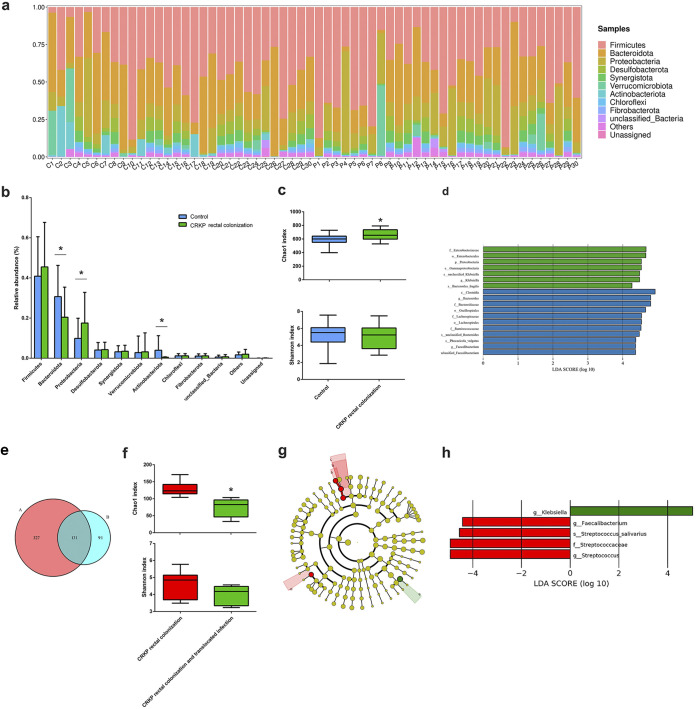
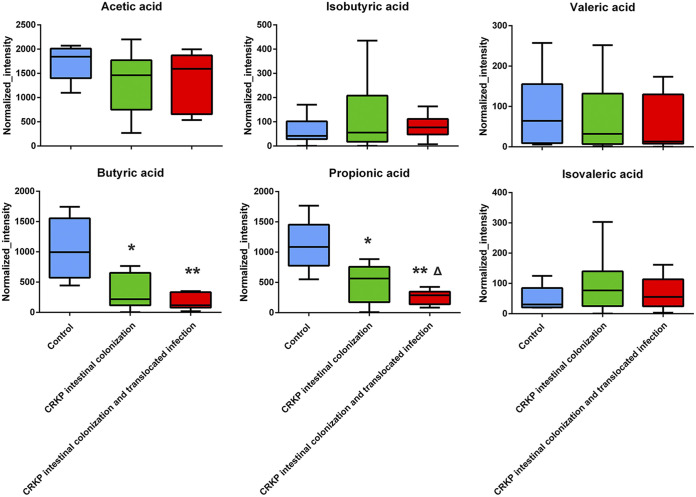
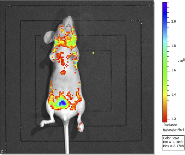
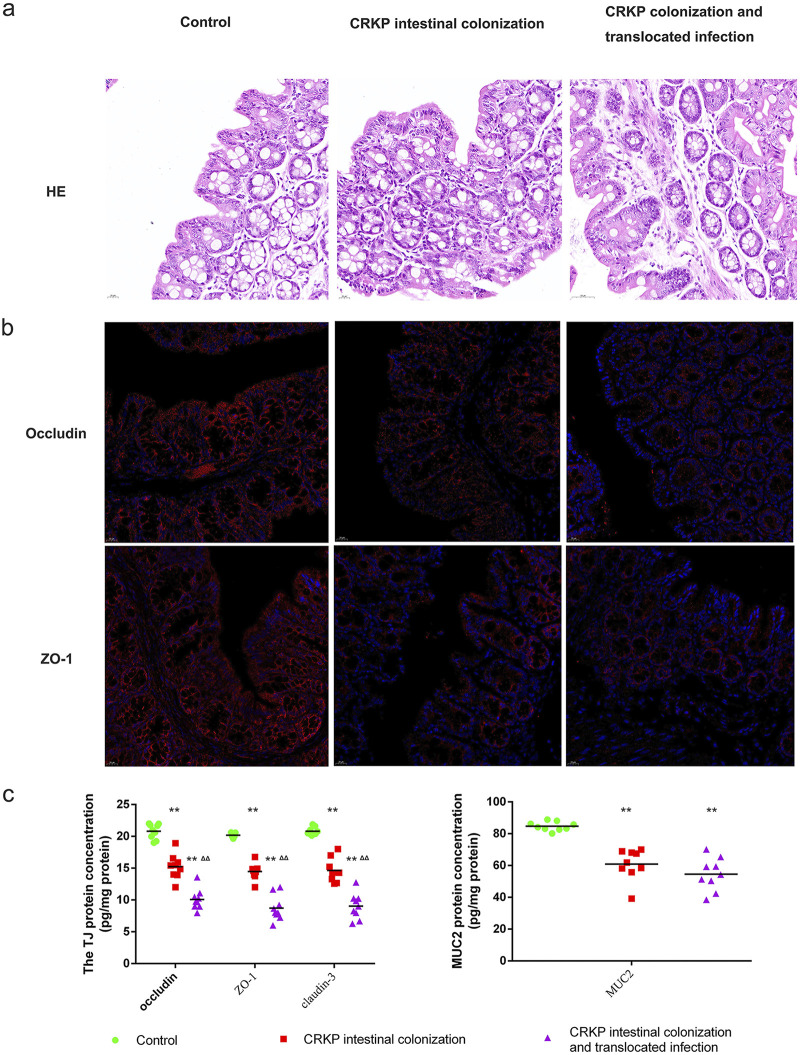
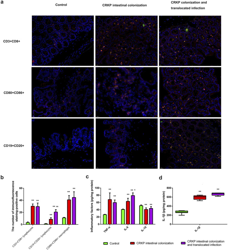
Gastrointestinal colonization with carbapenem-resistant Enterobacteriaceae (CRE) is always a prerequisite for the development of translocated infections. Here, we sought to screen for fecal carriage of CRE and identify the risk factors for CRE colonization as well as subsequent translocated pneumonia in critically ill patients admitted to the intensive care unit (ICU) of a university hospital in China. We further focused on the intestinal flora composition and fecal metabolic profiles in CRE rectal colonization and translocated infection patients. Animal models of gastrointestinal colonization with a carbapenemase-producing Klebsiella pneumoniae (carbapenem-resistant K. pneumoniae [CRKP]) clinical isolate expressing green fluorescent protein (GFP) were established, and systemic infection was subsequently traced using an in vivo imaging system (IVIS). The intestinal barrier, inflammatory factors, and infiltrating immune cells were further investigated. In this study, we screened 54 patients hospitalized in the ICU with CRE rectal colonization, and 50% of the colonized patients developed CRE-associated pneumonia, in line with the significantly high mortality rate. Upon multivariate analysis, risk factors associated with subsequent pneumonia caused by CRE in patients with fecal colonization included enteral feeding and carbapenem exposure. Furthermore, CRKP colonization and translocated infection influenced the diversity and community composition of the intestinal microbiome. Downregulated propionate and butyrate probably play important and multiangle roles in regulating immune cell infiltration, inflammatory factor expression, and mucus and intestinal epithelial barrier integrity. Although the risk factors and intestinal biomarkers for subsequent infections among CRE-colonized patients were explored, further work is needed to elucidate the complicated mechanisms. IMPORTANCE Carbapenem-resistant Enterobacteriaceae have emerged as a major threat to modern medicine, and the spread of carbapenem-resistant Enterobacteriaceae is a clinical and public health problem. Gastrointestinal colonization by potential pathogens is always a prerequisite for the development of translocated infections, and there is a growing need to assess clinical risk factors and microbiological and intestinal characteristics to prevent the development of clinical infection by carbapenem-resistant Enterobacteriaceae.

Keywords: carbapenem-resistant Enterobacteriaceae; intestinal flora; metabolism; rectal colonization; risk factor.


Conflict of interest statement

The authors declare no conflict of interest.

Figures

FIG 1
FIG 1
CRKP colonization and translocated infection influence the intestinal flora composition. (a) Compositional levels of fecal flora in the CRKP colonization group (n = 30) (C1 to C30) and the control group (n = 30) (P1 to P30) at the phylum level. (b) Statistics of the changes in relative abundance induced by CRKP colonization. (c) Alpha diversity indicated by the Chao1 index and Shannon index (*, P < 0.05 versus the control). (d) Distribution histogram based on linear discriminant analysis (LDA). The default criteria of an LDA score of >4 and a P value of <0.05 indicate different species and a higher abundance in one group than in the other. The histogram of LDA scores calculated for selected taxa shows significant differences in microbe types and abundances between the control group (blue) and the CRKP colonization group (green). LDA scores on a log10 scale are indicated at the bottom. The significance of the microbial marker increases with the LDA score. (e) Venn diagram displaying the operational taxonomic unit (OTU) overlap between the CRKP colonization group (A, left, pink) (n = 6) and the translocated infection group (B, right, powder blue) (n = 6). (f) Alpha diversity indicated by the Chao1 and Shannon indices between the CRKP colonization group (n = 6) and the translocated infection group (n = 6) (*, P < 0.05 versus the CRKP colonization group). (g) Linear discriminant analysis effect size (LEfSe) and LDA based on OTU characterizations of the microbiota of the CRKP colonization group (n = 6) and the translocated infection group (n = 6). The cladogram generated by the LEfSe method shows the phylogenetic distribution of fecal microbiomes associated with the two groups. Each filled circle represents one phylotype. The circle size is proportional to the phylotype abundance. By default, the taxonomic levels are arranged outward from phylum to genus. Red circles on the branches represent microbial communities playing pivotal roles in CRKP rectal colonization. Green circles represent microbial groups playing important roles in CRKP translocated infection. The default criteria of an LDA score of >4 and a P value of <0.05 indicate different species and a higher abundance in one group than in the other. a, s_Streptococcus_salivarius; b, g_Streptococcus; c, f_Streptococcaceae; d, g_Faecalibacterium; e, g_Klebsiella. (h) Histogram of LDA scores calculated for selected taxa showing significant differences in microbe types and abundances between the CRKP colonization group (red) and the translocated infection group (green). LDA scores on a log10 scale are indicated at the bottom. The significance of the microbial marker increases with the LDA score.
FIG 2
FIG 2
Results of statistical analysis of short-chain fatty acid metabolites changed in feces (n = 9) (**, P < 0.001 versus the control group; *, P < 0.05 versus the control group; Δ, P < 0.05 versus the CRKP rectal colonization group).
FIG 3
FIG 3
In vivo evaluation of CRKP colonization of the gastrointestinal tract and subsequent infection of C57BL/6J mice with immunosuppression and PPI use. Live-animal and ex vivo IVIS imaging confirmed CRKP colonization of the gastrointestinal tract and subsequent infection. The IVIS images were collected 25 days after oral gavage of GFP-labeled CRKP, and the nonspecific distribution of CRKP in multiple organs such as the lung, heart, and brain, in addition to the intestinal tract, is shown.
FIG 4
FIG 4
CRKP colonization and translocated infection disrupt the intestinal barrier. To evaluate the influences of CRKP rectal colonization and translocated infection on the intestinal barrier, we performed HE and fluorescence staining for occludin and ZO-1. The contents of tight junction (TJ) proteins (claudin-3, occludin, and ZO-1) and mucin2 (MUC2) were further analyzed by an ELISA. (a) HE staining was used to evaluate histopathological injury in three groups. (b) Expression of tight junction (occludin and ZO-1) proteins. The nucleus is shown in blue by DAPI staining. On the cell membrane, the tight junction marker is shown in red. (c) Statistics of the expression of tight junction proteins (claudin-3, occludin, and ZO-1) and MUC2 in the three groups. (**, P < 0.001 versus the control group; ΔΔ, P < 0.001 versus the CRKP rectal colonization group).
FIG 5
FIG 5
CRKP colonization and translocated infection induce obvious immune cell infiltration and inflammatory factor expression. To determine the features of immune cell infiltration in colon tissues subjected to CRKP colonization as well as translocated infection, double-immunofluorescence staining was performed in three groups of mice. We further evaluated the influence of CRKP colonization and translocated infection on inflammatory factor (IL-1β, TNF-α, IL-6, and IL-10) expression. (a) Expression of CD3+ CD8+ lymphocytes, CD19+ CD20+ lymphocytes, and CD80+ CD86+ macrophages in colon tissues. By fluorescence microscopy, the nucleus is shown in blue with DAPI staining. Merged staining is shown in yellow/orange on the membrane, which reveals CD3+ CD8+ lymphocytes, CD19+ CD20+ lymphocytes, and CD80+ CD86+ macrophages. The numbers of double-immunofluorescence-stained positive cells (yellow/orange on the membrane) were captured by fluorescence microscopy. (b) Statistics of CD3+ CD8+ lymphocytes, CD19+ CD20+ lymphocytes, and CD80+ CD86+ macrophages in tissues harvested from animal models (**, P < 0.001 versus the control group; ΔΔ, P < 0.001 versus the CRKP intestinal colonization group). (c) Statistics of expression of inflammatory factors (IL-1β, TNF-α, IL-6, and IL-10) in the three groups (**, P < 0.001 versus the control group; Δ, P < 0.05 versus the CRKP rectal colonization group).

References

    1. Bes T, Nagano D, Martins R, Marchi AP, Perdigão-Neto L, Higashino H, Prado G, Guimaraes T, Levin AS, Costa S. 2021. Bloodstream infections caused by Klebsiella pneumoniae and Serratia marcescens isolates co-harboring NDM-1 and KPC-2. Ann Clin Microbiol Antimicrob 20:57. doi: 10.1186/s12941-021-00464-5. - DOI - PMC - PubMed
    1. Ham DC, Mahon G, Bhaurla SK, Horwich-Scholefield S, Klein L, Dotson N, Rasheed JK, McAllister G, Stanton RA, Karlsson M, Lonsway D, Huang JY, Brown AC, Walters MS. 2021. Gram-negative bacteria harboring multiple carbapenemase genes, United States, 2012-2019. Emerg Infect Dis 27:2475–2479. doi: 10.3201/eid2709.210456. - DOI - PMC - PubMed
    1. Meletiadis J, Paranos P, Georgiou PC, Vourli S, Antonopoulou S, Michelaki A, Vagiakou E, Pournaras S. 2021. In vitro comparative activity of the new beta-lactamase inhibitor taniborbactam with cefepime and meropenem against Klebsiella pneumoniae and cefepime against Pseudomonas aeruginosa metallo-beta-lactamase producing clinical isolates. Int J Antimicrob Agents 58:106440. doi: 10.1016/j.ijantimicag.2021.106440. - DOI - PubMed
    1. Castanheira M, Doyle TB, Deshpande LM, Mendes RE, Sader HS. 2021. Activity of ceftazidime/avibactam, meropenem/vaborbactam, and imipenem/relebactam against carbapenemase-negative carbapenem-resistant Enterobacterales (CRE) isolates from US hospitals. Int J Antimicrob Agents 58:106439. doi: 10.1016/j.ijantimicag.2021.106439. - DOI - PubMed
    1. Chen Y, Chen Y. 2021. Clinical challenges with hypervirulent Klebsiella pneumoniae (hvKP) in China. J Transl Int Med 9:71–75. doi: 10.2478/jtim-2021-0004. - DOI - PMC - PubMed

Publication types

LinkOut - more resources