. 2022 Dec 21;10(6):e0273722.
doi: 10.1128/spectrum.02737-22. Epub 2022 Nov 29.

Oral Bacteriome and Mycobiome across Stages of Oral Carcinogenesis

Affiliations

Oral Bacteriome and Mycobiome across Stages of Oral Carcinogenesis

Weiwei Heng et al. Microbiol Spectr. .

Abstract

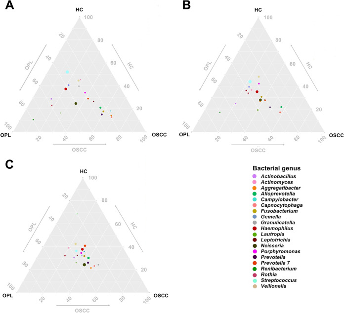
Oral microbial dysbiosis contributes to the development of oral squamous cell carcinoma (OSCC). Numerous studies have focused on variations in the oral bacterial microbiota of patients with OSCC. However, similar studies on fungal microbiota, another integral component of the oral microbiota, are scarce. Moreover, there is an evidence gap regarding the role that microecosystems play in different niches of the oral cavity at different stages of oral carcinogenesis. Here, we catalogued the microbial communities in the human oral cavity by profiling saliva, gingival plaque, and mucosal samples at different stages of oral carcinogenesis. We analyzed the oral bacteriome and mycobiome along the health-premalignancy-carcinoma sequence. Some species, including Prevotella intermedia, Porphyromonas endodontalis, Acremonium exuviarum, and Aspergillus fumigatus, were enriched, whereas others, such as Streptococcus salivarius subsp. salivarius, Scapharca broughtonii, Mortierella echinula, and Morchella septimelata, were depleted in OSCC. These findings suggest that an array of signature species, including bacteria and fungi, are closely associated with oral carcinogenesis. OSCC-associated diversity differences, species distinction, and functional alterations were most remarkable in mucosal samples, not in gingival plaque or saliva samples, suggesting an urgent need to define oral carcinogenesis-associated microbial dysbiosis based on the spatial microbiome. IMPORTANCE Abundant oral microorganisms constitute a complex microecosystem within the oral environment of the host, which plays a critical role in the adjustment of various physiological and pathological states of the oral cavity. In this study, we demonstrated that variations in the "core microbiome" may be used to predict carcinogenesis. In addition, sample data collected from multiple oral sites along the health-premalignancy-carcinoma sequence increase our understanding of the microecosystems of different oral niches and their specific changes during oral carcinogenesis. This work provides insight into the roles of bacteria and fungi in OSCC and may contribute to the development of early diagnostic assays and novel treatments.

Keywords: bacteriome; carcinogenesis; mycobiome; oral cavity; plaque; saliva.


Conflict of interest statement

The authors declare no conflict of interest.

Figures

FIG 1
FIG 1
Principal-component analysis of the buccal mucosal bacteriomes from HC, OPL, and OSCC individuals. (A) The buccal mucosal bacteriome of individuals with OSCC was statistically significantly different (P < 0.05) from that of HC and OPL individuals. (B) Box plots show the diversity and richness of the buccal mucosal bacteriomes from the HP, OPL, and OSCC groups at the OTU level.
FIG 2
FIG 2
Principal-component analysis of the buccal mucosal mycobiomes from HC, OPL, and OSCC individuals. (A) The buccal mucosal mycobiome of individuals with OSCC was statistically significantly different (P < 0.05) from that of HC and OPL individuals. (B) Box plots show the diversity and richness of the buccal mucosal mycobiomes from the HP, OPL, and OSCC groups at the OTU level.
FIG 3
FIG 3
Ternary plot of the oral bacteriome derived from all studied subjects in the HC, OPL, and OSCC groups. The plots represent different samples from the buccal mucosa (A), plaque (B), and saliva (C). The position of the bubble denotes a close correlation of the genera within each group. The arrows indicate the direction of oral carcinogenesis.
FIG 4
FIG 4
Ternary plot of the oral mycobiome derived from all studied subjects in the HC, OPL, and OSCC groups. The plots represent different samples from the buccal mucosa (A), plaque (B), and saliva (C). The position of the bubble denotes a close correlation of the genera within each group. The arrows indicate the direction of oral carcinogenesis.
FIG 5
FIG 5
Bubble plot of the buccal mucosal bacteriome (A) and mycobiome (B) from HC, OPL, and OSCC individuals. Each bubble represents one genus, and the bubble size represents the relative abundance of each genus. For the OSCC group, *, **, and *** denote P values of <0.05, <0.01, and <0.001 versus the HC group, respectively. For the OSCC group, #, ##, and ### denote P values of <0.05, <0.01, and <0.001 versus the OPL group, respectively. For the OPL group, $ and $$ denote P values of <0.05 and <0.01 versus the HC group, respectively.
FIG 6
FIG 6
Oral carcinogenesis-associated alterations in the abundance of the buccal mucosal bacteria examined using LEfSe. (A) Cladogram indicating the phylogenetic distribution of active bacteria that were remarkably enriched. (B) Bar plots at the species level with significant differences in abundance based on LEfSe. The colored bars show the LDA scores of species that were enriched in the indicated condition: purple bar, HC; green bar, OPL; pink bar, OSCC.
FIG 7
FIG 7
Oral carcinogenesis-associated alterations in the abundance of the buccal mucosal fungi examined using LEfSe. (A) Cladogram indicating the phylogenetic distribution of active fungi that were remarkably enriched. (B) Bar plots at the species level with significant differences in abundance based on LEfSe. The colored bars show the LDA scores of species that were enriched in the indicated condition: purple bar, HC; green bar, OPL; pink bar, OSCC.
FIG 8
FIG 8
AUC of the optimized models constructed with bacterial (A and B) and fungal (C and D) biomarkers and patient metadata of HC versus OPL and OPL versus OSCC. Mean AUCs and standard deviations of stratified 3-fold cross-validation are shown.
FIG 9
FIG 9
Functional alterations in the buccal mucosal bacteriome. The relative abundances of functional pathways were compared among HC, OPL, and OSCC individuals. Differentially abundant pathways were plotted, and the exact P values are presented in the heat map. The generalized fold change is indicated by color gradients. A generalized fold change of >0 means enriched in the latter; a generalized fold change of <0 means enriched in the former.
FIG 10
FIG 10
Functional alterations in the buccal mucosal mycobiome. The relative abundances of functional pathways were compared among HC, OPL, and OSCC individuals. Differentially abundant pathways were plotted, and the exact P values are presented in the heat map. A generalized fold change of >0 means enriched in the latter; a generalized fold change of <0 means enriched in the former.

References

    1. Ferlay J, Soerjomataram I, Dikshit R, Eser S, Mathers C, Rebelo M, Parkin DM, Forman D, Bray F. 2015. Cancer incidence and mortality worldwide: sources, methods and major patterns in GLOBOCAN 2012. Int J Cancer 136:E359–E386. doi: 10.1002/ijc.29210. - DOI - PubMed
    1. Kaur J, Jacobs R. 2016. Salivary and serum leptin levels in patients with squamous cell carcinoma of the buccal mucosa. Clin Oral Invest 20:39–42. doi: 10.1007/s00784-015-1472-x. - DOI - PubMed
    1. Hill SJ, D'Andrea AD. 2019. Predictive potential of head and neck squamous cell carcinoma organoids. Cancer Discov 9:828–830. doi: 10.1158/2159-8290.CD-19-0527. - DOI - PubMed
    1. Chen H, Liu X, Jin Z, Gou C, Liang M, Cui L, Zhao X. 2018. A three miRNAs signature for predicting the transformation of oral leukoplakia to oral squamous cell carcinoma. Am J Cancer Res 8:1403–1413. - PMC - PubMed
    1. Moutsopoulos NM, Konkel JE. 2018. Tissue-specific immunity at the oral mucosal barrier. Trends Immunol 39:276–287. doi: 10.1016/j.it.2017.08.005. - DOI - PMC - PubMed

Publication types

LinkOut - more resources