

Deubiquitinase OTUD1 Resolves Stalled Translation on polyA and Rare Codon Rich mRNAs
- PMID: 36445135
- PMCID: PMC9753717
- DOI: 10.1128/mcb.00265-22
Deubiquitinase OTUD1 Resolves Stalled Translation on polyA and Rare Codon Rich mRNAs
Abstract
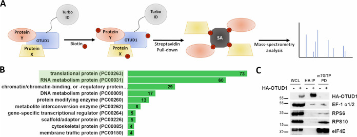
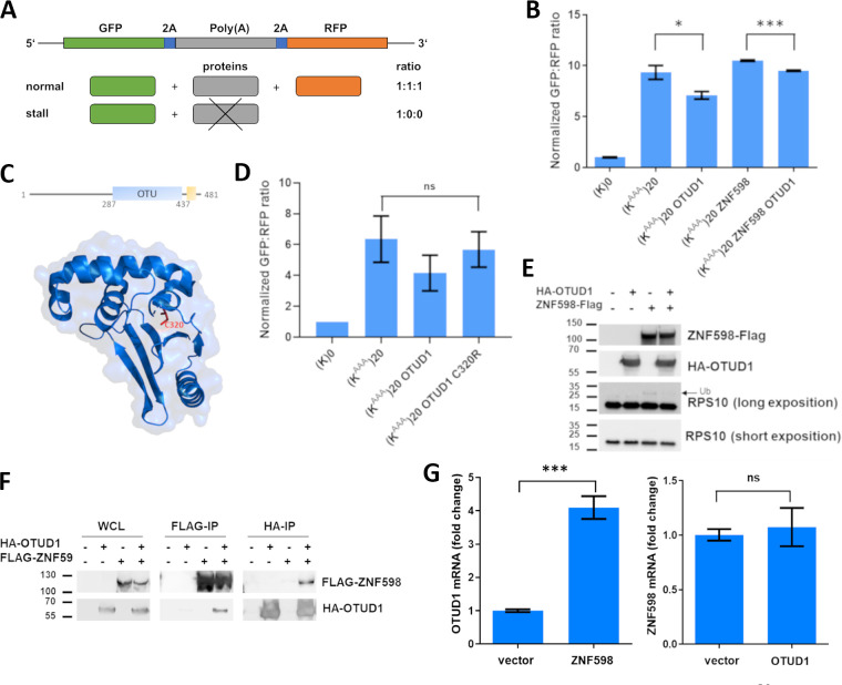
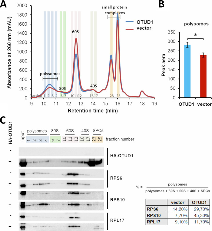
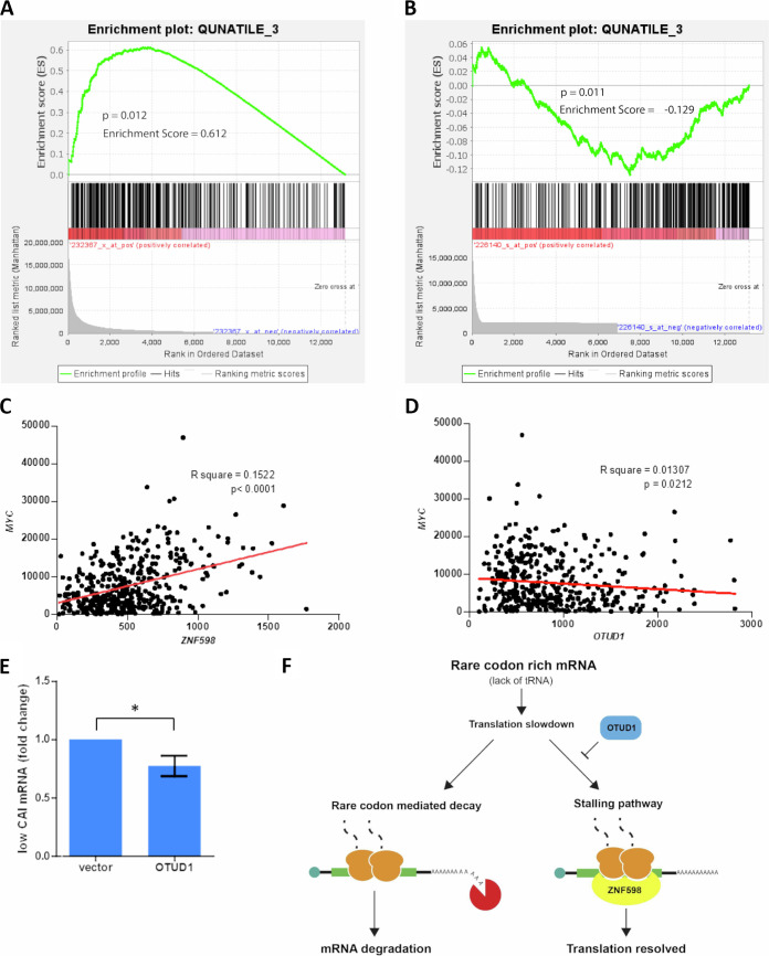
OTUD1 is a deubiquitinating enzyme involved in many cellular processes including cancer and innate, immune signaling pathways. Here, we perform a proximity labeling-based interactome study that identifies OTUD1 largely present in the translation and RNA metabolism protein complexes. Biochemical analysis validates OTUD1 association with ribosome subunits, elongation factors and the E3 ubiquitin ligase ZNF598 but not with the translation initiation machinery. OTUD1 catalytic activity suppresses polyA triggered ribosome stalling through inhibition of ZNF598-mediated RPS10 ubiquitination and stimulates formation of polysomes. Finally, analysis of gene expression suggests that OTUD1 regulates the stability of rare codon rich mRNAs by antagonizing ZNF598.
Keywords: ribosome stalling; translation; ubiquitination.
Conflict of interest statement
The authors declare no conflict of interest.
Figures
Similar articles
-
Pleiotropic Roles of a KEAP1-Associated Deubiquitinase, OTUD1.Antioxidants (Basel). 2023 Feb 1;12(2):350. doi: 10.3390/antiox12020350. Antioxidants (Basel). 2023. PMID: 36829909 Free PMC article. Review.
-
Initiation of Quality Control during Poly(A) Translation Requires Site-Specific Ribosome Ubiquitination.Mol Cell. 2017 Feb 16;65(4):743-750.e4. doi: 10.1016/j.molcel.2016.11.039. Epub 2017 Jan 5. Mol Cell. 2017. PMID: 28065601 Free PMC article.
-
ZNF598 Is a Quality Control Sensor of Collided Ribosomes.Mol Cell. 2018 Nov 1;72(3):469-481.e7. doi: 10.1016/j.molcel.2018.08.037. Epub 2018 Oct 4. Mol Cell. 2018. PMID: 30293783 Free PMC article.
-
The G3BP1-Family-USP10 Deubiquitinase Complex Rescues Ubiquitinated 40S Subunits of Ribosomes Stalled in Translation from Lysosomal Degradation.Mol Cell. 2020 Mar 19;77(6):1193-1205.e5. doi: 10.1016/j.molcel.2019.12.024. Epub 2020 Jan 24. Mol Cell. 2020. PMID: 31981475
-
Tales of poly(A): a review.Gene. 1990 Jul 16;91(2):151-8. doi: 10.1016/0378-1119(90)90082-3. Gene. 1990. PMID: 1976572 Review.
Cited by
-
Pleiotropic Roles of a KEAP1-Associated Deubiquitinase, OTUD1.Antioxidants (Basel). 2023 Feb 1;12(2):350. doi: 10.3390/antiox12020350. Antioxidants (Basel). 2023. PMID: 36829909 Free PMC article. Review.
-
OTUD1 downregulates PD-L1 expression by deubiquitinating STAT3 and promotes the immune response in CcRCC.Cell Oncol (Dordr). 2025 Jun 12. doi: 10.1007/s13402-025-01079-0. Online ahead of print. Cell Oncol (Dordr). 2025. PMID: 40506611
-
Znf598-mediated Rps10/eS10 ubiquitination contributes to the ribosome ubiquitination dynamics during zebrafish development.RNA. 2023 Dec;29(12):1910-1927. doi: 10.1261/rna.079633.123. Epub 2023 Sep 26. RNA. 2023. PMID: 37751929 Free PMC article.
-
Ribosomal collision is not a prerequisite for ZNF598-mediated ribosome ubiquitination and disassembly of ribosomal complexes by ASCC.Nucleic Acids Res. 2024 May 8;52(8):4627-4643. doi: 10.1093/nar/gkae087. Nucleic Acids Res. 2024. PMID: 38366554 Free PMC article.
-
Friend or foe? Reciprocal regulation between E3 ubiquitin ligases and deubiquitinases.Biochem Soc Trans. 2024 Feb 28;52(1):241-267. doi: 10.1042/BST20230454. Biochem Soc Trans. 2024. PMID: 38414432 Free PMC article. Review.
References
Publication types
MeSH terms
Substances
LinkOut - more resources
Full Text Sources
Molecular Biology Databases
Miscellaneous