

microbeSEG: A deep learning software tool with OMERO data management for efficient and accurate cell segmentation
- PMID: 36445903
- PMCID: PMC9707790
- DOI: 10.1371/journal.pone.0277601
microbeSEG: A deep learning software tool with OMERO data management for efficient and accurate cell segmentation
Abstract
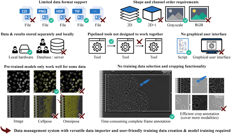
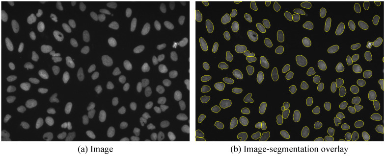
In biotechnology, cell growth is one of the most important properties for the characterization and optimization of microbial cultures. Novel live-cell imaging methods are leading to an ever better understanding of cell cultures and their development. The key to analyzing acquired data is accurate and automated cell segmentation at the single-cell level. Therefore, we present microbeSEG, a user-friendly Python-based cell segmentation tool with a graphical user interface and OMERO data management. microbeSEG utilizes a state-of-the-art deep learning-based segmentation method and can be used for instance segmentation of a wide range of cell morphologies and imaging techniques, e.g., phase contrast or fluorescence microscopy. The main focus of microbeSEG is a comprehensible, easy, efficient, and complete workflow from the creation of training data to the final application of the trained segmentation model. We demonstrate that accurate cell segmentation results can be obtained within 45 minutes of user time. Utilizing public segmentation datasets or pre-labeling further accelerates the microbeSEG workflow. This opens the door for accurate and efficient data analysis of microbial cultures.
Copyright: © 2022 Scherr et al. This is an open access article distributed under the terms of the Creative Commons Attribution License, which permits unrestricted use, distribution, and reproduction in any medium, provided the original author and source are credited.
Conflict of interest statement
The authors have declared that no competing interests exist.
Figures


Similar articles
-
DeepImageTranslator: A free, user-friendly graphical interface for image translation using deep-learning and its applications in 3D CT image analysis.SLAS Technol. 2022 Feb;27(1):76-84. doi: 10.1016/j.slast.2021.10.014. Epub 2021 Oct 25. SLAS Technol. 2022. PMID: 35058205
-
Segmentation, tracking and cell cycle analysis of live-cell imaging data with Cell-ACDC.BMC Biol. 2022 Aug 5;20(1):174. doi: 10.1186/s12915-022-01372-6. BMC Biol. 2022. PMID: 35932043 Free PMC article.
-
MIA is an open-source standalone deep learning application for microscopic image analysis.Cell Rep Methods. 2023 Jun 26;3(7):100517. doi: 10.1016/j.crmeth.2023.100517. eCollection 2023 Jul 24. Cell Rep Methods. 2023. PMID: 37533647 Free PMC article.
-
New open-source software for subcellular segmentation and analysis of spatiotemporal fluorescence signals using deep learning.iScience. 2022 Apr 21;25(5):104277. doi: 10.1016/j.isci.2022.104277. eCollection 2022 May 20. iScience. 2022. PMID: 35573197 Free PMC article.
-
Deep Learning Approaches Towards Skin Lesion Segmentation and Classification from Dermoscopic Images - A Review.Curr Med Imaging. 2020;16(5):513-533. doi: 10.2174/1573405615666190129120449. Curr Med Imaging. 2020. PMID: 32484086 Review.
Cited by
-
Software Tools for 2D Cell Segmentation.Cells. 2024 Feb 17;13(4):352. doi: 10.3390/cells13040352. Cells. 2024. PMID: 38391965 Free PMC article. Review.
-
A review of open-source image analysis tools for mammalian cell culture: algorithms, features and implementations.PeerJ Comput Sci. 2023 May 16;9:e1364. doi: 10.7717/peerj-cs.1364. eCollection 2023. PeerJ Comput Sci. 2023. PMID: 37346656 Free PMC article.
References
Publication types
MeSH terms
LinkOut - more resources
Full Text Sources
