

Oral Peroxisome Proliferator-Activated Receptor-α Agonist Enhances Corneal Nerve Regeneration in Patients With Type 2 Diabetes
- PMID: 36445944
- PMCID: PMC10281232
- DOI: 10.2337/db22-0611
Oral Peroxisome Proliferator-Activated Receptor-α Agonist Enhances Corneal Nerve Regeneration in Patients With Type 2 Diabetes
Abstract
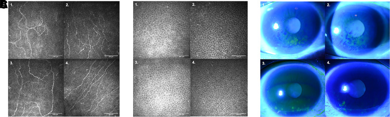
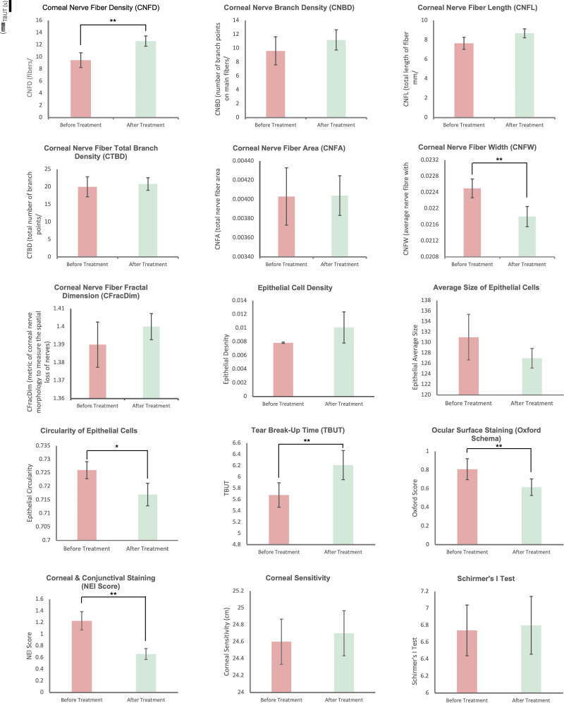
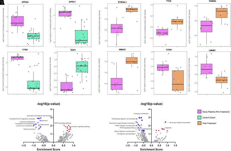
Diabetic corneal neuropathy (DCN) is a common complication of diabetes. However, there are very limited therapeutic options. We investigated the effects of a peroxisome proliferator-activated receptor-α (PPAR-α) agonist, fenofibrate, on 30 patients (60 eyes) with type 2 diabetes. On in vivo confocal microscopy evaluation, there was significant stimulation of corneal nerve regeneration and a reduction in nerve edema after 30 days of oral fenofibrate treatment, as evidenced by significant improvement in corneal nerve fiber density (CNFD) and corneal nerve fiber width, respectively. Corneal epithelial cell morphology also significantly improved in cell circularity. Upon clinical examination, fenofibrate significantly improved patients' neuropathic ocular surface status by increasing tear breakup time along with a reduction of corneal and conjunctival punctate keratopathy. Tear substance P (SP) concentrations significantly increased after treatment, suggesting an amelioration of ocular surface neuroinflammation. The changes in tear SP concentrations was also significantly associated with improvement in CNFD. Quantitative proteomic analysis demonstrated that fenofibrate significantly upregulated and modulated the neurotrophin signaling pathway and linolenic acid, cholesterol, and fat metabolism. Complement cascades, neutrophil reactions, and platelet activation were also significantly suppressed. Our results showed that fenofibrate could potentially be a novel treatment for patients with DCN.
Trial registration: ClinicalTrials.gov NCT03869931.
© 2023 by the American Diabetes Association.
Conflict of interest statement
Figures
Comment in
-
Fenofibrate for Treating Diabetic Eye Disease.Diabetes. 2023 Jul 1;72(7):838-840. doi: 10.2337/dbi22-0037. Diabetes. 2023. PMID: 37339356 No abstract available.
Similar articles
-
Topical and oral peroxisome proliferator-activated receptor-α agonist ameliorates diabetic corneal neuropathy.Sci Rep. 2024 Jun 11;14(1):13435. doi: 10.1038/s41598-024-64451-4. Sci Rep. 2024. PMID: 38862650 Free PMC article.
-
Pathogenic Role of PPARα Downregulation in Corneal Nerve Degeneration and Impaired Corneal Sensitivity in Diabetes.Diabetes. 2020 Jun;69(6):1279-1291. doi: 10.2337/db19-0898. Epub 2020 Mar 25. Diabetes. 2020. PMID: 32213513 Free PMC article.
-
In vivo confocal microscopy of corneal small fiber damage in diabetes mellitus.Graefes Arch Clin Exp Ophthalmol. 2010 Sep;248(9):1307-12. doi: 10.1007/s00417-010-1396-8. Epub 2010 May 21. Graefes Arch Clin Exp Ophthalmol. 2010. PMID: 20490534
-
Fibrates and microvascular complications in diabetes--insight from the FIELD study.Curr Pharm Des. 2009;15(5):537-52. doi: 10.2174/138161209787315701. Curr Pharm Des. 2009. PMID: 19199980 Review.
-
Corneal nerves in diabetes-The role of the in vivo corneal confocal microscopy of the subbasal nerve plexus in the assessment of peripheral small fiber neuropathy.Surv Ophthalmol. 2021 May-Jun;66(3):493-513. doi: 10.1016/j.survophthal.2020.09.003. Epub 2020 Sep 19. Surv Ophthalmol. 2021. PMID: 32961210 Review.
Cited by
-
A review of the application of in-vivo confocal microscopy on conjunctival diseases.Eye Vis (Lond). 2024 Nov 1;11(1):43. doi: 10.1186/s40662-024-00409-x. Eye Vis (Lond). 2024. PMID: 39482793 Free PMC article. Review.
-
Inhibition of miR-144-3p/FOXO1 Attenuates Diabetic Keratopathy Via Modulating Autophagy and Apoptosis.Invest Ophthalmol Vis Sci. 2024 Jan 2;65(1):1. doi: 10.1167/iovs.65.1.1. Invest Ophthalmol Vis Sci. 2024. PMID: 38165707 Free PMC article.
-
The role of PPAR in fungal keratitis.Front Immunol. 2024 Dec 23;15:1454463. doi: 10.3389/fimmu.2024.1454463. eCollection 2024. Front Immunol. 2024. PMID: 39763659 Free PMC article. Review.
-
Culture of Primary Neurons from Dissociated and Cryopreserved Mouse Trigeminal Ganglion.Tissue Eng Part C Methods. 2023 Aug;29(8):381-393. doi: 10.1089/ten.TEC.2023.0054. Epub 2023 Jun 27. Tissue Eng Part C Methods. 2023. PMID: 37212303 Free PMC article.
-
Potential therapeutic effects of peroxisome proliferator-activated receptors on corneal diseases.Exp Biol Med (Maywood). 2024 Jun 27;249:10142. doi: 10.3389/ebm.2024.10142. eCollection 2024. Exp Biol Med (Maywood). 2024. PMID: 38993197 Free PMC article. Review.
References
-
- Iqbal Z, Azmi S, Yadav R, et al. . Diabetic peripheral neuropathy: epidemiology, diagnosis, and pharmacotherapy. Clin Ther 2018;40:828–849 - PubMed
-
- Bodman MA, Varacallo M. Peripheral Diabetic Neuropathy. Treasure Island, FL, StatPearls Publishing, 2022 - PubMed
-
- Labetoulle M, Baudouin C, Calonge M, et al. . Role of corneal nerves in ocular surface homeostasis and disease. Acta Ophthalmol 2019;97:137–145 - PubMed
-
- Al-Aqaba MA, Dhillon VK, Mohammed I, Said DG, Dua HS. Corneal nerves in health and disease. Prog Retin Eye Res 2019;73:100762. - PubMed
Publication types
MeSH terms
Substances
Associated data
LinkOut - more resources
Full Text Sources
Medical
Miscellaneous
