. 2023 Jan 25;70(3):e220164.
doi: 10.1530/JME-22-0164. Print 2023 Apr 1.

Identification of prolactin receptor variants with diverse effects on receptor signalling

Affiliations

Identification of prolactin receptor variants with diverse effects on receptor signalling

Caroline M Gorvin et al. J Mol Endocrinol. .

Abstract

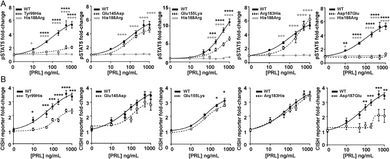
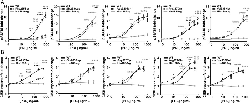
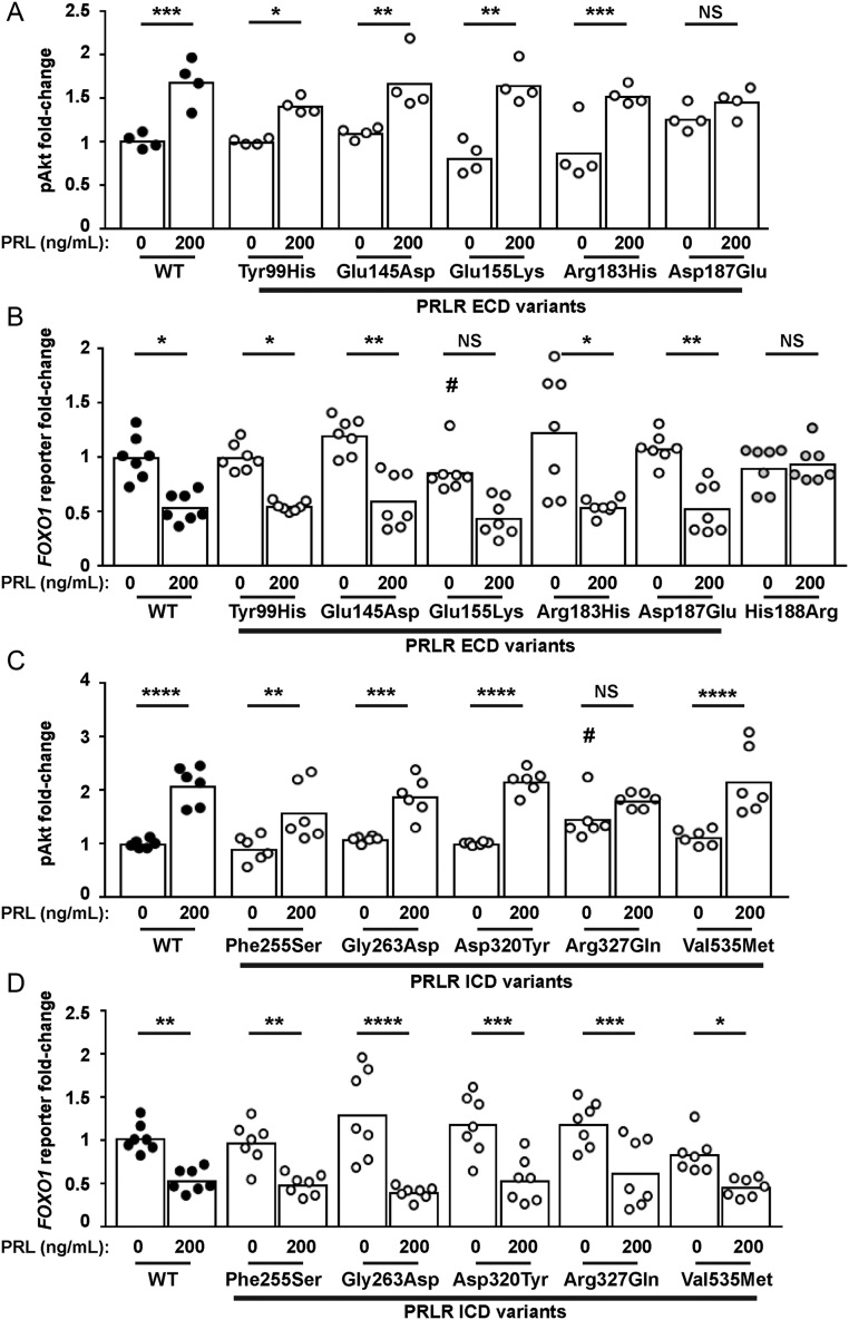
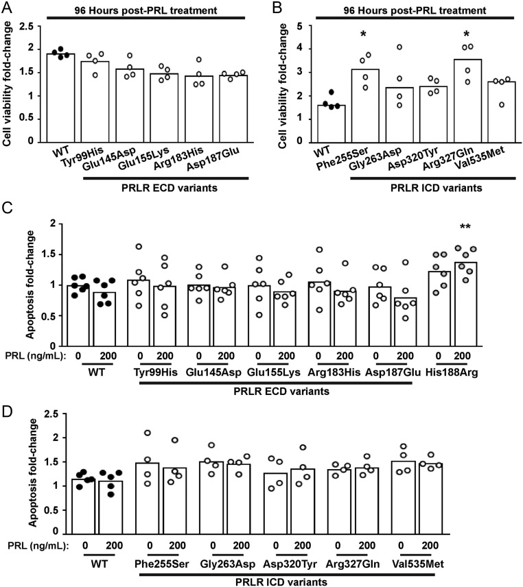
The prolactin receptor (PRLR) signals predominantly through the JAK2-STAT5 pathway regulating multiple physiological functions relating to fertility, lactation, and metabolism. However, the molecular pathology and role of PRLR mutations and signalling are incompletely defined, with progress hampered by a lack of reported disease-associated PRLR variants. To date, two common germline PRLR variants are reported to demonstrate constitutive activity, with one, Ile146Leu, overrepresented in benign breast disease, while a rare activating variant, Asn492Ile, is reported to be associated with an increased incidence of prolactinoma. In contrast, an inactivating germline heterozygous PRLR variant (His188Arg) was reported in a kindred with hyperprolactinaemia, while an inactivating compound heterozygous PRLR variant (Pro269Leu/Arg171Stop) was identified in an individual with hyperprolactinaemia and agalactia. We hypothesised that additional rare germline PRLR variants, identified from large-scale sequencing projects (ExAC and GnomAD), may be associated with altered in vitro PRLR signalling activity. We therefore evaluated >300 previously uncharacterised non-synonymous, germline PRLR variants and selected 10 variants for in vitro analysis based on protein prediction algorithms, proximity to known functional domains and structural modelling. Five variants, including extracellular and intracellular domain variants, were associated with altered responses when compared to the wild-type receptor. These altered responses included loss- and gain-of-function activities related to STAT5 signalling, Akt and FOXO1 activity, as well as cell viability and apoptosis. These studies provide further insight into PRLR structure-function and indicate that rare germline PRLR variants may have diverse modulating effects on PRLR signalling, although the pathophysiologic relevance of such alterations remains to be defined.

Keywords: JAK-STAT5; hyperprolactinaemia; prolactinoma; proliferation.


Conflict of interest statement

The authors declare that there is no conflict of interest.

Figures

Figure 1
Figure 1
Location of the PRLR ECD variants and structural characterisation of rare variants located in D1. (A) Crystal structure showing the two monomers (PRLR1 in pink and PRLR2 in light brown) of the rat PRLR extracellular domain (ECD) in complex with the PRL hormone (purple) (PDB 3NPZ (van Agthoven et al. 2010)). Each PRLR ECD monomer is comprised of two subdomains, designated D1 (residues 1–101) and D2 (residues 109–210), which are important in ligand binding and subsequent receptor activation (Svensson et al. 2008, Broutin et al. 2010, van Agthoven et al. 2010, Rao and Brooks 2011, Brooks 2012). Side chains of the potential deleterious variant residues from ExAC and GnomAD are shown in red. The His188 residue that is mutated in hyperprolactinaemia is shown in green. Residue 49 is labelled as Tyr in the rat structure, and in humans, this is His49. Residue 169 is labelled as Val in the rat structure, and in human, this is Ile169. Rare variants are shown in only one monomer. (B) Lys6, Pro7, and Ser88 are located in close proximity within the β6-β7-β8 region (Top). The variant Asn6 loses contact with Asp91 on the β8 strand, Ser7 gains contact with Val79 on the β6 strand, and Arg88 loses contact with Pro4 and gains a new contact with the adjacent Ser87 (all blue, bottom). (C) The Gly30 residue, located in the β2-β3 loop, forms a polar contact with the Ser2 residue (Top). When mutated to Arg30 (blue, bottom), the longer side chain is able to retain this contact and form a new hydrogen bond with Asn83 in the β6-β7 loop. (D) The Cys51 residue is located in the β4-β5 loop. Mutation in Tyr51 (blue, bottom) adds a more bulky residue within this tightly packed region, which is predicted to result in gain of two contacts with Asp53 and Phe63 of the β4-β5 loop.
Figure 2
Figure 2
Structural characterisation of rare variants located in the C-terminal D1 and D2 of PRLR ECD. (A) The Tyr99 residue is located close to the PRL-binding site with the PRLR. The hydroxyl group of the wild-type Tyr99 contacts Arg176 on the PRL protein (left). Mutation to His99 (blue, right) predicts retention of the contact, but at a more distal site, further away from the PRL α-helix and therefore is likely to increase the distance between the PRL and PRLR molecules which may affect binding and activation. (B) The Glu145 and Arg183 PRLR residues are located in adjacent β-strands and form polar contacts with each other. Mutation to His183 (middle) and Asp145 (bottom) is predicted to disrupt two of these contacts. Additionally, Asp145 is predicted to lose contact with the neighbouring Ile146, a residue previously demonstrated to be important for PRLR folding and stability (Dagil et al. 2012, Zhang et al. 2015). (C) The His183 also loses contact with Ala193, which forms part of the highly conserved WSxWS motif (red). (D) The wild-type Glu155 forms a contact with Lys114 on the opposite PRLR protomer. The Lys155 variant loses this contact and may disrupt homodimeric structural stability. (E) The wild-type Asp187 residue lies close to the PRL binding site and forms a contact with the His188 residue of PRLR, which has a critical role in ligand binding (Kulkarni et al. 2010) (Left). Mutation to Glu187 (blue, right) leads to loss of this contact, which may affect activation of the PRLR protein.
Figure 3
Figure 3
Structural characterisation of the PRLR variants located in the TMD and ICD. (A) A single α-helix forms the transmembrane domain (TMD) of the PRLR. Five rare PRLR variants (Ala222Gly, Ile227Thr, Trp230Cys, Val232Ala, Val232Met) are located within the TMD, and four of these are present within the published NMR structure of the PRLR TMD (PDB:2N7I (Bugge et al. 2016)). The four WT residues (black) are located at the cytoplasmic end of the TMD and each forms backbone contacts with adjacent residues within the α-helix. The mutant residues (blue) are not predicted to affect these backbone contacts. (B) Cartoon depicting the PRLR structure with the two monomers shown in brown and pink. The extracellular domain (ECD) contains two domains (D1 and D2) and is connected to the intracellular domain (ICD) via the TMD. The ICD is predicted to interact with the plasma membrane in at least three regions known as lipid-interacting domains (LID1-3). The ICD also interacts with the JAK2 proteins that activate signalling downstream of the PRLR. (C) Cartoon showing the known functional domains of the PRLR ICD, with the amino acid residues (236–300, 243–251, 324–329, 350–383, and 547–598) involved shown in parentheses. The LIDs are shown in green. Two regions, Box 1 and Box 2 (brown-shaded regions), are binding sites for JAK2 (Lebrun et al. 1995), and the DSGxxS region (blue-shaded region) acts as a degradation motif (Plotnikov et al. 2009). The PRLR residues investigated in this study are shown in brown above the cartoon and residues investigated in previous studies are shown in red below the cartoon (Bernard et al. 2016, Gorvin et al. 2018a). The location of the Asn492Ile gain-of-function PRLR variant that is associated with prolactinoma is indicated in orange.
Figure 4
Figure 4
Expression of the PRLR ECD and ICD variants. Western blot analyses of HEK293 cells expressing: (A) PRLR ECD rare variants and (B) PRLR ICD rare variants. Lysates show approximately equal expression levels of PRLR in cells transfected with each rare variant and the WT PRLR. Tubulin was used as a loading control. (C) Confocal microscopy images of the PRLR WT and variant proteins in transfected HEK293 cells. Bar indicates 10 µm.
Figure 5
Figure 5
Functional characterisation of the JAK-STAT signalling pathway by PRLR ECD variants. (A) pSTAT5 responses following prolactin (PRL) treatment in cells expressing wild-type (WT), mutant His188Arg, or ECD variants Tyr99His, Glu145Asp, Glu155Lys, Arg183His, Asp187Glu. PRL-induced pSTAT5 production was abolished in His188Arg- and Asp187Glu-expressing cells and significantly reduced in Tyr99His- and Glu155Lys-expressing cells compared to cells expressing WT. (B) CISH luciferase reporter activity in cells transfected with WT or the five ECD variant PRLRs. CISH reporter activity was significantly reduced in Tyr99His-, Glu155Lys-, and Asp187Glu-expressing cells compared to WT cells. Mean from four to five independent assays for all panels. Statistical analyses show comparisons between WT and the five ECD PRLR variants (black) and WT and the His188Arg mutant (grey) by two-way ANOVA with Dunnett’s or Sidak’s multiple comparisons tests. ****P < 0.0001, ***P < 0.001, **P < 0.01, *P < 0.05.
Figure 6
Figure 6
Functional characterisation of the JAK2-STAT5 signalling pathway by PRLR ICD variants. (A) pSTAT5 responses following prolactin (PRL) treatment in cells expressing wild-type (WT), mutant His188Arg, or ICD variants Phe255Ser, Gly263Asp, Asp320Tyr, Arg327Gln, Val535Met. PRL-induced pSTAT5 production was abolished in His188Arg expressing cells, significantly reduced in Phe255Ser and significantly increased in Arg327Gln expressing cells when compared to WT cells. Additionally, pSTAT5 was reduced in Gly263Asp- and Val535Met-expressing cells when treated with high (1000 ng/mL) PRL. (B) CISH luciferase reporter activity in cells transfected with WT, mutant His188Arg, or the five ICD variant PRLRs. CISH reporter activity was abolished in His188Arg-expressing cells, significantly reduced in Phe255Ser and significantly increased in Arg327Gln-expressing cells when compared to WT cells. Mean from four to five independent assays for all panels. Statistical analyses show comparisons between WT and the five ICD PRLR variants (black) and WT and the His188Arg mutant (grey) by two-way ANOVA with Dunnett’s or Tukey’s multiple comparisons tests. ****P < 0.0001, ***P < 0.001, **P < 0.01, *P < 0.05.
Figure 7
Figure 7
Functional characterisation of the Akt signalling pathway by PRLR rare variants. (A) pAkt responses following PRL treatment in cells transfected with wild-type (WT) or the five extracellular domain (ECD) variant PRLRs. Asp187Glu had impaired pAkt responses when compared to WT-expressing cells. (B) FOXO1 luciferase reporter activity in cells transfected with WT, mutant His188Arg, or the five ECD variant PRLRs. Addition of 200 ng/mL PRL reduces FOXO1 luciferase activity in WT and four variant cell lines. However, no response was observed in His188Arg cells and Glu155Lys had reduced basal FOXO1 activity and impaired PRL-induced responses. (C) pAkt responses following PRL treatment in cells transfected with WT or the five intracellular domain (ICD) variant PRLRs. Arg327Gln-expressing cells had elevated basal pAkt activity. (D) FOXO1 luciferase activity in cells transfected with WT or the five ICD variant PRLRs. Addition of 200 ng/mL prolactin to WT and variant PRLRs reduces FOXO1 luciferase activity similarly in all cell lines. Data in all panels were expressed relative to WT cells treated with 0 ng/mL PRL. Mean from 4 to 6 independent assays for pAkt assays. Mean (panel D) or median (panel B) from seven independent assays for luciferase reporter assays. Statistical analyses performed by one-way ANOVA with Sidak’s or Dunnett’s multiple comparisons tests for panels A, C, and D. Statistical analysis performed by Kruskal–Wallis test with Dunn’s multiple comparisons tests for panel B. Comparisons show 0 vs 200 nM PRL (asterisks) or between WT and variant (#). ****P < 0.0001, ***P < 0.001, **P < 0.01, *P < 0.05, NS, not significant.
Figure 8
Figure 8
Effect of the PRLR rare variants on cell viability and apoptosis. (A-B) Effect of PRL (200 ng/mL) on viability in cells expressing WT, or (A) the extracellular domain (ECD) or (B) the intracellular domain (ICD) variant PRLRs. Cell viability was increased in cells expressing the Phe255Ser and Arg327Gln variant PRLRs at 96 h post-treatment with PRL, when compared to WT cells. (C-D) Effect of PRL (200 ng/mL) on apoptosis in cells expressing WT, mutant His188Arg, or (C) the ECD or (D) the ICD variant PRLRs. Each point shows one biological replicate (derived from the mean of four technical replicates) performed on independent occasions. Statistical analyses performed by one-way ANOVA with Dunnett’s multiple comparisons tests for panels A, C, and D. Statistical analysis performed by Kruskal–Wallis test with Dunn’s multiple comparisons tests for panel B. Comparisons show WT vs variant (asterisks). ****P < 0.0001, ***P < 0.001, **P < 0.01, *P < 0.05.

Similar articles

Cited by

References

    1. Acosta JJ, Munoz RM, Gonzalez L, Subtil-Rodriguez A, Dominguez-Caceres MA, Garcia-Martinez JM, Calcabrini A, Lazaro-Trueba I, Martin-Perez J.2003Src mediates prolactin-dependent proliferation of T47D and MCF7 cells via the activation of focal adhesion kinase/ERK1/2 and phosphatidylinositol 3-kinase pathways. Molecular Endocrinology 172268–2282. (10.1210/me.2002-0422) - DOI - PubMed
    1. Adzhubei I, Jordan DM, Sunyaev SR.2013Predicting functional effect of human missense mutations using PolyPhen-2. Current Protocols in Human Genetics 761–7. (10.1002/0471142905.hg0720s76) - DOI - PMC - PubMed
    1. Adzhubei IA, Schmidt S, Peshkin L, Ramensky VE, Gerasimova A, Bork P, Kondrashov AS, Sunyaev SR.2010A method and server for predicting damaging missense mutations. Nature Methods 7248–249. (10.1038/nmeth0410-248) - DOI - PMC - PubMed
    1. Agarwala V, Flannick J, Sunyaev SGoT2D Consortium & Altshuler D.2013Evaluating empirical bounds on complex disease genetic architecture. Nature Genetics 451418–1427. (10.1038/ng.2804) - DOI - PMC - PubMed
    1. Ali S, Ali S.1998Prolactin receptor regulates Stat5 tyrosine phosphorylation and nuclear translocation by two separate pathways. Journal of Biological Chemistry 2737709–7716. (10.1074/jbc.273.13.7709) - DOI - PubMed

Publication types