. 2022 Dec 6;119(49):e2208904119.
doi: 10.1073/pnas.2208904119. Epub 2022 Nov 29.

The N-Myc-responsive lncRNA MILIP promotes DNA double-strand break repair through non-homologous end joining

Affiliations

The N-Myc-responsive lncRNA MILIP promotes DNA double-strand break repair through non-homologous end joining

Pei Lin Wang et al. Proc Natl Acad Sci U S A. .

Erratum in

Abstract

The protooncoprotein N-Myc, which is overexpressed in approximately 25% of neuroblastomas as the consequence of MYCN gene amplification, has long been postulated to regulate DNA double-strand break (DSB) repair in neuroblastoma cells, but experimental evidence of this function is presently scant. Here, we show that N-Myc transcriptionally activates the long noncoding RNA MILIP to promote nonhomologous end-joining (NHEJ) DNA repair through facilitating Ku70-Ku80 heterodimerization in neuroblastoma cells. High MILIP expression was associated with poor outcome and appeared as an independent prognostic factor in neuroblastoma patients. Knockdown of MILIP reduced neuroblastoma cell viability through the induction of apoptosis and inhibition of proliferation, retarded neuroblastoma xenograft growth, and sensitized neuroblastoma cells to DNA-damaging therapeutics. The effect of MILIP knockdown was associated with the accumulation of DNA DSBs in neuroblastoma cells largely due to decreased activity of the NHEJ DNA repair pathway. Mechanistical investigations revealed that binding of MILIP to Ku70 and Ku80 increased their heterodimerization, and this was required for MILIP-mediated promotion of NHEJ DNA repair. Disrupting the interaction between MILIP and Ku70 or Ku80 increased DNA DSBs and reduced cell viability with therapeutic potential revealed where targeting MILIP using Gapmers cooperated with the DNA-damaging drug cisplatin to inhibit neuroblastoma growth in vivo. Collectively, our findings identify MILIP as an N-Myc downstream effector critical for activation of the NHEJ DNA repair pathway in neuroblastoma cells, with practical implications of MILIP targeting, alone and in combination with DNA-damaging therapeutics, for neuroblastoma treatment.

Keywords: DNA repair; MYCN; N-Myc; lncRNA; neuroblastoma.


Conflict of interest statement

The authors declare no competing interest.

Figures
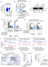
Fig. 1.
Fig. 1.
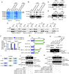
N-Myc regulates MILIP expression that is associated with poor patient outcome. (A) Regression analysis of the relationship between MILIP and N-Myc mRNA expression (RPM) in the RNA-seq SEQC-RPM-seqcnb1 neuroblastoma dataset. (B) MILIP expression in neuroblastoma cell lines with or without MYCN amplification. (C) N-Myc (Lower panel) bound to the MILIP promoter (Upper panel). (D) N-Myc knockdown down-regulated MILIP expression. (E) Luciferase reporter assays showing that the transcriptional activity of a MILIP promoter reporter construct containing the Myc-BR was reduced by N-Myc knockdown, whereas a MILIP promoter reporter construct with the Myc-BR deleted displayed decreased activity that was not affected by N-Myc knockdown. (F) Kaplan–Meier analysis of the probability of patient overall survival (OS). (G) Representative microscopic photographs of ISH analysis of MILIP expression in FFPE tissues (n = 27 tumors). (H) Quantitation of MILIP expression as detected in G. (I) Regression analysis of the relationship between MILIP expression (G) and N-Myc protein expression (SI Appendix, Fig. S2C). RS: reactive score; IRS: immunoreactive score. Data shown are mean ± SEM (B, bar charts in D and E) or representative (C and western blotting panels in D) of 3 independent experiments. One-way ANOVA followed by Tukey’s multiple comparison test (B, D, and E) or two-tailed Student’s t test (H).
Fig. 2.
Fig. 2.
MILIP promotes neuroblastoma tumorigenicity. (AE) MILIP knockdown (A) reduced cell viability (B), induced caspase-3 activation and PARP cleavage (C), caused G0/G1 cell cycle arrest (D), and reduced clonogenicity (E). (F) Quantitation of relative clonogenicity as shown in E. (G and H) MILIP expression (G) and the clonogenicity in cells with induced MILIP knockdown were recovered upon withdrawal of doxycycline (Dox). (Scale bar, 1 cm.) (I) Quantitation of relative clonogenicity as shown in H. (J and K) Photographs (J) and growth curves (K) of BE(2)-C.shMILIP.2 xenografts in nu/nu mice with or without cessation of Dox treatment. (Scale bar, 1 cm.) Data shown are mean ± SEM (A, B, D, F, G, and I) or representative (C, E, and H) of 3 independent experiments. One-way ANOVA followed by Tukey’s multiple comparison test.
Fig. 3.
Fig. 3.
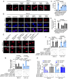
MILIP facilitates Ku70–Ku80 heterodimerization. (A) RPD followed by mass spectrometry analysis showing that Ku70 and Ku80 bound to MILIP (n = 2 independent experiments). S: sense; AS: antisense. (B) Endogenous Ku70 and Ku80 were copulled down by MILIP. (C and D) MILIP was coprecipitated with endogenous Ku70 (C) and Ku80 (D). (E) MILIP knockdown reduced the amount of endogenous Ku70 coprecipitated with Ku80. (F) Quantitation of the relative amount of Ku70 associated with Ku80 as shown in E. (G) Schematic illustration of full-length MILIP (MILIP-FL) and MILIP mutants used for deletion mapping. (H) MILIP Δ-1488/-1895 but not Δ-991/-1895 pulled down Ku70, whereas MILIP Δ-991/-1895 but not ΔE2 pulled down Ku80 in a cell-free system. (I) Schematic illustration of full-length Ku70 and Ku80 (Ku70-FL and Ku80-FL, respectively) and Ku70 or Ku80 mutants with its N terminus, the central DNA-binding domain, or C terminus deleted (K70/80-△NTD, K70/80-△DBD, or K70/80-△CTD). (J) RIP assays showing that in vitro–synthesized MILIP was coprecipitated with purified Ku70-△NTD and Ku70-△DBD but not Ku70-△CTD in a cell-free system. (K) RIP assays showing that in vitro–synthesized MILIP was coprecipitated with Ku80-△CTD and Ku80-△DBD but not Ku80-△NTD in a cell-free system. Data shown are mean ± SEM (F) or representative (B, C, D, E, H, J, and K) of 3 independent experiments. A two-tailed Student’s t test.
Fig. 4.
Fig. 4.
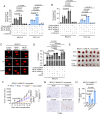
MILIP promotes the NHEJ pathway. (A and B) MILIP knockdown induced the appearance of comet tails (A) and the formation of γH2A.X foci (red) (B). (C and D) Quantitation of the relative tail DNA content of the comets and γH2A.X foci as shown in A (C) and B (D), respectively. (E) Introduction of MILIP-sh.R but not MILIP-△-617/-990 or MILIP-△-991/-1487 reduced the comet tails caused by MILIP knockdown. (F) Quantitation of the relative tail DNA content as shown in E. (G) Introduction of MILIP-sh.R but not MILIP-△-617/-990 or MILIP-△-991/-1487 diminished the inhibition of cell viability caused by knockdown of endogenous MILIP. (H) MILIP knockdown caused reductions in NHEJ but not HR activity measured using NHEJ and HR GFP reporters, respectively. Data shown are mean ± SEM (C, D, F, G, and H) or representative (A, B, and E) of 3 independent experiments. One-way ANOVA followed by Tukey’s multiple comparison test.
Fig. 5.
Fig. 5.
MILIP protects against DNA-damaging therapeutics. (A and B) MILIP knockdown and IR (A) or CDDP (B) cooperatively induced apoptosis. (C) MILIP nockdown and IR (A) or CDDP (B) cooperatively induced comet tails. (D) Quantitation of the relative tail DNA content as shown in C. (E and F) Photographs (E) and growth curves (F) of BE(2)-C.shMILIP.2 xenografts in nu/nu mice with or without treatment with Dox, CDDP, or Dox plus CDDP (n = 6 mice per group). (Scale bar, 1 cm.) (G) Representative microscopic photographs of TUNEL staining on randomly selected BE(2)-C.shMILIP.2 tumors (n = 3 tumors). (H) Quantitation of TUNEL staining as shown in G (n = 3 tumors). IRS: immunoreactive score. Data shown are mean ± SEM (A, B, and D) or representative (C) of 3 independent experiments. One-way ANOVA followed by Tukey’s multiple comparison test.
Fig. 6.
Fig. 6.
MILIP Gapmers inhibit, and in cooperation with CDDP to inhibit, neuroblastoma xenograft growth. (A) MILIP expression in cells with or without transfection of Gapmer.MILIP. (B and C) Introduction of Gapmer.MILIP reduced cell viability (B) and clonogenicity (C). (Scale bar, 1 cm.) (D) Quantitation of relative clonogenicity as shown in C. (E and F) Photographs (E) and growth curves (F) of BE(2)-C xenografts in nu/nu mice treated as indicated (n = 6 mice per group). (Scale bar, 1 cm.) (G) Quantitation of tumor weights as shown in E and F showing that cotreatment with Gapmer.MILIP (10 mg/kg, i.v. injection) and CDDP (1 mg/kg, i.p. injection) induced greater inhibition of BE(2)-C tumor growth than treatment with Gapmer.MILIP or CDDP alone in nu/nu mice (n = 6 mice per group, mean ± SEM, one-way ANOVA followed by Tukey’s multiple comparison test). (H) MILIP expression in representative BE(2)-C tumors (n = 3 tumors per group). (I) Representative microscopic photographs of TUNEL staining on randomly selected tumor tissues from mice treated in E and F (n = 3 tumors per group). (J) Quantitation of TUNEL staining as shown in I (n = 3 tumors per group). IRS: immunoreactive score. Data shown are mean ± SEM (A, B, D, and G) or representative (C) of 3 independent experiments. One-way ANOVA followed by Tukey’s multiple comparison test.

References

    1. Brodeur G. M., Neuroblastoma: Biological insights into a clinical enigma. Nat. Rev. Cancer 3, 203–216 (2003). - PubMed
    1. Liu P. Y., Atmadibrata B., Mondal S., Tee A. E., Liu T., NCYM is upregulated by lncUSMycN and modulates N-Myc expression. Int. J. Oncol. 49, 2464–2470 (2016). - PubMed
    1. Papadopoulos D., et al. , MYCN recruits the nuclear exosome complex to RNA polymerase II to prevent transcription-replication conflicts. Mol. Cell 82, 159–176.e112 (2022). - PubMed
    1. Luoto K. R., et al. , Tumor cell kill by c-MYC depletion: Role of MYC-regulated genes that control DNA double-strand break repair. Cancer Res. 70, 8748–8759 (2010). - PubMed
    1. Mehta A., Haber J. E., Sources of DNA double-strand breaks and models of recombinational DNA repair. Cold Spring Harb Perspect Biol. 6, a016428 (2014). - PMC - PubMed

Publication types