

Sentinel Lymph Node Identification in Patients With Breast Cancer Using Lymphosonography
- PMID: 36446688
- PMCID: PMC9943072
- DOI: 10.1016/j.ultrasmedbio.2022.10.020
Sentinel Lymph Node Identification in Patients With Breast Cancer Using Lymphosonography
Abstract
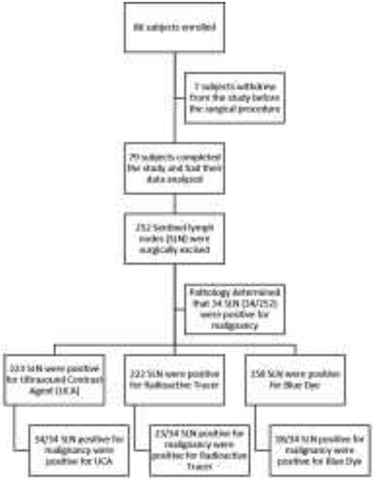
The objective of the work described here was to evaluate the efficacy of lymphosonography in identifying sentinel lymph nodes (SLNs) in patients with breast cancer undergoing surgical excision. Of the 86 individuals enrolled, 79 completed this institutional review board-approved study. Participants received subcutaneous 1.0-mL injections of ultrasound contrast agent (UCA) around the tumor. An ultrasound scanner with contrast-enhanced ultrasound (CEUS) capabilities was used to identify SLNs. Participants were administered with blue dye and radioactive tracer to guide SLN excision as standard-of-care. Excised SLNs were classified as positive or negative for the presence of blue dye, radioactive tracer and UCA, and sent for pathology. Two hundred fifty-two SLNs were excised; 158 were positive for blue dye, 222 were positive for radioactive tracer and 223 were positive for UCA. Comparison with blue dye revealed accuracies of 96.2% for radioactive tracer and 99.4% for lymphosonography (p > 0.15). Relative to radioactive tracer, blue dye had an accuracy of 68.5%, and lymphosonography achieved 86.5% (p < 0.0001). Of 252 SLNs excised, 34 were determined to be malignant by pathology; 18 were positive for blue dye (detection rate = 53%), 23 for radioactive tracer (detection rate = 68%) and 34 for UCA (detection rate = 100%) (p < 0.0001). Lymphosonography was similar in accuracy to radioactive tracer and higher in accuracy than blue dye in identifying SLNs. All 34 malignant SLNs were identified by lymphosonography.
Keywords: Breast cancer; Contrast-enhanced ultrasound; Lymphatic mapping; Lymphatic tracer; Lymphosonography; Sentinel lymph node; Ultrasound; Ultrasound contrast agent.
Copyright © 2022 World Federation for Ultrasound in Medicine & Biology. Published by Elsevier Inc. All rights reserved.
Conflict of interest statement
Conflict of interest disclosure P.M., J.B.L., L.N., M.L., A.I.W., K.B., S.N. and A.B. have nothing to disclose. F.F. received grant and equipment support from GE.
Figures
References
-
- Boughey Judy C., Suman Vera J., Mittendorf Elizabeth A., Ahrendt Gretchen M., Wilke Lee G., Taback Bret, Leitch A. Marilyn, Flippo-Morton Teresa S., Kuerer Henry M., and Bowling Monet. 2015. 'Factors affecting sentinel lymph node identification rate after neoadjuvant chemotherapy for breast cancer patients enrolled in ACOSOG Z1071 (Alliance)', Annals of surgery, 261: 547. - PMC - PubMed
-
- Cabioğlu Neslihan, Karanlik Hasan, Kangal Dilek, Özkurt Enver, Öner Gizem, Sezen Fatma, Yilmaz Ravza, Tükenmez Mustafa, Önder Semen, and İğci Abdullah. 2018. 'Improved false-negative rates with intraoperative identification of clipped nodes in patients undergoing sentinel lymph node biopsy after neoadjuvant chemotherapy', Annals of Surgical Oncology, 25: 3030–36 - PubMed
-
- Camp ER, Cendan JC, Feezor R, and Lind DS. 2004. 'The hottest sentinel lymph node is not always the positive node', The American surgeon, 70: 475. - PubMed
-
- Cho Nariya, Moon Woo Kyung, Han Wonshik, Park In Ae, Cho Jihyoung, and Noh Dong-Young. 2009. 'Preoperative sonographic classification of axillary lymph nodes in patients with breast cancer: node-to-node correlation with surgical histology and sentinel node biopsy results', American Journal of Roentgenology, 193: 1731–37 - PubMed
-
- Cox K, Sever A, Jones S, Weeks J, Mills P, Devalia H, Fish D, and Jones P. 2013. 'Validation of a technique using microbubbles and contrast enhanced ultrasound (CEUS) to biopsy sentinel lymph nodes (SLN) in pre-operative breast cancer patients with a normal grey-scale axillary ultrasound', European Journal of Surgical Oncology (EJSO), 39: 760–65 - PubMed
Publication types
MeSH terms
Substances
Grants and funding
LinkOut - more resources
Full Text Sources
Medical
