Review
. 2023 Feb;30(2):304-312.
doi: 10.1038/s41418-022-01094-w. Epub 2022 Nov 29.

Mitochondria and cell death-associated inflammation

Affiliations
Review

Mitochondria and cell death-associated inflammation

Esmee Vringer et al. Cell Death Differ. 2023 Feb.

Abstract

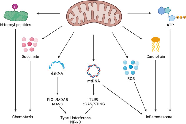
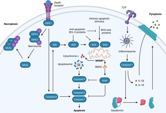
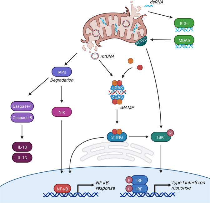
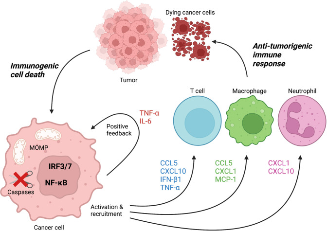
Mitochondria have recently emerged as key drivers of inflammation associated with cell death. Many of the pro-inflammatory pathways activated during cell death occur upon mitochondrial outer membrane permeabilization (MOMP), the pivotal commitment point to cell death during mitochondrial apoptosis. Permeabilised mitochondria trigger inflammation, in part, through the release of mitochondrial-derived damage-associated molecular patterns (DAMPs). Caspases, while dispensable for cell death during mitochondrial apoptosis, inhibit activation of pro-inflammatory pathways after MOMP. Some of these mitochondrial-activated inflammatory pathways can be traced back to the bacterial ancestry of mitochondria. For instance, mtDNA and bacterial DNA are highly similar thereby activating similar cell autonomous immune signalling pathways. The bacterial origin of mitochondria suggests that inflammatory pathways found in cytosol-invading bacteria may be relevant to mitochondrial-driven inflammation after MOMP. In this review, we discuss how mitochondria can initiate inflammation during cell death highlighting parallels with bacterial activation of inflammation. Moreover, we discuss the roles of mitochondrial inflammation during cell death and how these processes may potentially be harnessed therapeutically, for instance to improve cancer treatment.


Conflict of interest statement

The authors declare no competing interests.

Figures

Fig. 1
Fig. 1. Overview of mitochondrial-derived DAMPs.
Mitochondria contain DAMPs that can be exposed upon mitochondrial stress and damage. These DAMPs include succinate, N-formyl peptides, dsRNA, mtDNA, ROS, cardiolipin, and ATP. Loss or exposure of these DAMPs activates several immune pathways including transcription of type I interferons and NF-κB target genes, inflammasome activation, and the recruitment of immune cells.
Fig. 2
Fig. 2. Overview of necroptotic, apoptotic and pyroptotic signalling pathways.
Binding of ligands to one of the death receptors (e.g. TNF or Fas) initiates pleiotropic signalling leading to cell survival, inflammation, apoptosis or necroptosis, mediated by the key signalling protein RIPK1. Upon caspase-8 inhibition, RIPK1 forms the necrosome with RIPK3 leading to phosphorylation and activation of MLKL causing membrane permeabilization and necroptosis. Activation of initiator caspases (caspase-8) by death receptors leads to cleavage and activation of executioner caspases (caspase-3 and -7) causing apoptosis. The intrinsic pathway requires an intrinsic apoptotic stimulus which activates pro-apoptotic BCL-2 family members BAX and BAK. Upon their activation pores are formed in the mitochondrial outer membrane leading to the release of intermembrane space proteins (including cytochrome c and SMAC). Release of cytochrome c allows apoptosome formation which recruits and activates caspase-9 followed by the activation of the executioner caspases. SMAC binds to XIAP, thereby blocking the caspase-inhibiting potential of XIAP. MOMP is also initiated by the extrinsic apoptotic pathway through BID cleavage by caspase-8. Furthermore, cells can die via pyroptosis through DAMP recognition by TLRs leading to inflammasome activation and subsequently caspase-1 activation. Caspase-1 cleaves gasdermins (GSDM) of which the N-terminal cleavage fragments form pores in the plasma membrane. In addition, caspase-1 cleaves pro-IL-1β and pro-IL-18 into their mature forms that are released via GSDM pores.
Fig. 3
Fig. 3. MOMP-induced inflammation.
MOMP activates several pro-inflammatory pathways. (1) Under caspase-inhibited conditions MOMP causes IAP degradation which subsequently leads to NIK stabilisation and accumulation followed by the transcription of NF-κB target genes. In addition, degradation of IAPs activates caspase-1 and caspase-8 leading to processing and release of IL-1β and IL-18. (2) Cytosolic release of mtDNA leads to recognition of cGAS which subsequently forms cGAMP out of GTP and ATP. cGAMP is a second messenger for ER-resident STING initiating its activating and the subsequent transcription of NF-κB target genes and type I interferons. (3) Release of cytosolic dsRNA leads to its recognition by RIG-I and MDA5, followed by activation of mitochondria-localised MAVS and a type I interferon response.
Fig. 4
Fig. 4. Anti-tumorigenic immune response upon MOMP-induced immunogenic cell death in tumours.
Induction of immunogenic cell death in cancer cells can be achieved through MOMP in combination with caspase inhibition. Activation of several cell autonomous immune signalling pathways leads to the transcription and release of type I interferons and NF-κB target genes. Release of cytokines and chemokines recruits and activates anti-tumour T cells, macrophages and neutrophils thereby enhancing cancer cell death and tumour regression.

References

    1. Wallace DC. Mitochondria and cancer. Nat Rev Cancer. 2012;12:685–98. doi: 10.1038/nrc3365. - DOI - PMC - PubMed
    1. Friedman JR, Nunnari J. Mitochondrial form and function. Nature. 2014;505:335–43. doi: 10.1038/nature12985. - DOI - PMC - PubMed
    1. Rodriguez-Nuevo A, Zorzano A. The sensing of mitochondrial DAMPs by non-immune cells. Cell Stress. 2019;3:195–207. doi: 10.15698/cst2019.06.190. - DOI - PMC - PubMed
    1. Martin WF, Garg S, Zimorski V. Endosymbiotic theories for eukaryote origin. Philos Trans R Soc Lond B Biol Sci. 2015;370:20140330. doi: 10.1098/rstb.2014.0330. - DOI - PMC - PubMed
    1. Roger AJ, Munoz-Gomez SA, Kamikawa R. The origin and diversification of mitochondria. Curr Biol. 2017;27:R1177–92. doi: 10.1016/j.cub.2017.09.015. - DOI - PubMed

Publication types