Review
. 2023 Feb;25(1):46-57.
doi: 10.1007/s11307-022-01789-z. Epub 2022 Nov 29.

Current and Future Applications of Fluorescence Guidance in Orthopaedic Surgery

Affiliations
Review

Current and Future Applications of Fluorescence Guidance in Orthopaedic Surgery

Samuel S Streeter et al. Mol Imaging Biol. 2023 Feb.

Abstract

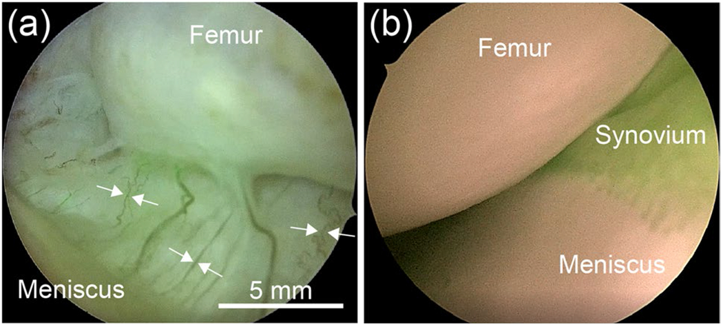
Fluorescence-guided surgery (FGS) is an evolving field that seeks to identify important anatomic structures or physiologic phenomena with helpful relevance to the execution of surgical procedures. Fluorescence labeling occurs generally via the administration of fluorescent reporters that may be molecularly targeted, enzyme-activated, or untargeted, vascular probes. Fluorescence guidance has substantially changed care strategies in numerous surgical fields; however, investigation and adoption in orthopaedic surgery have lagged. FGS shows the potential for improving patient care in orthopaedics via several applications including disease diagnosis, perfusion-based tissue healing capacity assessment, infection/tumor eradication, and anatomic structure identification. This review highlights current and future applications of fluorescence guidance in orthopaedics and identifies key challenges to translation and potential solutions.

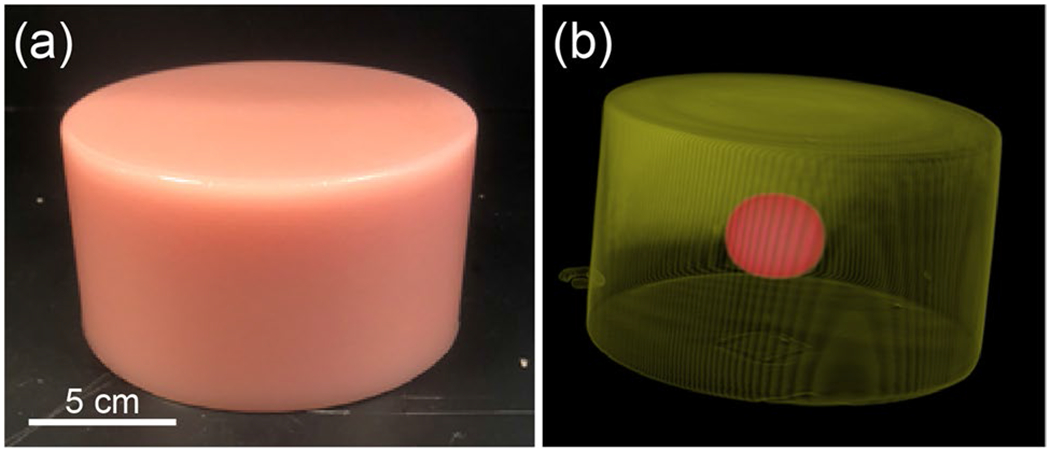
Keywords: Fluorescence-guided surgery; Indocyanine green imaging; Meniscus tear; Necrotizing soft tissue infection; Nerve imaging; Tissue-simulating phantoms; Tumor imaging.


Conflict of interest statement

S. Streeter is a part-time employee of QUEL Imaging LLC, a Small Business Innovation Research-funded start-up that focuses on the commercialization of optical targets for fluorescence guided surgical systems. S. Gibbs is a co-founder and stockholder of Trace Biosciences, a start-up company focused on the development and clinical translation of nerve-specific probes for FGS. S. Gibbs also has three patents pending related to this work (US No. 62/711,465, US No. 62/729,932, and US No. 62/956,614). E. Henderson is an educational consultant for Stryker Orthopaedics. All other authors declare no competing interests.

Figures

Fig. 1
Fig. 1
Overview of the three general applications of fluorescence guidance in orthopaedic surgery discussed in this review article: tissue infection management, perfusion-based assessment of tissue healing capacity and joint repairs, and nerve and tumor tissue identification.
Fig. 2
Fig. 2
Representative patient presenting with confirmed necrotizing fasciitis (NF) in the lower leg. a White light color image shows minimal contrast to disease extent. b Wide-field ICG image at two-min post-injection reveals approximate extent of disease (yellow dashed line) with disease represented by ICG signal void; quantitative fluorescence standard (QUEL Imaging, White River Junction, VT) is shown on left; regions of interest depict deep NF-affected tissue (red), near-boarder NF-affected tissue (blue), and unaffected tissue (yellow). c Time-resolved fluorescence kinetic curve parameterization yields four features: max intensity (Imax), time-to-peak (TTP), ingress slope (IS), and egress slope (ES). d Kinetic curves extracted from the three regions of interest defined in (B). e Imax parameter map. f TTP parameter map. g IS parameter map. h ES parameter map. Scale bar in a also applies to b, e–h.
Fig. 3
Fig. 3
Two representative knee joints imaged using the Image1 S RU Rubina OPAL 1 arthroscope (Karl Storz, Tuttlingen, Germany). a ICG signal intensity enables high-contrast visualization of joint microvascular (white arrows). b ICG signal visualizes well-perfused synovial tissue and highlights regions of low and potentially jeopardized perfusion corresponding to fluorescence signal voids. The 5-mm scale bar in a also applies to b.
Fig. 4
Fig. 4
Preliminary data of shoulder arthroplasty tendon perfusion assessment in six patients using dynamic, contrast-enhanced ICG imaging. a The peak fluorescence intensity pre- and post-procedure for each of six study patients. b Distribution of fluorescence intensities extracted from a region of interest delineating the exposed sub-scapularis tendon, before and after the procedure. c Mean region of interest fluorescence intensity curves for each patient during ICG wash-in and wash-out, before and after the procedure.
Fig. 5
Fig. 5
Resected (i.e., ex vivo) benign-aggressive bone lesion tissue (white dashed line) exhibits increased ICG fluorescence relative to healthy resected tissues (white arrows). Quantitative fluorescence standard is shown on the left (QUEL Imaging, White River Junction, VT). Surgery and imaging performed 24 h post-injection.
Fig. 6
Fig. 6
Representative fluorescence images demonstrating positive contrast using the nerve-specific, NIR fluorophore LGW16-03 in a human limb perfusion model. a Fluorescence highlights in vivo deep peroneal nerve surrounded by adipose tissue; the intensity profile (red dashed line) is plotted in b. c Ex vivo tissues imaged highlight common peroneal nerve fluorescence relative to muscle tissue.
Fig. 7
Fig. 7
Gelatin-based, tissue-simulating phantom for assessing conventional medical imaging (i.e., CT and MRI) and fluorescence imaging for surgical guidance. a White light image of a representative phantom. b Volumetric rendering of the same phantom showing tumor-simulating MRI contrast agent inclusion (red). Scale bar in a also applies to b.

References

    1. Weissleder R, Mahmood U (2001) Molecular imaging. Radiol 219:316–333. 10.1148/radiology.219.2.r01ma19316 - DOI - PubMed
    1. van Leeuwen FWB, Hardwick JCH, van Erkel AR (2015) Luminescence-based imaging approaches in the field of interventional molecular imaging. Radiology 276:12–29. 10.1148/radiol.2015132698 - DOI - PubMed
    1. Paraboschi I, Farneti F, Jannello L, Manzoni G, Berrettini A, Mantica G (2022) Narrative review on applications of fluorescence guided surgery in adult and pediatric urology. AME Med J 7:15. 10.21037/amj-20-194 - DOI
    1. Vahrmeijer AL, Hutteman M, van der Vorst JR et al. (2013) Image-guided cancer surgery using near-infrared fluorescence. Nat Rev Clin Oncol 10:507–518. 10.1038/nrclinonc.2013.123 - DOI - PMC - PubMed
    1. Schouw HM, Huisman LA, Janssen YF et al. (2021) Targeted optical fluorescence imaging: a meta-narrative review and future perspectives. Eur J Nucl Med Mol Imaging 48:4272–4292. 10.1007/s00259-021-05504-y - DOI - PMC - PubMed

Publication types

Substances