. 2022 Nov 29;28(1):143.
doi: 10.1186/s10020-022-00566-6.

Surgical implantation of human adipose derived stem cells attenuates experimentally induced hepatic fibrosis in rats

Affiliations

Surgical implantation of human adipose derived stem cells attenuates experimentally induced hepatic fibrosis in rats

Masateru Nomura et al. Mol Med. .

Abstract

Background: Mesenchymal stem cells (MSCs) are multipotent stromal cells and could exert hepatoprotective effects against acute liver injury, steatohepatitis, and fibrogenesis. Here, we evaluated the effects of human adipose derived stem cells (hADSCs) to attenuate experimentally induced hepatic fibrosis and early cirrhosis in rats.

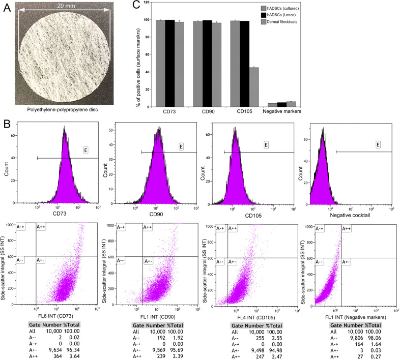
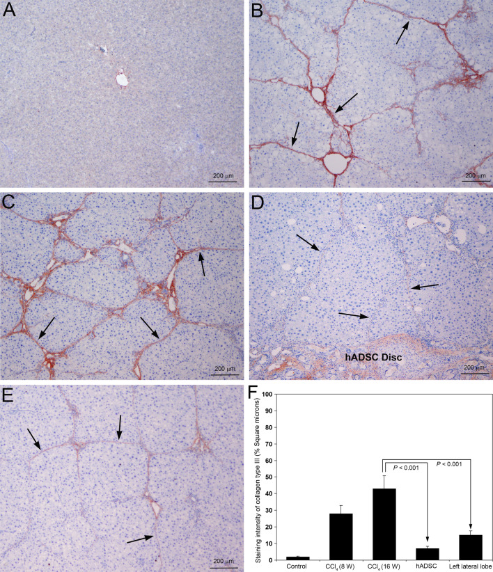
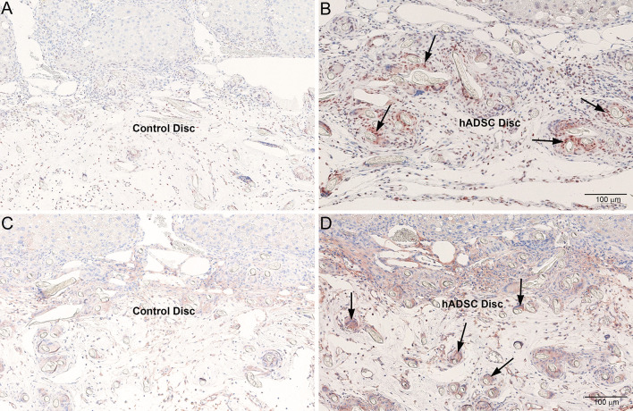
Methods: Hepatic fibrosis was induced by intraperitoneal injections of CCl4 (0.1 ml/100 g body weight) twice a week for 8 weeks. hADSCs were isolated and cultured on polyethylene discs coated with hydroxyapatite and 2 cm diameter disc was surgically implanted on the right lateral lobe of the liver. Discs implanted without hADSCs served as control. The animals were injected again with CCl4 once a week for another 8 weeks. All the animals were sacrificed at the end of 16th week.

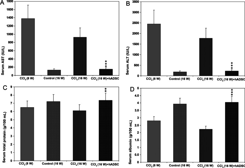
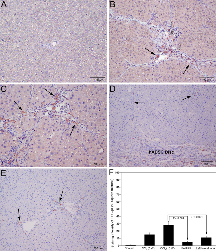
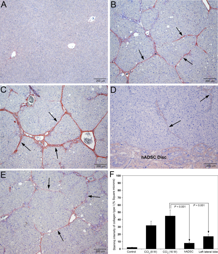
Results: Serial administrations of CCl4 resulted in well developed fibrosis and early cirrhosis at 8th week which maintained until the 16th week. Animals treated with hADSC discs depicted over 50% decrease of collagen with significant increase in serum albumin and total protein levels. Immunohistochemical staining for TGF-β1, α-smooth muscle actin, and collagen type I and type III demonstrated marked decrease compared to the animals without hADSC treatment.

Conclusions: Treatment with hADSCs improved liver functions, markedly reduced hepatic fibrosis and early cirrhosis. Various pleiotropic and paracrine factors secreted from the hADSCs seem to serve as reparative functions in the attenuation of liver cirrhosis. The data demonstrated that treatment with hADSCs can be successfully used as a potent therapeutic method to prevent progression of hepatic fibrosis and related adverse events.

Keywords: Adipose derived stem cells; Carbon tetrachloride; Hepatic fibrosis; Liver cirrhosis; MSCs; Mesenchymal stem cells; hADSCs.


Conflict of interest statement

The authors do not have any conflicts of interest to declare in connection with this manuscript.

Figures

Fig. 1
Fig. 1
A Photographic image of non-woven fabric polyethylene-polypropylene (PE-PP) disc coated with hydroxyapatite used for isolation and culture of human adipose tissue derived stem cells (hADSCs) without enzymatic digestion. B Representative histogram and dot plot images of the flow cytometric analysis of isolated and cultured hADSCs (2nd passage) for the expression of CD73, CD90, and CD105 and a cocktail of negative markers (CD34, CD11b, CD19, CD45, and HLA-DR) after harvested using Accutase cell detachment solution and treatment with respective anti-human mAbs conjugated with various fluorescent tags. The gated cells in A +—column indicates they are positive for the respective surface marker and the cells in A– column indicates that the negative markers are not expressed in hADSCs. Practically no cells are present in A +—column with the expression of negative markers. C Graphical presentation of the flow cytometry data for the expression of surface markers in cultured hADSCs, human ADSCs from Lonza, and human dermal fibroblast cells. The data are mean ± S.D. of 5 independent experiments
Fig. 2
Fig. 2
Differentiation of hADSCs into adipocytes and osteocytes. The hADSCs were cultured with respective differentiation medium for 17 days. A hADSCs cultured with and without adipogneic differentiation factors and stained with AdipoRed. B hADSCs cultured with and without osteogenic differentiation factors and stained with Alizarin Red. Original magnification, × 200
Fig. 3
Fig. 3
Aspartate aminotransferase (AST), alanine aminotransferase (ALT), total protein (TP), and albumin levels in the serum following serial administrations of CCl4 and after treatment with hADSC disc. A AST levels B ALT levels C Serum total protein D Serum albumin. Treatment with hADSC disc resulted in restoration of normal levels of AST, ALT, TP, and albumin in the serum. The data are mean ± S.D. of 5 rats in each group. **P < 0.01 and ***P < 0.001 CCl4 (16 W) treated group versus CCl4 (16 W) + hADSC group
Fig. 4
Fig. 4
Azan trichrome staining for collagen in rat liver sections following serial administrations of CCl4 and after treatment with hADSC disc. A Staining for collagen was absent in the control livers treated with olive oil for 16 weeks. B Serial administrations of CCl4 for 8 weeks produced well developed fibrosis and early cirrhosis with deposition of mature collagen fibers (arrows). C At 16th week of CCl4 administration, the collagen fibers became thick and more prominent with well developed cirrhosis (arrows). D Treatment with hADSC disc for 8 weeks during CCl4 administration resulted in more than 50% decrease of collagen fibers and incomplete fibrous septa (arrows). The hADSC disc which is surgically implanted on right lateral lobe of the liver is visible at the bottom portion. E Marked reduction of cirrhosis on left lateral lobe of the liver in which the animal is treated with hADSC disc on right lateral lobe. The collagen fibers were thin with incomplete fibrous septa and partial bridging (arrows). Original magnification, × 40. F The staining intensity of total collagen was quantified using Image-Pro Discovery software. The data are mean ± S.D. of 10 randomly selected microscopic fields from five rats per group
Fig. 5
Fig. 5
Immunohistochemical staining for transforming growth factor-β1 (TGF-β1) in rat liver sections following serial administrations of CCl4 and after treatment with hADSC disc. A Staining for TGF-β1 was completely absent in the control livers treated with olive oil for 16 weeks. B Marked staining for TGF-β1 was present in the fibrotic areas of rat livers treated with CCl4 for 8 weeks (arrows). C Prominent and conspicuous staining for TGF-β1 was present at 16th week of CCl4 administration, especially in the fibrotic zone (arrows). D Treatment with hADSC disc for 8 weeks showed marked reduction in staining for TGF-β1 in the hepatic parenchyma (arrows). The implanted hADSC disc is visible at the bottom portion of the image with positive staining for TGF-β1. E There was significant decrease in staining of TGF-β1 in the left lateral lobe, where the right lateral lobe is implanted with hADSC disc (arrows). AC Original magnification, × 100. D, E Original magnification, × 40. F The staining intensity of TGF-β1 was quantified using Image-Pro Discovery software. The data are mean ± S.D. of 10 randomly selected microscopic fields from five rats per group
Fig. 6
Fig. 6
Immunohistochemical staining of α-smooth muscle actin (α-SMA) in rat liver sections following serial administrations of CCl4 and after treatment with hADSC disc. A Staining for α-SMA was completely absent in the control livers treated with olive oil for 16 weeks. B Remarkable staining for α-SMA was present in the fibrotic areas of rat livers treated with CCl4 for 8 weeks (arrows). C At 16th week of CCl4 administration, enormous and obvious staining for α-SMA was present in the periphery of thick collagen fibers (arrows). D Treatment with hADSC disc for 8 weeks demonstrated significant reduction in staining for α-SMA in the fibrotic zone (arrows). The implanted hADSC disc is clearly visible at the bottom portion of the image. E Marked decrease in staining of α-SMA in the left lateral lobe, where the right lateral lobe is implanted with hADSC disc (arrows). AC Original magnification, × 100. D, E Original magnification, × 40. F Quantification of the staining intensity of α-SMA as square microns. The intensity of α-SMA staining was quantified using Image-Pro Discovery software. The data are mean ± S.D. of 10 randomly selected microscopic fields from five rats per group
Fig. 7
Fig. 7
Immunohistochemical staining for collagen type I in rat liver sections following serial administrations of CCl4 and after treatment with hADSC disc. A Staining for collagen type I was absent in the hepatic parenchyma of control livers treated with olive oil for 16 weeks. B At 8th week of CCl4 administration, there was marked staining for collagen type I in the fibrotic zone indicating deposition of newly formed collagen (arrows). C At 16th week, the staining for collagen type I became more prominent depicting thick collagen fibers (arrows). D Treatment with hADSC disc for 8 weeks resulted in marked decrease of collagen type I staining indicating decreased synthesis and deposition of collagen type I (arrows). The hADSC disc with weak staining for collagen type I is visible at the bottom portion. E Marked reduction of staining for collagen type I in the left lateral lobe, where the right lateral lobe is implanted with hADSC disc. The collagen type I fibers were thin with incomplete fibrous septa and partial bridging (arrows). Original magnification, × 40. F The staining intensity of collagen type I was quantified using Image-Pro Discovery software. The data are mean ± S.D. of 10 randomly selected microscopic fields from five rats per group
Fig. 8
Fig. 8
Immunohistochemical staining for collagen type III in rat liver sections following serial administrations of CCl4 and after treatment with hADSC disc. A Staining for collagen type III was completely absent in the control livers treated with olive oil for 16 weeks. B Prominent and marked staining for collagen type III was present in the fibrotic septa of all the rats treated with CCl4 for 8 weeks (arrows). C At 16th week of CCl4 administration, remarkable and conspicuous staining for collagen type III with thick fibers were present (arrows). D Treatment with hADSC disc for 8 weeks depicted marked reduction in staining for collagen type III with feeble and incomplete collagen fibers (arrows). E Significant decrease of staining for collagen type III in the left lateral lobe, where the right lateral lobe is implanted with hADSC disc. Collagen fibers were thin with incomplete fibrous septa and partial bridging (arrows). Original magnification, × 40. F The intensity of collagen type III staining was quantified using Image-Pro Discovery software. The data are mean ± S.D. of 10 randomly selected microscopic fields from five rats per group
Fig. 9
Fig. 9
Immunohistochemical staining for human lamin B1 and CD73 at 8 weeks after implantation of hADSC disc on rat livers. A Implantation of disc alone without hADSCs. Staining for lamin B1 was completely absent. B Positive staining for human lamin B1, a characteristic highly conserved marker to demonstrate that the cells are human origin (arrows). C Implantation of disc alone without hADSCs. Since CD73 is expressed on subsets of T and B lymphocytes, biliary epithelial cells, and sinusoidal endothelial cells, there was focalized moderate staining for CD73 in the hepatic parenchyma. D Adipose-derived stem cells were stained positive for CD73 (arrows)

References

    1. Acharya P, Chouhan K, Weiskirchen S, Weiskirchen R. Cellular mechanisms of liver fibrosis. Front Pharmacol. 2021;12:671640. doi: 10.3389/fphar.2021.671640. - DOI - PMC - PubMed
    1. Ajami M, Soleimani M, Abroun S, Atashi A. Comparison of cord blood CD34 + stem cell expansion in coculture with mesenchymal stem cells overexpressing SDF-1 and soluble /membrane isoforms of SCF. J Cell Biochem. 2019;120(9):15297–15309. doi: 10.1002/jcb.28797. - DOI - PubMed
    1. Bianchi F, Maioli M, Leonardi E, Olivi E, Pasquinelli G, Valente S, Mendez AJ, Ricordi C, Raffaini M, Tremolada C, Ventura C. A new nonenzymatic method and device to obtain a fat tissue derivative highly enriched in pericyte-like elements by mild mechanical forces from human lipoaspirates. Cell Transplant. 2013;22(11):2063–2077. doi: 10.3727/096368912X657855. - DOI - PubMed
    1. Chen Z, Wan L, Jin X, Wang W, Li D. Transforming growth factor-β signaling confers hepatic stellate cells progenitor features after partial hepatectomy. J Cell Physiol. 2020;235(3):2655–2667. doi: 10.1002/jcp.29169. - DOI - PubMed
    1. Christ B, Brückner S, Winkler S. The therapeutic promise of mesenchymal stem cells for liver restoration. Trends Mol Med. 2015;21(11):673–686. doi: 10.1016/j.molmed.2015.09.004. - DOI - PubMed

Publication types

LinkOut - more resources