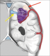
Atypical slow-flow paramedian AVM with venous varix
- PMID: 36447861
- PMCID: PMC9699874
- DOI: 10.25259/SNI_920_2022
Atypical slow-flow paramedian AVM with venous varix
Abstract
Background: Cerebral arteriovenous malformations (CAVMs) are either clinically silent or symptomatic. The most common presentation in more than half of all CAVMs presenting patients is hemorrhage which is accompanied by long-standing neurological morbidity and mortality. This report presents a case of an atypical large, slow-flow paramedian AVM with a dilated venous varix managed with surgery. The impact of the intraoperative findings on the diagnosis and the operative technique will be discussed.
Case description: In otherwise, healthy 26-year-old male complained of repeated episodes of generalized seizures and loss of consciousness. Brain magnetic resonance imaging (MRI) revealed a right parietal paramedian arteriovenous malformation (AVM) with signs of an old hemorrhagic cavity beneath it. Digital subtraction angiography demonstrated a slow-filling AVM with dilated venous varix drains into the superior sagittal sinus. However, the exact point of drainage cannot be appreciated. The filling of the AVM occurred precisely with the beginning of the venous phase. Intraoperatively, we noticed a whitish spherical mass, thick hemosiderin tissue, and a large cavity below the nidus; then, a complication-free complete microsurgical resection of this high-grade AVM was performed. Postoperatively, the patient suffered two attacks of seizures in the first few hours after the surgery, for which he received antiepileptics. MRI was clear during follow-up, and the patient was seizure-free and neurologically intact.
Conclusion: Parietal convexity AVMs are challenging lesions to tackle. However, the chronicity and the slow-filling of the AVM, in this case, can render the surgical pathway more direct and accessible.
Keywords: Cerebral arteriovenous malformation; Microsurgical resection; Venous varix.
Copyright: © 2022 Surgical Neurology International.
Conflict of interest statement
There are no conflicts of interest.
Figures


References
-
- Bass DI, Walker M, Ferreira M, Ghodke B. A case report of an unruptured tectal AVM presenting with obstructive hydrocephalus that resolved upon spontaneous obliteration of the venous varix. J Clin Neurosci. 2019;65:157–60. - PubMed
-
- Brown RD, Jr, Wiebers DO, Forbes G, O’Fallon WM, Piepgras DG, Marsh WR, et al. The natural history of unruptured intracranial arteriovenous malformations. J Neurosurgery. 1988;68:352–7. - PubMed
Publication types
LinkOut - more resources
Full Text Sources
