

The functional anatomy of the foramina of Luschka revisited
- PMID: 36447888
- PMCID: PMC9699837
- DOI: 10.25259/SNI_931_2022
The functional anatomy of the foramina of Luschka revisited
Abstract
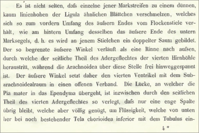
Background: The German Anatomist Hubert Von Luschka first described the foramina of Luschka (FOL) in 1855 as lateral holes in the fourth ventricle. By his discovery, he refuted previous beliefs about the lateral recess as blind ends of the fourth ventricle, proving the continuity of the ventricular system with the central canal of the spinal cord. In this paper, we question the outline variations of the patent parts of FOL and their consistency, drawing attention to the apparent query of the valvular mechanism of FOL.
Methods: We conducted a literature review in PubMed and Google Scholar databases to review the existing literature describing the history, pertinent anatomy, and function of FOL. In addition, we reviewed the original German book written by Luschka.
Results: While reading the available articles and original works regarding FOL, we noticed the developmental phases through which FOL was discovered, tracking the process from Aristotle till Luschka's discovery. We also discussed controversies and opinions about FOL's existence and function.
Conclusion: FOL is halved into two compartments: choroidal and patent. The function of FOL resembles a oneway valve mechanism, and it depends on the patent slit-like part. Luschka had discovered over 20 anatomical structures, including several foramina, confusion in a debate may result from eponyms.
Keywords: Choroid plexus; Foramen of Luschka; Fourth ventricle; Lateral openings; Patency.
Copyright: © 2022 Surgical Neurology International.
Conflict of interest statement
There are no conflicts of interest.
Figures


References
-
- Barany L, Baksa G, Patonay L, Ganslandt O, Buchfelder M, Kurucz P. Morphometry and microsurgical anatomy of Bochdalek’s flower basket and the related structures of the cerebellopontine angle. Acta Neurochir (Wien) 2017;159:1539–45. - PubMed
-
- Barany L, Baksa G, Patonay L, Racz G, Ganslandt O, Buchfelder M, et al. Primary obstruction of the foramen of Luschka: Anatomy, histology, and clinical significance. World Neurosurg. 2018;112:e288–97. - PubMed
-
- Boonstra EA, Lorenz K, Porte RJ. The quest for Luschka’s duct: An eponym leading a life of its own? Dig Surg. 2014;31:104–7. - PubMed
-
- De MeloMussi AC, da Luz de Oliveira EP. Comprehensive Overview of Modern Surgical Approaches to Intrinsic Brain Tumors. Ch. 5. Amsterdam: Elsevier Science; 2019. Ventricular anatomy; pp. 107–18.
-
- Duque-Parra JE, Barco-Ríos J, García-Aguirre JF. A historical approach to the ventricular system of the brain. Rev Fac Med. 2017;65:473–7.
Publication types
LinkOut - more resources
Full Text Sources