Myofibroblasts Are Not Characteristic Features of Keloid Lesions
- PMID: 36448015
- PMCID: PMC9699581
- DOI: 10.1097/GOX.0000000000004680
Myofibroblasts Are Not Characteristic Features of Keloid Lesions
Abstract
Keloids are disfiguring, scar-like lesions that are challenging to treat, with low response rates to current interventions and frequent recurrence. It has been widely reported that keloids are characterized by myofibroblasts, specialized contractile fibroblasts that express alpha-smooth muscle actin (α-SMA). However, evidence supporting a role for myofibroblasts in keloid pathology is inconclusive, with conflicting reports in the literature. This complicates development of more effective therapies, as the benefit of interventions targeting myofibroblasts is unclear. This study was undertaken to determine whether myofibroblasts can be considered characteristic of keloids.
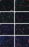
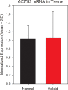
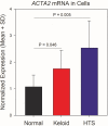
Methods: Myofibroblasts in tissue sections from keloids, hypertrophic scars (HTSs), and normal skin were localized by α-SMA immunostaining. Expression of α-SMA mRNA (ACTA2 gene) in normal skin and keloid tissue, and in fibroblasts from normal skin, keloid, and HTSs, was measured using quantitative polymerase chain reaction.
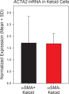
Results: Normal skin did not exhibit α-SMA-expressing myofibroblasts, but myofibroblasts were identified in 50% of keloids and 60% of HTSs. No significant differences in ACTA2 expression between keloid and normal skin tissue were observed. Mean ACTA2 expression was higher in HTS (2.54-fold, P = 0.005) and keloid fibroblasts (1.75-fold, P = 0.046) versus normal fibroblasts in vitro. However, α-SMA expression in keloids in vivo was not associated with elevated ACTA2 in keloid fibroblasts in vitro.
Conclusions: Despite elevated ACTA2 in cultured keloid fibroblasts, myofibroblast presence is not a consistent feature of keloids. Therefore, therapies that target myofibroblasts may not be effective for all keloids. Further research is required to define the mechanisms driving keloid formation for development of more effective therapies.
Copyright © 2022 The Authors. Published by Wolters Kluwer Health, Inc. on behalf of The American Society of Plastic Surgeons.
Figures


References
-
- Glass DA, II. Current understanding of the genetic causes of keloid formation. J Investig Dermatol Symp Proc. 2017;18:S50–S53. - PubMed
-
- Motoki THC, Isoldi FC, Filho AG, et al. . Keloid negatively affects body image. Burns. 2018;45:610–614. - PubMed
-
- Tan A, Glass DA, II. Patient-reported outcomes for keloids: a systematic review. G Ital Dermatol Venereol. 2019;154:148–165. - PubMed
-
- Bock O, Schmid-Ott G, Malewski P, et al. . Quality of life of patients with keloid and hypertrophic scarring. Arch Dermatol Res. 2006;297:433–438. - PubMed
LinkOut - more resources
Full Text Sources
Miscellaneous
