Review
. 2022 Dec 21;35(4):e0008619.
doi: 10.1128/cmr.00086-19. Epub 2022 Nov 30.

Osteoarticular Mycoses

Affiliations
Review

Osteoarticular Mycoses

Maria N Gamaletsou et al. Clin Microbiol Rev. .

Abstract

Osteoarticular mycoses are chronic debilitating infections that require extended courses of antifungal therapy and may warrant expert surgical intervention. As there has been no comprehensive review of these diseases, the International Consortium for Osteoarticular Mycoses prepared a definitive treatise for this important class of infections. Among the etiologies of osteoarticular mycoses are Candida spp., Aspergillus spp., Mucorales, dematiaceous fungi, non-Aspergillus hyaline molds, and endemic mycoses, including those caused by Histoplasma capsulatum, Blastomyces dermatitidis, and Coccidioides species. This review analyzes the history, epidemiology, pathogenesis, clinical manifestations, diagnostic approaches, inflammatory biomarkers, diagnostic imaging modalities, treatments, and outcomes of osteomyelitis and septic arthritis caused by these organisms. Candida osteomyelitis and Candida arthritis are associated with greater events of hematogenous dissemination than those of most other osteoarticular mycoses. Traumatic inoculation is more commonly associated with osteoarticular mycoses caused by Aspergillus and non-Aspergillus molds. Synovial fluid cultures are highly sensitive in the detection of Candida and Aspergillus arthritis. Relapsed infection, particularly in Candida arthritis, may develop in relation to an inadequate duration of therapy. Overall mortality reflects survival from disseminated infection and underlying host factors.

Keywords: antifungal therapy; aspergillosis; candidiasis; coccidioidomycosis; cryptococcosis; histoplasmosis; mucormycosis; mycoses; osteomyelitis; phaeohyphomycosis.


Conflict of interest statement

The authors declare no conflict of interest.

Figures

FIG 1
FIG 1
Candida osteomyelitis/discitis with psoas abscess. A 62-year-old male with diffuse large B cell lymphoma (stage 4) and decompensated cirrhosis developed Candida albicans fungemia and chorioretinitis after the first cycle of chemotherapy and was treated with fluconazole, micafungin, and intravitreal antifungals. After 2 additional cycles of chemotherapy, he developed worsening back pain, tenderness at the lumbar spine, and limited mobility. MRI showed L3-L4 osteomyelitis/discitis with psoas abscess (left, T1-weighted scan; right, T2-weighted scan). Cultures of specimens from CT-guided biopsy of the vertebral body and soft tissue specimens grew C. albicans, and histology showed yeast. TSE, turbo spin echo.
FIG 2
FIG 2
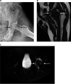
Aspergillus osteomyelitis and arthritis. A 52-year-old male with lymphoblastic lymphoma and a history of pulmonary aspergillosis developed pain in the left hip. MRI showed a femoral head lesion and avascular necrosis. After another cycle of chemotherapy, he was admitted to the hospital with a worsening of chronic left-hip pain and limited range of motion. (Left) A plain radiograph showed bone destruction, osteolysis of the left femoral head, and a decrease in the left articular space. (Middle and right) MRI showed a 3.8- by 2.1-cm oval mass in the anterior medial inferior left femoral head with a low T1 intensity (middle) and a high signal intensity with T2 (right), extending into the neck of the femur, with a well-defined border. There was moderate left-hip effusion and abnormal T2 hyperintensity in muscles around the left hip. A left-hip aspirate smear demonstrated hyphae by microscopy and grew Aspergillus flavus in culture.
FIG 3
FIG 3
Fusarium osteomyelitis of the head of the 4th and 5th metacarpal bones with soft tissue involvement and septic arthritis of the 4th and 5th metacarpophalangeal (MCP) joints. A 68-year-old leukemic male presented with left-shin ulceration and multiple subcutaneous nodules while on posaconazole prophylaxis after two cycles of chemotherapy for acute myelogenous leukemia. Skin biopsy specimen and blood cultures grew Fusarium spp. He then developed pain/swelling/tenderness of the left 5th MCP joint and left metacarpal. MRI showed bony destruction of the head of the 5th metacarpal and soft tissue involvement with extension into the left 5th MCP. Intraoperative findings were consistent with osteomyelitis and septic arthritis.
FIG 4
FIG 4
Hyalohyphomycosis-related osteomyelitis. Shown is the histopathology of the left lateral malleolus depicted in Fig. 6 in a patient with osteomyelitis caused by Scedosporium spp. (A) Gomori methenamine-stained fungal balls and fungal hyphae branched at 45° invading bone tissue (magnification, ×400). (B) Periodic acid-Schiff-stained fungal balls with peripheral zonation and septate hyphae (magnification, ×400).
FIG 5
FIG 5
Hyalohyphomycosis-related septic arthritis. Shown is a histological section of the articular cartilage within a lesion containing multiple septate hyaline hyphae from a trans-metatarsal amputation in a patient with Fusarium osteoarticular infection of the left foot, some of which are indicated by arrows (H&E staining) (magnification, ×600). (Reproduced from reference with permission from Oxford University Press.)
FIG 6
FIG 6
Hyalohyphomycosis-related osteomyelitis. A plain radiograph shows osteolytic lesions of the left lateral malleolus in a patient with Scedosporium osteomyelitis (arrows).
FIG 7
FIG 7
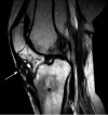
Scedosporium osteomyelitis. An MRI coronal T2 image shows the progressive signal alteration of the tibial cancellous bone in the metaphyseal region (black asterisk) associated with an abnormal cystic lesion (grains) in the soft tissue (white arrow) from an immunocompetent patient with tsunami-related Scedosporium apiospermum osteomyelitis. (Reproduced from reference with permission from the International Society for Infectious Diseases.)
FIG 8
FIG 8
Phaeohyphomycotic osteomyelitis. Shown are hyphae with intercalary and terminal chlamydoconidium swellings as seen by the direct preparation of infected tissue using Blankophor P fluorescent stain (magnification, ×400).
FIG 9
FIG 9
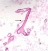
Osteoarticular mucormycosis. Mucorales hyphae are seen in a bone biopsy specimen from the tibial bone shown in Fig. 10. Typical hyphae are broad, thin walled, and pleomorphic (arrow). They vary in caliber and produce irregular branches that often arise from parent hyphae at right angles. Hematoxylin and eosin stain was used (magnification, ×400). (Reproduced from reference with permission from the Association of Bone and Joint Surgeons.)
FIG 10
FIG 10
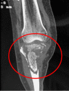
Tibial mucormycotic osteomyelitis. A coronal view CT scan of the knee before radical debridement shows destruction of cortical and cancellous bone. (Reproduced from reference with permission from the Association of Bone and Joint Surgeons.)
FIG 11
FIG 11
Vertebral mucormycosis. A 22-year-old female after allogeneic HCT for relapsed AML and a history of pulmonary and CNS mucormycosis presented with new-onset back pain. MRI scan of the spine showed a pathological fracture of the L3 body, paravertebral enhancement, a complex nondisplaced fracture of S1, and a right-psoas abscess (FRFSE, fast recovery fast spin echo). The psoas abscess was drained, and hyphae were seen by microscopy. L1-L3 laminectomy and L3-L4 corpectomy were performed, and pathology showed osteomyelitis and hyphae.

References

    1. Gamaletsou MN, Kontoyiannis DP, Sipsas NV, Moriyama B, Alexander E, Roilides E, Brause B, Walsh TJ. 2012. Candida osteomyelitis: analysis of 207 pediatric and adult cases (1970-2011). Clin Infect Dis 55:1338–1351. 10.1093/cid/cis660. - DOI - PMC - PubMed
    1. Gamaletsou MN, Rammaert B, Bueno MA, Sipsas NV, Moriyama B, Kontoyiannis DP, Roilides E, Zeller V, Taj-Aldeen SJ, Miller AO, Petraitiene R, Lortholary O, Walsh TJ. 2016. Candida arthritis: analysis of 112 pediatric and adult cases. Open Forum Infect Dis 3:ofv207. 10.1093/ofid/ofv207. - DOI - PMC - PubMed
    1. Fuzibet JG, Squara P, Verdier JM, Lapalus P, Gratecos N, Cassuto JP, Chichmanian RM, Dujardin P. 1982. Candida albicans spondylitis—a case report with a study of bone penetration of 5-fluorocytosine and a review of the literature. Ann Med Interne (Paris) 133:410–415. - PubMed
    1. Brass C, Galgiani JN, Blaschke TF, Defelice R, O’Reilly RA, Stevens DA. 1982. Disposition of ketoconazole, an oral antifungal, in humans. Antimicrob Agents Chemother 21:151–158. 10.1128/AAC.21.1.151. - DOI - PMC - PubMed
    1. Denes E, Boumediene A, Durox H, Oksman A, Saint-Marcoux F, Darde M-L, Gaulier J-M. 2007. Voriconazole concentrations in synovial fluid and bone tissues. J Antimicrob Chemother 59:818–819. 10.1093/jac/dkm023. - DOI - PubMed

Publication types

Substances