. 2022 Dec 21;96(24):e0138822.
doi: 10.1128/jvi.01388-22. Epub 2022 Nov 30.

Different Mechanisms Are Utilized by Coronavirus Transmissible Gastroenteritis Virus To Regulate Interferon Lambda 1 and Interferon Lambda 3 Production

Affiliations

Different Mechanisms Are Utilized by Coronavirus Transmissible Gastroenteritis Virus To Regulate Interferon Lambda 1 and Interferon Lambda 3 Production

Mei Xue et al. J Virol. .

Abstract

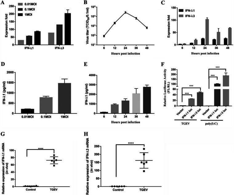
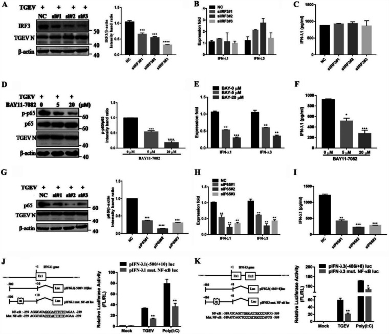
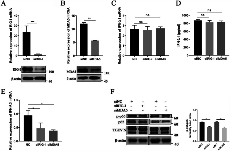
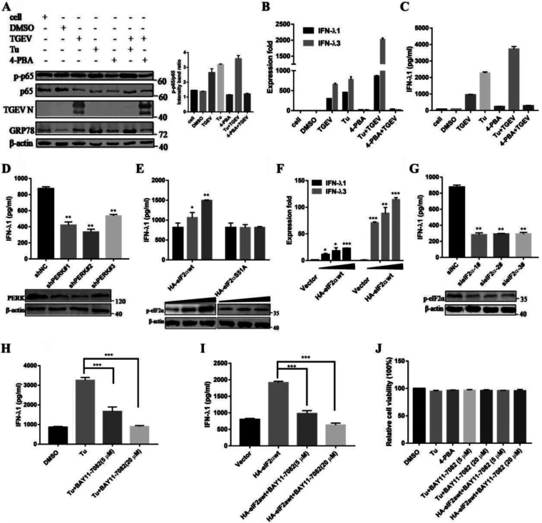
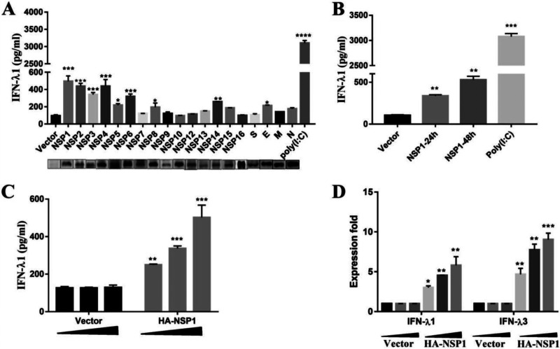
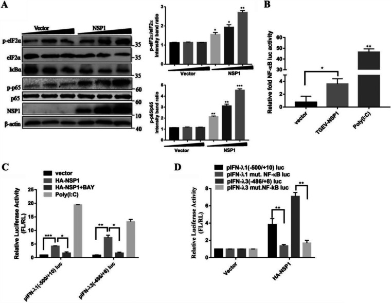
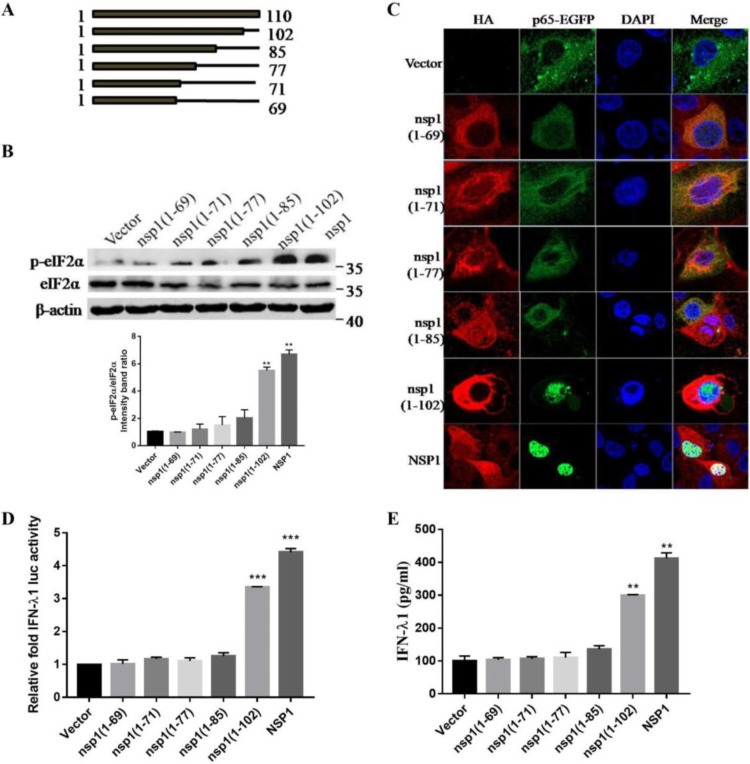
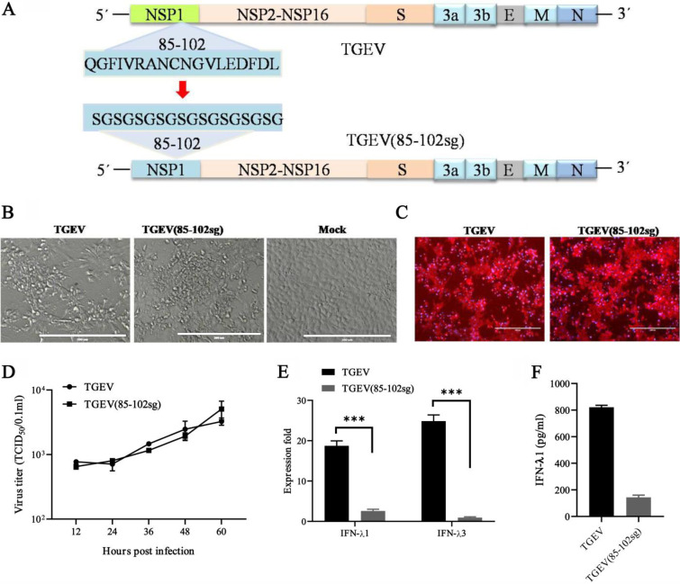
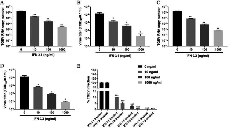
Type III interferons (IFN-λ) are shown to be preferentially produced by epithelial cells, which provide front-line protection at barrier surfaces. Transmissible gastroenteritis virus (TGEV), belonging to the genus Alphacoronavirus of the family Coronaviridae, can cause severe intestinal injuries in porcine, resulting in enormous economic losses for the swine industry, worldwide. Here, we demonstrated that although IFN-λ1 had a higher basal expression, TGEV infection induced more intense IFN-λ3 production in vitro and in vivo than did IFN-λ1. We explored the underlying mechanism of IFN-λ induction by TGEV and found a distinct regulation mechanism of IFN-λ1 and IFN-λ3. The classical RIG-I-like receptor (RLR) pathway is involved in IFN-λ3 but not IFN-λ1 production. Except for the signaling pathways mediated by RIG-I and MDA5, TGEV nsp1 induces IFN-λ1 and IFN-λ3 by activating NF-κB via the unfolded protein responses (UPR) PERK-eIF2α pathway. Furthermore, functional domain analysis indicated that the induction of IFN-λ by the TGEV nsp1 protein was located at amino acids 85 to 102 and was dependent on the phosphorylation of eIF2α and the nuclear translocation of NF-κB. Moreover, the recombinant TGEV with the altered amino acid motif of nsp1 85-102 was constructed, and the nsp1 (85-102sg) mutant virus significantly reduced the production of IFN-λ, compared with the wild strain. Compared to the antiviral activities of IFN-λ1, the administration of IFN-λ3 showed greater antiviral activity against TGEV infections in IPEC-J2 cells. In summary, our data point to the significant role of IFN-λ in the host innate antiviral responses to coronavirus infections within mucosal organs and in the distinct mechanisms of IFN-λ1 and IFN-λ3 regulation. IMPORTANCE Coronaviruses cause infectious diseases in various mammals and birds and exhibit an epithelial cell tropism in enteric and respiratory tracts. It is critical to explore how coronavirus infections modulate IFN-λ, a key innate cytokine against mucosal viral infection. Our results uncovered the different processes of IFN-λ1 and IFN-λ3 production that are involved in the classical RLR pathway and determined that TGEV nsp1 induces IFN-λ1 and IFN-λ3 production by activating NF-κB via the PERK-eIF2α pathway in UPR. These studies highlight the unique regulation of antiviral defense in the intestine during TGEV infection. We also demonstrated that IFN-λ3 induced greater antiviral activity against TGEV replication than did IFN-λ1 in IPEC-J2 cells, which is helpful in finding a novel strategy for the treatment of coronavirus infections.

Keywords: NF-κB; RLR-mediated signaling; coronavirus; nonstructural protein 1 (nsp1); protein kinase R-like ER kinase; transmissible gastroenteritis virus; type III IFN.


Conflict of interest statement

The authors declare no conflict of interest.

Figures

FIG 1
FIG 1
TGEV infection induces IFN-λ production in vitro and in vivo. (A) IPEC-J2 cells were mock-infected or infected with TGEV H87 for 24 h at an MOI of 0.01, 0.1, or 1. The IFN-λ1 and IFN-λ3 expression was measured via RT-qPCR. (B) IPEC-J2 cells were infected with TGEV H87 at an MOI of 1. The samples were collected at 0, 6, 12, 24, 36, and 48 hpi. One-step growth curve of TGEV in IPEC-J2 cells. (C) A time-dependent increase of IFN-λ1 and IFN-λ3 expression was revealed via RT-qPCR in IPEC-J2 cells. (D) The IFN-λ1 expression at different MOI values was confirmed via ELISA. (E) The expression of IFN-λ1 in the supernatants in TGEV infected cells at different time points was tested via ELISA. (F) The IFN-λ1 and IFN-λ3 promoters were activated in TGEV-infected IPEC-J2 cells. Poly (I:C) (1 μg/mL) was used as a positive-control for IFN-λ activation. (G and H) TGEV infection induces IFN-λ1 and IFN-λ3 production in vivo. 12 two-day-old SPF piglets were orally inoculated with TGEV H87 strain or DMEM to serve as uninfected controls. All of the piglets were euthanized by the end of the study, which was terminated at 48 hpi. The expression of IFN-λ1 and IFN-λ3 expression in the ileum tissues was detected via RT-qPCR.
FIG 2
FIG 2
NF-κB, but not IRF3, is critical to producing IFN-λ during a TGEV infection. (A) The IRF3 knockdown efficiency was confirmed in IRF3-knockdown cells via Western blotting. (B) The IFN-λ1 and IFN-λ3 expression in IRF3-knockdown cells was analyzed via RT-qPCR after the TGEV infection. (C) The protein levels of IFN-λ1 were analyzed in IRF3-knockdown cells. (D) The effect of the NF-κB inhibitor BAY-11-7082 on NF-κB activation and on TGEV N expression was confirmed via Western blotting. (E) TGEV-induced IFN-λ1 and IFN-λ3 was reduced in a dose-dependent manner after treatment with BAY11-7082. (F) The protein levels of IFN-λ1 were analyzed after IPEC-J2 cells were pretreated with BAY11-7082. (G) The p65 knockdown efficiency was confirmed in the p65-knockdown cells via Western blotting. (H) The expression levels of IFN-λ1 and IFN-λ3 in the p65-knockdown cells were analyzed via RT-qPCR after the TGEV infection. (I) The protein levels of IFN-λ1 were analyzed in p65-knockdown cells. (J) Schematic representation of the mutation of the binding sites for NF-κB (underlined) in the pig IFN-λ1 promoter. IPEC-J2 cells were transfected with empty vector pIFN-λ1 (−500/+10) Luc and its mutation construct for 6 h, and this was followed by either TGEV infection or stimulation with poly (I·C) for 24 h. The cell lysates were then prepared for dual-luciferase reporter assays. (K) Schematic representation of the mutation of the binding sites for NF-κB (underlined) in the pig IFN-λ3 promoter. IPEC-J2 cells were transfected with empty vector pIFN-λ3 (−486/+8) Luc and its mutation construct for 6 h, and this was followed by TGEV infection or stimulation with poly (I·C) for 24 h. The cell lysates were then prepared for dual-luciferase reporter assays.
FIG 3
FIG 3
The RLR signaling pathway is involved in IFN-λ3 production but not in IFN-λ1 production. The IPEC-J2 cells were transfected with 50 nM specific siRNAs targeting RIG-I, MDA5, or negative-control siRNA for 24 h, and the cells were infected with TGEV (MOI = 1). (A) At 24 hpi, cells were collected for the analysis of the RIG-I mRNA levels via RT-qPCR assays and protein expression via Western blotting. (B) Cells transfected with specific siRNAs targeting MDA5 were collected for the analysis of the MDA5 mRNA levels via RT-qPCR assays and protein expression via Western blotting. (C) Cells transfected with specific siRNAs targeting RIG-I or MDA5 were collected for the analysis of IFN-λ1 mRNA levels via RT-qPCR assays. (D) Cell supernatants transfected with specific siRNAs targeting RIG-I or MDA5 were harvested and subjected to ELISA to assess the protein level of IFN-λ1. (E) Cells transfected with specific siRNAs targeting RIG-I or MDA5 were collected for the analysis of the IFN-λ3 mRNA levels via RT-qPCR assays. (F) Western blotting was performed to test p-p65, p65, and TGEV N in RIG-I or MDA5-knockdown cells. β-actin was used as a sample loading control. The ratio of p-p65 to p65 (p-p65/p65) was calculated.
FIG 4
FIG 4
TGEV-induced, PERK-specific ER stress is critical to producing IFN-λ through activating NF-κB. (A) IPEC-J2 cells were either uninfected or pretreated with Tu (2 μg/mL), 4-PBA (2 mM), or DMSO carrier control for 2 h, and this was followed by infection with TGEV (MOI = 1) and maintainance at that concentration after infection. Western blotting was performed to test the activation of NF-κB and GRP78, as well as the viral infection. β-actin was used as a sample loading control. The ratio of p-p65 to p65 (p-p65/p65) was calculated. (B) At 24 hpi, cells were collected for the analysis of the IFN-λ1 and IFN-λ3 mRNA levels via RT-qPCR assays. (C) Cell supernatants were harvested and subjected to ELISA to assess the protein level of IFN-λ1. (D) IPEC-J2 cells were transfected with shRNA targeting PERK or control shRNA for 24 h, and then the cells were challenged with TGEV. After 24 hpi, the cell supernatants were harvested and subjected to ELISA to assess the protein level of IFN-λ1. The PERK knockdown efficiency was analyzed via Western blotting. (E) IPEC-J2 cells were transfected with HA-eIF2αwt or HA-eIF2αS51A for 24 h and were then infected with TGEV H87. At 24 hpi, the cell supernatants were harvested and subjected to ELISA to assess the protein level of IFN-λ1. The phosphorylation of eIF2α was subjected to Western blotting with antibodies against p-eIF2α. β-actin was used as a loading control. (F) Cells transfected with HA-eIF2αwt were collected for the analysis of the IFN-λ1 and IFN-λ3 mRNA levels via RT-qPCR assays. (G) IPEC-J2 cells were transfected with siRNA of eIF2α for 24 h and were then infected with TGEV H87. After 24 hpi, cell supernatants were harvested and subjected to ELISA to assess the protein level of IFN-λ1. The eIF2α knockdown efficiency was analyzed via Western blotting. (H) IPEC-J2 cells were pretreated with Tu (2 μg/mL) or DMSO carrier control 2 h before infection and maintained at that concentration after infection. Then, cells were treated or nontreated (exposed to equal amounts of DMSO) with 5 or 20 μM BAY11-7082. At 24 hpi, cell supernatants were harvested and subjected to ELISA to assess the protein level of IFN-λ. (I) IPEC-J2 cells were transfected with HA-eIF2αwt for 24 h and then infected with TGEV H87. Then, cells were treated or nontreated (exposed to equal amounts of DMSO) with 5 or 20 μM BAY11-7082. At 24 hpi, the cell supernatants were harvested and subjected to ELISA to assess the protein level of IFN-λ1. (J) Effect of Tu and 4-PBA on cell viability. IPEC-J2 cells were treated with Tu and 4-PBA or Tu plus 5 or 20 μM BAY11-7082 and HA-eIF2αwt plus 5 or 20 μM BAY11-7082. Cell cytotoxicity was analyzed with a CCK-8 system.
FIG 5
FIG 5
TGEV nsp1 induces IFN-λ. (A) IPEC-J2 cells were transfected with various TGEV protein expression vectors. Cell supernatants were subjected to ELISA to assess the production of IFN-λ1. (B) IPEC-J2 cells were transfected with pcaggs or pcaggs-TGEV-nsp1-HA (TGEV nsp1) at 24 h and 48 h posttransfection. Cell supernatants were harvested and subjected to ELISA to assess the production of IFN-λ1. (C and D) IPEC-J2 cells were transfected with different concentrations of pcaggs or pcaggs-TGEV-nsp1-HA (TGEV nsp1). After 24 hpi the cell supernatants were subjected to ELISA to assess the production of IFN-λ1. Cells were harvested and subjected to RT-qPCR to assess the expression of IFN-λ1 and IFN-λ3.
FIG 6
FIG 6
TGEV nsp1 induces IFN-λ via eIF2α phosphorylation-mediated NF-κB activation. (A) IPEC-J2 cells were transfected with different concentrations of pcaggs or pcaggs-TGEV-nsp1-HA (TGEV nsp1). Cell extracts were subjected to Western blotting using an anti-p-eIF2α antibody, anti-eIF2α antibody, anti-HA antibody, anti-IκBα antibody, anti-p-p65 antibody, anti-p65 antibody, or anti-β-actin antibody. The ratios of p-eIF2α/eIF2α and p-p65/p65 were calculated. (B) 293T cells were cotransfected with pNF-κB-Luc (NF-κB luciferase reporter plasmid) and pcaggs or pcaggs-TGEV-nsp1-HA (TGEV nsp1). Cells transfected with poly (I:C) were used as the positive control. At 24 h posttransfection, cell lysates were prepared and subjected to luciferase assays. Error bars show the standard deviations (SDs) of the results from three independent experiments. (C) 293T cells were cotransfected with pIFN-λ1 (−500/+10) Luc or pIFN-λ3 (−486/+8) Luc and pcaggs or pcaggs-TGEV-nsp1-HA (TGEV nsp1). Then, cells were treated or nontreated (exposed to equal amounts of DMSO) with 20 μM BAY11-7082. Cells transfected with poly (I:C) were used as the positive control. At 24 h posttransfection, cell lysates were prepared and subjected to luciferase assays. (D) 293T cells were transfected with pIFN-λ 1(−500/+10)Luc, pIFN-λ1 mut. NF-κB Luc, pIFN-λ3 (−486/+8)Luc, or pIFN-λ3 mut. NF-κB Luc, along with either an empty vector or pcaggs-TGEV-nsp1-HA (TGEV nsp1). At 24 h posttransfection, cell lysates were prepared and subjected to luciferase assays.
FIG 7
FIG 7
TGEV nsp1 (amino acids 85 to 102) is important in activating NF-κB and in inducing IFN-λ production. (A) The truncations (at the C terminus) of TGEV nsp1 are shown below the bars. (B) IPEC-J2 cells were transfected with TGEV nsp1 truncation mutants. At 24 h posttransfection, cell extracts were subjected to Western blotting, using an anti-p-eIF2α antibody, anti-eIF2α antibody, or anti-β-actin antibody. The ratio of p-eIF2α to eIF2α (p-eIF2α/eIF2α) was calculated. (C) IPEC-J2 cells were cotransfected with the truncation mutants of TGEV nsp1 and pEGFP-p65. The location of NF-κB p65 was detected via immunofluorescence and was observed under laser confocal microscopy. (D) 293T cells were cotransfected with pIFN-λ1 (−500/+10) Luc and pcaggs or with the truncation mutants of TGEV nsp1. At 24 h posttransfection, the cell lysates were prepared and subjected to luciferase assays. (E) IPEC-J2 cells were transfected with the truncation mutants of TGEV nsp1. At 24 h posttransfection, cell supernatants were harvested and subjected to ELISA to assess the production of IFN-λ1.
FIG 8
FIG 8
The nsp1 (85-102) mutants (TGEV [85-102sg]) induced lower IFN-λ responses. (A) DNA-based reverse genetics BAC system for TGEV and TGEV (85-102sg). The amino acid replacements in position 85 to 102 of NSP1 are indicated. (B and C) The recombinant virus was successfully used to infect ST cells. TGEV-BAC and TGEV (85-102sg)-BAC were transfected into ST cells with Lipo3000, and then the virus was passaged once in ST cells. The CPE was observed after 36 h (B), and the indirect immunofluorescence assay was used to detect the TGEV N protein (C). (D) Multistep growth curves for TGEV and TGEV (85-102sg) in ST cells at a MOI of 1. The virus titers at different time points, as indicated, were determined via an endpoint dilution assay. The data are represented as the mean ± the standard deviation, with n = 3. (E) TGEV (85-102sg) induced lower IFN-λ1 and IFN-λ3 expression than did WT TGEV in the IPEC-J2 cells. (F) The IFN-λ1 production of the cells infected with WT TGEV and TGEV (85-102sg) was confirmed via ELISA.
FIG 9
FIG 9
IFN-λ3 exhibits greater antiviral activity against TGEV than that of IFN-λ1. (A and B) IPEC-J2 cells were pretreated with the indicated concentration of rpIFN-λ1 2 h before infection and were maintained at that concentration after infection. TGEV infection was determined at 24 hpi via either RT-qPCR (A) or titration (B). (C and D) IPEC-J2 cells were pretreated with the indicated concentration of rpIFN-λ3 2 h before infection and were maintained at that concentration after infection. TGEV infection was determined at 24 hpi via either RT-qPCR (C) or titration (D). (E) The infection percentage of TGEV treated with rpIFN-λ1 and rpIFN-λ3 was calculated.
FIG 10
FIG 10
Modulation of the IFN-λ response by TGEV. TGEV infection leads to the phosphorylation of initiation factor eIF2α through the action of the kinase PERK. Once active, eIF2α inhibits translation initiation, thereby reducing the synthesis of IκBα. The reduction in IκBα leads to the activation of NF-κB and binds DNA to promote IFN-λ1 and IFN-λ3 expression. TGEV nsp1 leads to the phosphorylation of eIF2α, which in turn suppresses IκBs. The reduced IκB synthesis reduces the inhibition of NF-κB and increases nuclear NF-κB and IFN-λ1 and IFN-λ3 expression. The RLR signaling pathway is involved in TGEV-induced IFN-λ3 production but not in IFN-λ1 production. The arrows indicate activation, and the blunt-ended lines indicate inhibition.

Similar articles

Cited by

References

    1. Lazear HM, Nice TJ, Diamond MS. 2015. Interferon-lambda: immune functions at barrier surfaces and beyond. Immunity 43:15–28. 10.1016/j.immuni.2015.07.001. - DOI - PMC - PubMed
    1. Mordstein M, Neugebauer E, Ditt V, Jessen B, Rieger T, Falcone V, Sorgeloos F, Ehl S, Mayer D, Kochs G, Schwemmle M, Gunther S, Drosten C, Michiels T, Staeheli P. 2010. Lambda interferon renders epithelial cells of the respiratory and gastrointestinal tracts resistant to viral infections. J Virol 84:5670–5677. 10.1128/JVI.00272-10. - DOI - PMC - PubMed
    1. Pott J, Mahlakoiv T, Mordstein M, Duerr CU, Michiels T, Stockinger S, Staeheli P, Hornef MW. 2011. IFN-lambda determines the intestinal epithelial antiviral host defense. Proc Natl Acad Sci USA 108:7944–7949. 10.1073/pnas.1100552108. - DOI - PMC - PubMed
    1. Zhou JH, Wang YN, Chang QY, Ma P, Hu Y, Cao X. 2018. Type III interferons in viral infection and antiviral immunity. Cell Physiol Biochem 51:173–185. 10.1159/000495172. - DOI - PubMed
    1. Sommereyns C, Paul S, Staeheli P, Michiels T. 2008. IFN-lambda (IFN-lambda) is expressed in a tissue-dependent fashion and primarily acts on epithelial cells in vivo. PLoS Pathog 4:e1000017. 10.1371/journal.ppat.1000017. - DOI - PMC - PubMed

Publication types

MeSH terms