. 2022 Dec 21;96(24):e0157822.
doi: 10.1128/jvi.01578-22. Epub 2022 Nov 30.

Duck Enteritis Virus Inhibits the cGAS-STING DNA-Sensing Pathway To Evade the Innate Immune Response

Affiliations

Duck Enteritis Virus Inhibits the cGAS-STING DNA-Sensing Pathway To Evade the Innate Immune Response

Li Gao et al. J Virol. .

Abstract

Cyclic GMP-AMP synthase (cGAS), a key DNA sensor, detects cytosolic viral DNA and activates the adaptor protein stimulator of interferon genes (STING) to initiate interferon (IFN) production and host innate antiviral responses. Duck enteritis virus (DEV) is a duck alphaherpesvirus that causes an acute and contagious disease with high mortality in waterfowl. In the present study, we found that DEV inhibits host innate immune responses during the late phase of viral infection. Furthermore, we screened DEV proteins for their ability to inhibit the cGAS-STING DNA-sensing pathway and identified multiple viral proteins, including UL41, US3, UL28, UL53, and UL24, which block IFN-β activation through this pathway. The DEV tegument protein UL41, which exhibited the strongest inhibitory effect, selectively downregulated the expression of interferon regulatory factor 7 (IRF7) by reducing its mRNA accumulation, thereby inhibiting the DNA-sensing pathway. Ectopic expression of UL41 markedly reduced viral DNA-triggered IFN-β production and promoted viral replication, whereas deficiency of UL41 in the context of DEV infection increased the IFN-β response to DEV and suppressed viral replication. In addition, ectopic expression of IRF7 inhibited the replication of the UL41-deficient virus, whereas IRF7 knockdown facilitated its replication. This study is the first report identifying multiple viral proteins encoded by a duck DNA virus, which inhibit the cGAS-STING DNA-sensing pathway. These findings expand our knowledge of DNA sensing in ducks and reveal a mechanism through which DEV antagonizes the host innate immune response. IMPORTANCE Duck enteritis virus (DEV) is a duck alphaherpesvirus that causes an acute and contagious disease with high mortality, resulting in substantial economic losses in the commercial waterfowl industry. The evasion of DNA-sensing pathway-mediated antiviral innate immunity is essential for the persistent infection and replication of many DNA viruses. However, the mechanisms used by DEV to modulate the DNA-sensing pathway remain poorly understood. In the present study, we found that DEV encodes multiple viral proteins to inhibit the cGAS-STING DNA-sensing pathway. The DEV tegument protein UL41 selectively diminished the accumulation of interferon regulatory factor 7 (IRF7) mRNA, thereby inhibiting the DNA-sensing pathway. Loss of UL41 potently enhanced the IFN-β response to DEV and impaired viral replication in ducks. These findings provide insights into the host-virus interaction during DEV infection and help develop new live attenuated vaccines against DEV.

Keywords: DNA sensing; cGAS-STING; duck enteritis virus; innate immunity.


Conflict of interest statement

The authors declare no conflict of interest.

Figures
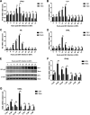
FIG 1
FIG 1
DEV inhibits the production of IFN-β and IFN-stimulated genes (ISGs) during infection in DEFs and in ducks. (A to D) DEFs were infected with the virulent DEV CV strain at a multiplicity of infection (MOI) of 0.01. The mRNA levels of IFN-β (A), IL-6 (B), and duck ISGs Mx (C) and OASL (D) were measured by real-time qPCR from 4 to 72 hpi. (E) The expression of DEV protein gE during viral infection in DEFs was monitored by Western blotting. (F to G) 2-week-old specific pathogen-free ducks were inoculated intramuscularly with 10 TCID50 of DEV CV strain, and the mRNA levels of IFN-β (F) and OASL (G) in the spleen samples were measured by real-time qPCR. The relative amounts of IFN-β, IL-6, Mx, and OASL mRNA were normalized to the β-actin mRNA level in each sample, and the fold differences were compared with those in the mock samples. All controls and treated groups were performed and examined in triplicate. *, P < 0.05; **, P < 0.01; ns, no significant difference.
FIG 2
FIG 2
Screening of DEV viral proteins that modulate the cGAS-STING pathway. (A) DEFs were cotransfected with the IFN-β Luc reporter, pRL-TK, and the empty vector or cGAS-HA and STING-HA combined. The luciferase activity was measured at 36 h posttransfection. (B) DEFs were cotransfected with the IFN-β Luc reporter, cGAS-HA, and STING-HA expression plasmids and then inoculated with DEV (MOI = 0.01). The luciferase activity was measured at 12 h and 24 h postinfection. (C) DEFs were cotransfected with cGAS-HA and STING-HA expression plasmids and then inoculated with DEV (MOI = 0.01). The IFN-β mRNA level was measured by real-time qPCR at 12 h and 24 h postinfection. (D) DEFs were transfected with the IFN-β Luc reporter and pRL-TK, together with cGAS-HA, STING-HA, and an DEV ORF expression plasmid or the empty vector. The IFN-β luciferase activity was measured at 36 h posttransfection. The data of DEV ORFs and the empty vector from three independent tests are presented as a heat map. Higher IFN-β Luc activation levels are indicated by red, whereas lower levels are indicated by green, which corresponds to a higher degree of inhibition. (E) Various doses of the top five DEV viral protein inhibitors and the empty vector were cotransfected with cGAS-HA and STING-HA expression plasmids into DEFs, and IFN-β Luc activity was measured at 36 h posttransfection. (F) The top five DEV viral protein inhibitors and the empty vector were cotransfected with cGAS-HA and STING-HA expression plasmids into DEFs. The IFN-β mRNA levels were measured by real-time qPCR at 36 h posttransfection. The fold changes were compared with those of the empty vector controls. All controls and treated groups were performed and examined in triplicate. *, P < 0.05; **, P < 0.01; ***, P < 0.001.
FIG 3
FIG 3
DEV UL41 inhibits IFN-β production induced by interferon-stimulatory and viral DNA. (A) DEFs were transfected with UL41-Flag expression plasmid or empty vector for 24 h and then transfected with interferon-stimulatory DNA (ISD); the mRNA levels of IFN-β, IL-6, Mx, and OASL were measured by qPCR 6 h and 12 h later. UL41 expression was examined by Western blotting at 12 h after ISD transfection. (B) DEFs were transfected with UL41-Flag expression plasmid or empty vector for 24 h and then transfected with poly(dA·dT); the mRNA levels of IFN-β, IL-6, Mx, and OASL were measured by qPCR 6 h and 12 h later. UL41 expression was examined by Western blotting at 12 h after poly(dA·dT) transfection. (C) DEFs were transfected with UL41-Flag expression plasmid or empty vector and then left uninfected or infected with DEV (MOI = 0.01). The mRNA levels of IFN-β and Mx in these cells were measured by qPCR at 12 h postinfection, and UL41 expression was detected by Western blotting. (D) DEFs were transfected with UL41-Flag expression plasmid or empty vector and then left uninfected or infected with HVT (MOI = 0.01). The mRNA levels of IFN-β and Mx in these cells were measured by qPCR at 12 h postinfection, and UL41 expression was detected by Western blotting. (E) DEFs transfected with UL41-Flag plasmid or empty vector were infected with DEV at various MOIs (0.01 or 0.001). The viral titer was tested by qPCR at 24 and 48 hpi, and UL41 expression was examined by Western blotting at 48 hpi. (F) DEFs transfected with UL41-Flag plasmid or empty vector were infected with HVT at various MOIs (0.01 or 0.001). The viral titer was tested by qPCR at 24 and 48 hpi, and UL41 expression was examined by Western blotting at 48 hpi. The relative amount of IFN-β, IL-6, Mx, and OASL mRNA was normalized to β-actin mRNA levels in each sample, and the relative amount of DEV and HVT genomic DNA was normalized to GAPDH levels in each sample. All controls and treated groups were performed and examined in triplicate. *, P < 0.05; **, P < 0.01; ns, no significant difference.
FIG 4
FIG 4
UL41 deficiency enhances DEV-triggered induction of IFN-β and downstream antiviral genes. (A) Cytopathic effects (bar length = 400 μm) and indirect immunofluorescence (bar length = 200 μm) analysis of the DEFs infected with DEV-WT or DEV-dUL41 using rabbit anti-UL41 and anti-rabbit IgG-TRITC antibodies. (B and C) Effects of UL41 deficiency on transcription of IFN-β, IL-6, Mx, and OASL in DEFs. DEFs were infected with DEV-WT or DEV-dUL41 (MOI = 0.01) for 24 h before analysis of IFN-β, IL-6, Mx, and OASL mRNA levels. The amounts of IFN-β, IL-6, Mx, and OASL mRNA were normalized to the β-actin mRNA level in each sample, and the fold difference relative to the mock controls was determined. (D) The growth properties of DEV-WT and DEV-dUL41 in DEFs. Data are presented as means ± SDs from three independent experiments. *, P < 0.05; **, P < 0.01; ns, no significant difference.
FIG 5
FIG 5
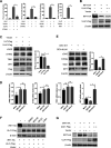
DEV UL41 inhibits the cGAS-STING pathway by downregulating IRF7 expression. (A) DEFs were cotransfected with IFN-β-luc reporter, pRL-TK, and cGAS-HA, STING-HA, TBK1-HA, or IRF7-HA along with empty vector or UL41-Flag expression plasmid as indicated. Cells were harvested 36 h after transfection and subjected to dual-luciferase reporter assays. All controls and treated groups were performed and examined in triplicate. (B) 293T cells were cotransfected with IRF7-HA and UL41-Flag plasmids. Twenty-four hours after transfection, cells were harvested and subjected to Western blot analysis. (C) DEFs were transfected with UL41-Flag, UL7-Flag plasmid, or the empty vector. Twenty-four hours after transfection, cells were harvested and subjected to Western blot analysis with the indicated antibodies. (D) DEFs were infected with DEV-WT or DEV-dUL41 (MOI = 0.01). Twenty-four hours after infection, cells were harvested and analyzed with qRT-PCR. (E) DEFs were infected with DEV-WT or DEV-dUL41 (MOI = 0.01). Twenty-four hours after infection, cells were harvested and subjected to Western blot analysis with the indicated antibodies. (F) 293T cells were cotransfected with IRF7-HA and UL41-Flag plasmids. Twenty-four hours after transfection, cells were treated with dimethyl sulfoxide (DMSO), MG132 (10 μM), NH4Cl (20 mM), and 3-MA (10 mM) for 12 h. Protein expression was measured using Western blotting. (G) Vero cells were cotransfected with IRF7-HA and UL41-Flag plasmids, and cells were subjected to Western blot analysis 36 h after transfection. *, P < 0.05; **, P < 0.01; ***, P < 0.001; ns, no significant difference.
FIG 6
FIG 6
IRF7 mediates the defense against the replication of UL41-deficient DEV. (A, B) DEFs were transfected with the empty vector (DEF-EV) or with the IRF7-HA plasmid (DEF-IRF7). Twenty-four hours after transfection, the cells were infected with DEV-WT or DEV-dUL41 (MOI = 0.01), and then cells were harvested at 24 and 48 hpi for viral titration (A) and Western blot (B) analyses. (C and D) DEFs were transfected with negative-control siRNA (DEF-NC) or with siRNA specific to IRF7 (DEF-IRF7KD). Twenty-four hours after transfection, the cells were infected with DEV-WT or DEV-dUL41 (MOI = 0.01), and then cells were harvested at 24 and 48 hpi and subjected to viral titration (C) and Western blot (D) analyses. Data are presented as means ± SDs from three independent experiments. *, P < 0.05; **, P < 0.01; ns, no significant difference.
FIG 7
FIG 7
UL41 deficiency facilitated IFN-β induction and attenuated DEV replication in ducks. (A to C) Ducks were infected intramuscularly with 100 TCID50 of DEV-WT or DEV-dUL41, and IFN-β (A), Mx (B), and OASL (C) production was measured by qPCR at the indicated time points. (D) Ducks were infected intramuscularly with 100 TCID50 of DEV-WT or DEV-dUL41, and virus genome copy numbers in the spleen were monitored by qPCR at the indicated time points. All samples from the mock control and DEV-infected groups were examined in triplicate. *, P < 0.05; **, P < 0.01; ns, no significant difference.

Similar articles

Cited by

References

    1. Dhama K, Kumar N, Saminathan M, Tiwari R, Karthik K, Kumar MA, Palanivelu M, Shabbir MZ, Malik YS, Singh RK. 2017. Duck virus enteritis (duck plague)–a comprehensive update. Vet Q 37:57–80. 10.1080/01652176.2017.1298885. - DOI - PubMed
    1. Li N, Hong T, Li R, Guo M, Wang Y, Zhang J, Liu J, Cai Y, Liu S, Chai T, Wei L. 2016. Pathogenicity of duck plague and innate immune responses of the Cherry Valley ducks to duck plague virus. Sci Rep 6:32183. 10.1038/srep32183. - DOI - PMC - PubMed
    1. Qi X, Yang X, Cheng A, Wang M, Zhu D, Jia R. 2008. The pathogenesis of duck virus enteritis in experimentally infected ducks: a quantitative time-course study using TaqMan polymerase chain reaction. Avian Pathol 37:307–310. 10.1080/03079450802043775. - DOI - PubMed
    1. Wu J, Chen ZJ. 2014. Innate immune sensing and signaling of cytosolic nucleic acids. Annu Rev Immunol 32:461–488. 10.1146/annurev-immunol-032713-120156. - DOI - PubMed
    1. Brubaker SW, Bonham KS, Zanoni I, Kagan JC. 2015. Innate immune pattern recognition: a cell biological perspective. Annu Rev Immunol 33:257–290. 10.1146/annurev-immunol-032414-112240. - DOI - PMC - PubMed

Publication types