. 2022 Dec 2;8(48):eadd2536.
doi: 10.1126/sciadv.add2536. Epub 2022 Nov 30.

Molecular architecture of the Chikungunya virus replication complex

Affiliations

Molecular architecture of the Chikungunya virus replication complex

Yaw Bia Tan et al. Sci Adv. .

Abstract

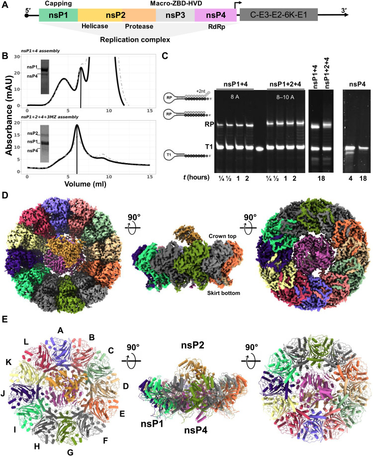
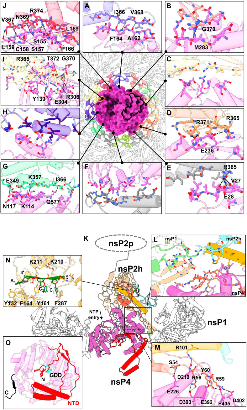
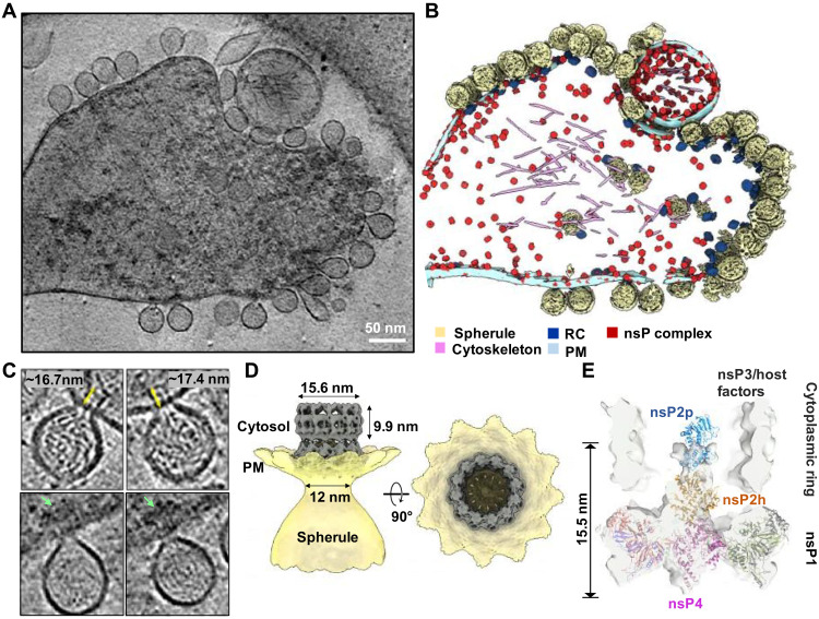
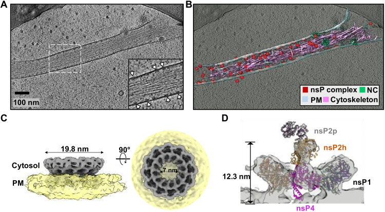
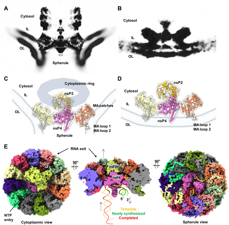
To better understand how positive-strand (+) RNA viruses assemble membrane-associated replication complexes (RCs) to synthesize, process, and transport viral RNA in virus-infected cells, we determined both the high-resolution structure of the core RNA replicase of chikungunya virus and the native RC architecture in its cellular context at subnanometer resolution, using in vitro reconstitution and in situ electron cryotomography, respectively. Within the core RNA replicase, the viral polymerase nsP4, which is in complex with nsP2 helicase-protease, sits in the central pore of the membrane-anchored nsP1 RNA-capping ring. The addition of a large cytoplasmic ring next to the C terminus of nsP1 forms the holo-RNA-RC as observed at the neck of spherules formed in virus-infected cells. These results represent a major conceptual advance in elucidating the molecular mechanisms of RNA virus replication and the principles underlying the molecular architecture of RCs, likely to be shared with many pathogenic (+) RNA viruses.


Figures

Fig. 1.
Fig. 1.. Recombinant nsPs were in vitro reconstituted via tandem purification and activated their RNA synthesis ability.
(A) The alphavirus genome is partitioned into two open-reading frames (ORFs) where the first ORF encodes for four nsPs (nsP1 to 4) to assemble into an RC for the viral RNA replication process, while the second ORF is regulated by a 26S promoter (black arrow) and encodes for structural proteins (C-E3-E2-6K-E1) for virion assembly. (B) Top: Anion-exchange chromatography profile of nsP1 + 4 assembled from CHIKV nsP1 mutant (H37A; amino acids 1 to 535) and ONNV nsP4 (amino acids 1904 to 2514), eluted at 32 mS/cm (elution peak marked with vertical line). Bottom: Anion-exchange chromatography profile of RC core (nsP1 + 2 + 4 + 3 M) assembled from CHIKV nsP1 mutant (H37A; amino acids 1 to 535), CHIKV nsP2 (nsP2; amino acids 536 to 1333), CHIKV nsP3 macrodomain (nsP3MZ; amino acids 1334 to 1659), and ONNV nsP4 (amino acids 1904 to 2514), eluted at 29 mS/cm (elution peak marked with vertical line). mAU, milli–absorbance unit. (C) The RNA polymerase activity of the RCs was observed with elongation of the T1 RNA template to its RNA products (RPs; the number of ATP incorporation labeled in white letters for each RC) at several time points (0.25 to 18 hours) along with the nsP4 recombinant protein as a control. (D and E) The graphical illustrations of RC core (nsP1 + 2 + 4) architecture in map representation in (D) and molecular structure in (E) at their top view, side view, and bottom view (90° rotations, from left to right). The coloring in (D) and (E) was assigned according to their chain numbers in the RC core (nsP1 chain coloring: violet, A; salmon, B; light green, C; tan, D; orange, E; light gray, F; green, G; dark gray, H; spring green, I; indigo, J; light yellow, K; and crimson, L; nsP4 chain coloring: magenta, X; nsP2 chain coloring: peru, Y; RNA of nsP2 chain coloring: green, Z).
Fig. 2.
Fig. 2.. The macromolecular architecture of the RC displays a multiple interface network.
(A to J) The interaction network of RC (made of nsP1 + 2 + 4 at the top view) is presented with each colored by each subunit chain for nsP1 dodecameric ring and nsP4 (surface), based on Fig. 1 chain coloring. For clarity of the interaction overview, nsP2 and its RNA ligand are hidden. Instead, the nsP2:nsP4 interface area is outlined with a thick black line. The hydrogen bonds (black dotted lines) between the individual chain of nsP1 and nsP4 are shown here in the interface blown-up view with each residue involved named and colored according to the chain. (K) The overall side-view impression of interfaces spanning across CHIKV RC (cartoon representations) where their three-way interactions between nsP1, nsP2, and nsP4 are shown in (L) a blown-up window (solid line and box). The nsP2h (amino acids 1 to 465) region is colored according to its subdomain: NTD in orange, STALK in gold, 1B in cyan, and RecA1-A2 in peru, while chains C and D of nsP1 are respectively colored in light green and tan. The unbuilt nsP2 protease (nsP2p; after amino acids 466) region is drawn here at the C terminus of nsP2h for visual guidance for (K). NTP entry site at the nsP4 motif D within the palm subdomain (magenta region) is annotated. (M) The hydrogen bonds (dotted lines) between the nsP2:nsP4 interface are listed on another blown-up window (dotted line and box). (N) The interacting residues from nsP2h and RNA (green) are labeled at a zoomed-in view (dotted line and box). (O) The bottom view of nsP4 showcases the spatial coordinates of its C terminus (C; black; amino acids 600 to 611) and N-terminal domain (NTD; red; amino acids 1 to 105) and the active site (named GDD; cyan stick).
Fig. 3.
Fig. 3.. CHIKV RNA replication spherule structures revealed by cryo-ET.
(A) Tomographic slice of cell periphery depicting CHIKV RNA replication spherules at the PM. Scale bar, 50 nm. (B) Corresponding 3D segmentation of cellular features. See also movie S1. (C) Snapshot of the individual spherules. Yellow arrows measure the ordered density within the center of the cytoplasmic ring. Green arrows mark the additional associated density proximal to the RC cytoplasmic ring. (D) CHIKV spherule 3D volume is determined by subtomogram averaging with imposed C12 symmetry. (E) The RC core complex (nsP1 + 2 + 4) is fitted into the C1 subtomogram average map of the RC. A cytoplasmic ring as observed in (C), likely made of nsP3, RNA, and host factors remain loosely connected to the nsP1 ring, which is bound at the neck of the spherule. The extra density above the nsP2h region is likely to be the C-terminal protease of nsP2, stabilized or restrained by the cytoplasmic ring.
Fig. 4.
Fig. 4.. Inactive nsP complex induced membrane protrusion revealed by cryoET.
(A) Tomographic slice of cell periphery depicting a filopodia-like membrane protrusion structure extended from the PM. White arrows point to the membrane-associated nsP complexes. Scale bar, 100 nm. (B) Corresponding 3D segmentation of cellular features. See also movie S2. (C) CHIKV nsP complex 3D volume determined by subtomogram averaging with imposed C12 symmetry. (D) The RC core complex (nsP1 + 2 + 4) is fitted into the C1 subtomogram average map of the nsP complex. Note that the cytoplasmic ring as observed in Fig. 3C is absent in this inactive nsP complex. Consequently, there is no density observed for the C-terminal protease of nsP2.
Fig. 5.
Fig. 5.. Remodeling of the host membrane is coupled to assembly of active viral RC.
(A) A central slice view of the tomography volume at the active viral RC spherule highlights the curved PM at the nsP1 contact sites [membrane association loops 1 and 2 (MA loops 1 and 2) from the bottom of the nsP1 ring and newly identified MA patches from the nsP1 upper ring] as shown in (C). In contrast, (B) shows the central slice view of the tomography volume at the inactive viral nsP complex, which is simply docked to the inner leaflet of the PM, using the MA loops 1 and 2 on nsP1 ring in (D). (E) Molecular model of how the viral RC core complex facilitates viral (+) RNA replication. The dsRNA replication fork is modeled and colored. Template RNA, i.e., antigenomic (−) RNA is in yellow, newly synthesized progeny RNA (or subgenomic RNA transcript) is in green, and the RNA in red is the completed product RNA to be exported to the cytoplasm for translation and virion assembly. NTP entry tunnel on nsP4 polymerase and the putative RNA exit tunnel are labeled (also refer to fig. S2, E and F).

References

    1. Gasque P., Bandjee M. C., Reyes M. M., Viasus D., Chikungunya pathogenesis: From the clinics to the bench. J. Infect. Dis. 214, S446–S448 (2016). - PubMed
    1. Albulescu I. C., Tas A., Scholte F. E. M., Snijder E. J., van Hemert M. J., An in vitro assay to study chikungunya virus RNA synthesis and the mode of action of inhibitors. J. Gen. Virol. 95, 2683–2692 (2014). - PubMed
    1. Pietila M. K., van Hemert M. J., Ahola T., Purification of highly active alphavirus replication complexes demonstrates altered fractionation of multiple cellular membranes. J. Virol. 92, e01852-17 (2018). - PMC - PubMed
    1. Pietilä M. K., Hellström K., Ahola T., Alphavirus polymerase and RNA replication. Virus Res. 234, 44–57 (2017). - PubMed
    1. Romero-Brey I., Bartenschlager R., Membranous replication factories induced by plus-strand RNA viruses. Viruses 6, 2826–2857 (2014). - PMC - PubMed