. 2022 Dec 2;8(48):eadd4150.
doi: 10.1126/sciadv.add4150. Epub 2022 Nov 30.

The SARS-CoV-2 spike protein binds and modulates estrogen receptors

Affiliations

The SARS-CoV-2 spike protein binds and modulates estrogen receptors

Oscar Solis et al. Sci Adv. .

Abstract

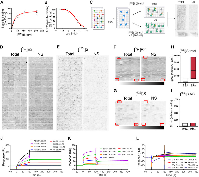
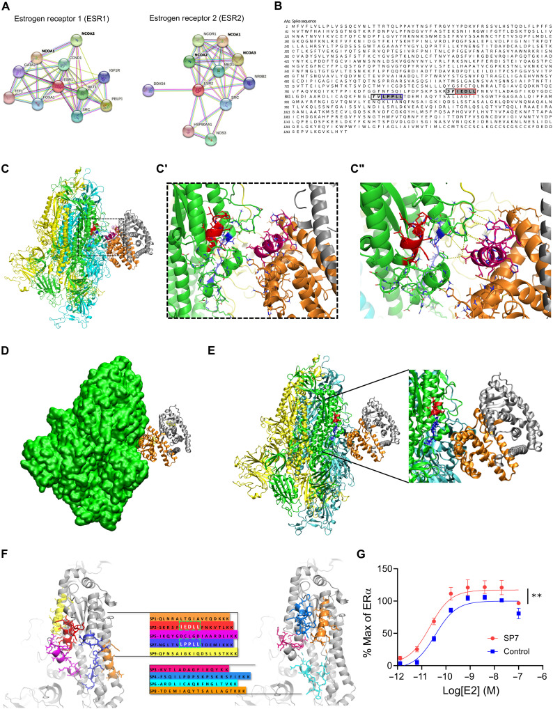
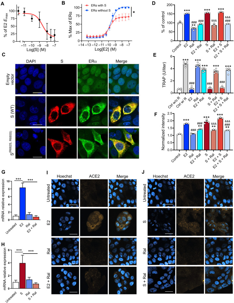
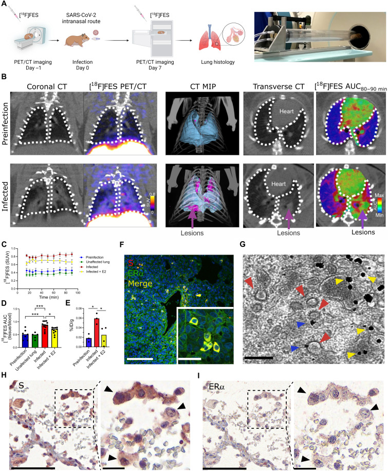
The severe acute respiratory syndrome coronavirus 2 (SARS-CoV-2) spike (S) protein binds angiotensin-converting enzyme 2 as its primary infection mechanism. Interactions between S and endogenous proteins occur after infection but are not well understood. We profiled binding of S against >9000 human proteins and found an interaction between S and human estrogen receptor α (ERα). Using bioinformatics, supercomputing, and experimental assays, we identified a highly conserved and functional nuclear receptor coregulator (NRC) LXD-like motif on the S2 subunit. In cultured cells, S DNA transfection increased ERα cytoplasmic accumulation, and S treatment induced ER-dependent biological effects. Non-invasive imaging in SARS-CoV-2-infected hamsters localized lung pathology with increased ERα lung levels. Postmortem lung experiments from infected hamsters and humans confirmed an increase in cytoplasmic ERα and its colocalization with S in alveolar macrophages. These findings describe the discovery of a S-ERα interaction, imply a role for S as an NRC, and advance knowledge of SARS-CoV-2 biology and coronavirus disease 2019 pathology.


Figures

Fig. 1.
Fig. 1.. S binds ERα with high affinity.
(A) [125I]S saturation and (B) competition binding to recombinant ACE2. (C) Schematic of ProtoArray experimental design. (D) Positive control ProtoArray autoradiograms showing total and nonspecific (NS) binding of [3H]E2. (E) ProtoArray autoradiograms (experiment 1 of 3) showing total and nonspecific binding of [125I]S. (F and G) Representative array blocks showing total and nonspecific [3H]E2 and [125I]S binding. Red rectangles show location of ERα proteins. (H and I) Quantification of total and nonspecific [125I]S binding at ERα and bovine serum albumin (BSA) (control). Data are representative of three independent experiments. (J to L) Representative SPR sensorgrams showing kinetic and equilibrium binding analyses of immobilized S exposed to increasing concentrations of ACE2, NRP1, and ERα protein (Kon = 2.03 × 10⁵, Koff = 1.96 × 10⁻³, and KD = 9.7 nM). In (A) and (B), data are represented as means ± SEM. In (H) and (I), data are presented as median ± min and max limits.
Fig. 2.
Fig. 2.. S and ER interact at conserved LXD NRC motifs.
(A) ER interaction network showing known and predicted protein associations. (B) LXD-like patterns in the S sequence. The LXXLL motif and a homologous region are highlighted in blue and red boxes, respectively, with dark gray background. Positions (−1 and −2) are reported in italic and light gray background, respectively. AAs, amino acids. (C and C′) The LPPLL and IEDLL residues of the two motifs are shown in the 3D x-ray S structure [Protein Data Bank (PDB) ID 6VYB] with blue and red colors, respectively. The ER dimer is in orange and gray, while the helix-12 is reported in magenta. (C″) The image shows favorable interactions between ER and the S’s regions containing the LXXLL motifs. The interacting residues and the predicted interactions are reported in stick and yellow dots, respectively. (D) S-ER motif–oriented docking. The best 3D docking hypothesis is shown. The ER dimer is in orange and gray, and S is green. (E) Alignment between the best-pose and the 3OLL model tied with NCOA1. The region occupied by S’s α helix interacts in the area where the NCOA fragment was crystallized. (F) S protein peptides and their location with respect to the S 3D structure. (G) The SP7 peptide containing the LPPLL motif significantly increased ERα activation [F(1,48) = 30.38, **P < 0.01, two-way analysis of variance (ANOVA); peptide treatment main effect]. Data are shown as means ± SEM.
Fig. 3.
Fig. 3.. S modulates ER-dependent biological functions.
S inhibits (A) E2-induced ERα DNA binding in MCF-7 nuclear extracts and (B) transcriptional activation in an ERα reporter cell line [F(1,28) = 21.73, *P = 0.01, two-way ANOVA; S treatment × E2 concentration interaction effect]. (C) Immunofluorescent staining of S and endogenous ERα in MCF-7 cells transfected with empty vector, WT, or the furin cleavage site mutant S(R682S, R685S). Scale bars, 16 mm. (D) S increases MCF-7 cell proliferation in an ER-dependent manner (**P < 0.01 and ***P < 0.001 versus control; ###P < 0.001 versus E2; &&&P < 0.001 versus S; one-way ANOVA with Tukey post hoc test). (E) S decreases osteoclast differentiation in an ER-dependent manner (***P < 0.001 versus control without RANKL; ###P < 0.001 versus control with RANKL; one-way ANOVA with Tukey post hoc test). (F) S and E2 increase ACE2 protein levels in MCF-7 cells in an ER-dependent manner (***P < 0.001 versus control; ###P < 0.001 versus E2; &&&P < 0.001 versus S; one-way ANOVA with Tukey post hoc test). (G and H) E2 and S increase ACE2 mRNA (***P < 0.001, one-way ANOVA with Tukey post hoc test) and (I and J) protein in the Calu-3 lung cell line in an ER-dependent manner. Scale bars, 30 mm. All data are shown as means ± SEM.
Fig. 4.
Fig. 4.. SARS-CoV-2 infection increases cytoplasmic ERα accumulation and S-ERα colocalization in pulmonary macrophages.
(A) Schematic showing experimental design of SARS-CoV-2 hamster studies and BSL-3 imaging compartment (Created with BioRender). (B) CT, [18F]FES PET, and area under the curve (AUC) heatmap overlay images from hamsters at preinfection (day −1) and infection (day 7). MIP, maximum intensity projection. (C) Time activity curves showing standard uptake value ratio (SUVr; tissue/blood [18F]FES content) in each experimental group. n = 4 to 5 per group. ***P < 0.001 versus preinfection (D) [18F]FES uptake expressed as area under the curve ratio (AUCr; tissue/blood [18F]FES content). F(3,32) = 12.15, ***P < 0.001 and *P < 0.05. (E) [18F]FES uptake expressed as % injected dose (%ID)/gram of body weight in postmortem hamster lung (harvested 110 min after injection; n = 3 to 4 per group). F(2,7) = 7.161, *P < 0.05. (F) Hamster lung IHC showing colocalization of S and ERα immunoreactivity (experiment 1 of 2). (G) Immunogold electron microscopy showing SARS-CoV-2 particles (red arrowheads) and ERα-bound gold nanoparticles (blue arrowheads) in a hamster alveolar macrophage. Scale bars, 200 nm. Yellow arrowheads correspond to cytoplasmic ERα accumulation. (H) S and (I) ERα immunostaining in SARS-CoV-2–infected human lung showing S-ERα colocalization in macrophages (black arrowheads). Scale bars, 100 nm (low mag) and 25 nm (high mag). All data are shown as means ± SEM.

Update of

References

    1. Zhou X., Cheng Z., Luo L., Zhu Y., Lin W., Ming Z., Chen W., Hu Y., Incidence and impact of disseminated intravascular coagulation in COVID-19 a systematic review and meta-analysis. Thromb. Res. 201, 23–29 (2021). - PMC - PubMed
    1. Mitrovic M., Sabljic N., Cvetkovic Z., Pantic N., Zivkovic Dakic A., Bukumiric Z., Libek V., Savic N., Milenkovic B., Virijevic M., Vucinic V., Milosevic I., Pravdic Z., Suvajdzic N., Fareed J., Antic D., Rotational thromboelastometry (ROTEM) profiling of COVID-19 patients. Platelets 32, 690–696 (2021). - PubMed
    1. La Vignera S., Cannarella R., Condorelli R. A., Torre F., Aversa A., Calogero A. E., Sex-specific SARS-CoV-2 mortality: Among hormone-modulated ACE2 expression, risk of venous thromboembolism and hypovitaminosis D. Int. J. Mol. Sci. 21, 2978 (2020). - PMC - PubMed
    1. Montopoli M., Zorzi M., Cocetta V., Prayer-Galetti T., Guzzinati S., Bovo E., Rugge M., Calcinotto A., Clinical outcome of SARS-CoV-2 infection in breast and ovarian cancer patients who underwent antiestrogenic therapy. Ann. Oncol. 32, 676–677 (2021). - PMC - PubMed
    1. Chen Y., Liu Q., Guo D., Emerging coronaviruses: Genome structure, replication, and pathogenesis. J. Med. Virol. 92, 418–423 (2020). - PMC - PubMed

Substances