Review
. 2023 Nov 1;47(6):fuac047.
doi: 10.1093/femsre/fuac047.

Mitochondrial genome maintenance-the kinetoplast story

Affiliations
Review

Mitochondrial genome maintenance-the kinetoplast story

Simona Amodeo et al. FEMS Microbiol Rev. .

Abstract

Mitochondrial DNA replication is an essential process in most eukaryotes. Similar to the diversity in mitochondrial genome size and organization in the different eukaryotic supergroups, there is considerable diversity in the replication process of the mitochondrial DNA. In this review, we summarize the current knowledge of mitochondrial DNA replication and the associated factors in trypanosomes with a focus on Trypanosoma brucei, and provide a new model of minicircle replication for this protozoan parasite. The model assumes the mitochondrial DNA (kinetoplast DNA, kDNA) of T. brucei to be loosely diploid in nature and the replication of the genome to occur at two replication centers at the opposing ends of the kDNA disc (also known as antipodal sites, APS). The new model is consistent with the localization of most replication factors and in contrast to the current model, it does not require the assumption of an unknown sorting and transport complex moving freshly replicated DNA to the APS. In combination with the previously proposed sexual stages of the parasite in the insect vector, the new model provides a mechanism for maintenance of the mitochondrial genetic diversity.

Keywords: kDNA; kinetoplast; mitochondrial DNA; mitochondrial DNA replication; trypanosomes.


Conflict of interest statement

The authors declare no conflict of interest.

Figures
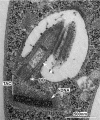
Figure 1.
Figure 1.
The kinetoplast of T. brucei. (A) Transmission electron microscopy image of a thin section through the basal body and kDNA network of T. brucei. In this orientation, the kDNA disc is cut orthogonal to its surface. BB, basal body; F, flagellum; TAC, tripartite attachment complex; and kDNA, mitochondrial genome.
Figure 2.
Figure 2.
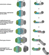
Overview of kDNA replication and segregation in T. brucei. (A) Dorsoventral view of the posterior region of a cell depicting the flagellar pocket, the basal bodies, and the mitochonrial membranes surrounding the kDNA at the beginning of kDNA replication. (B) and (C) Minicircles are released into the kinetoflagellar zone (KFZ), replicated, and reattached at the APS. Maxicircles are replicated, while remaining inside the growing kDNA network. Simultaneously to the kDNA replication process, the pro-basal body (pBB) tilts to face the mitochondrial membrane, and via the exclusion zone filaments (EZF), sets the base for growth of the new tripartite attachment complex (TAC). (C) and (D) During pBB maturation, the pBB rotates around the mature basal body (BB), and assembles the TAC proteins of the differentiated membranes (DM) and the unilateral filaments (ULF). (D) After completion of minicircle replication, the replicated kinetoplasts remain attached to each other by the maxicircles that accumulate between the two kDNA discs. Microtubule quartet (MTQ). The Images in this figure are stills from an animated model (see supplementary material).
Figure 3.
Figure 3.
The diploid model of minicircle replication in the kDNA. Minicircles are released from the disc into the KFZ, bind to some replication factors, and move to the APS (green), where replication is initiated and proceeds via theta intermediates. Replication leaves the minicircle created from the leading strand with a single nick, i.e. rapidly repaired at the APS before the minicircle is reattached to the growing kDNA network. The lagging strand minicircle is left with multiple nicks and gaps to be repaired. Therefore, it will remain in the APS for a prolonged period of time (where the nicks and gaps are being repaired). As the disc is growing, the APS, and thus the site of minicircle reattachment, will move away from the position where the leading strand minicircle was reattached, leading to a spatial separation of the daughter minicircles. The depiction in this figure focuses on the processes in the disc on the left (depicted in cyan). The same process also applies to the disc growing on the right (depicted in gray). The APS of the disc in focus, are depicted in green, the network bound minicircles of the diploid genome are depicted in cyan; before DNA synthesis, free minicircles are shown in black; leading strand daughter circles are then depicted in red, while lagging strand daughter circles are shown in yellow. For simplicity we omitted the replication of the maxicircles in this figure.

References

    1. Abu-Elneel K, Kapeller I, Shlomai J. Universal minicircle sequence-binding protein, a sequence-specific DNA-binding protein that recognizes the two replication origins of the kinetoplast DNA minicircle. J Biol Chem. 1999;274:13419–26. - PubMed
    1. Abu-Elneel K, Robinson DR, Drew MEet al. . Intramitochondrial localization of universal minicircle sequence-binding protein, a trypanosomatid protein that binds kinetoplast minicircle replication origins. J Cell Biol. 2001;153:725–34. - PMC - PubMed
    1. Amodeo S, Jakob M, Ochsenreiter T. Characterization of the novel mitochondrial genome replication factor MiRF172 in Trypanosoma brucei. J Cell Sci. 2018;131:jcs211730. - PMC - PubMed
    1. Aphasizhev R, Aphasizheva I. Uridine insertion/deletion editing in trypanosomes: a playground for RNA-guided information transfer. WIREs RNA. 2011;2:669–85. - PMC - PubMed
    1. Aphasizheva I, Alfonzo J, Carnes Jet al. . Lexis and grammar of mitochondrial RNA processing in trypanosomes. Trends Parasitol. 2020;36:337–55. - PMC - PubMed

Publication types

MeSH terms