. 2023 Jan 6;22(1):138-151.
doi: 10.1021/acs.jproteome.2c00551. Epub 2022 Nov 30.

Multiomics of Colorectal Cancer Organoids Reveals Putative Mediators of Cancer Progression Resulting from SMAD4 Inactivation

Affiliations

Multiomics of Colorectal Cancer Organoids Reveals Putative Mediators of Cancer Progression Resulting from SMAD4 Inactivation

Jelmer J Dijkstra et al. J Proteome Res. .

Abstract

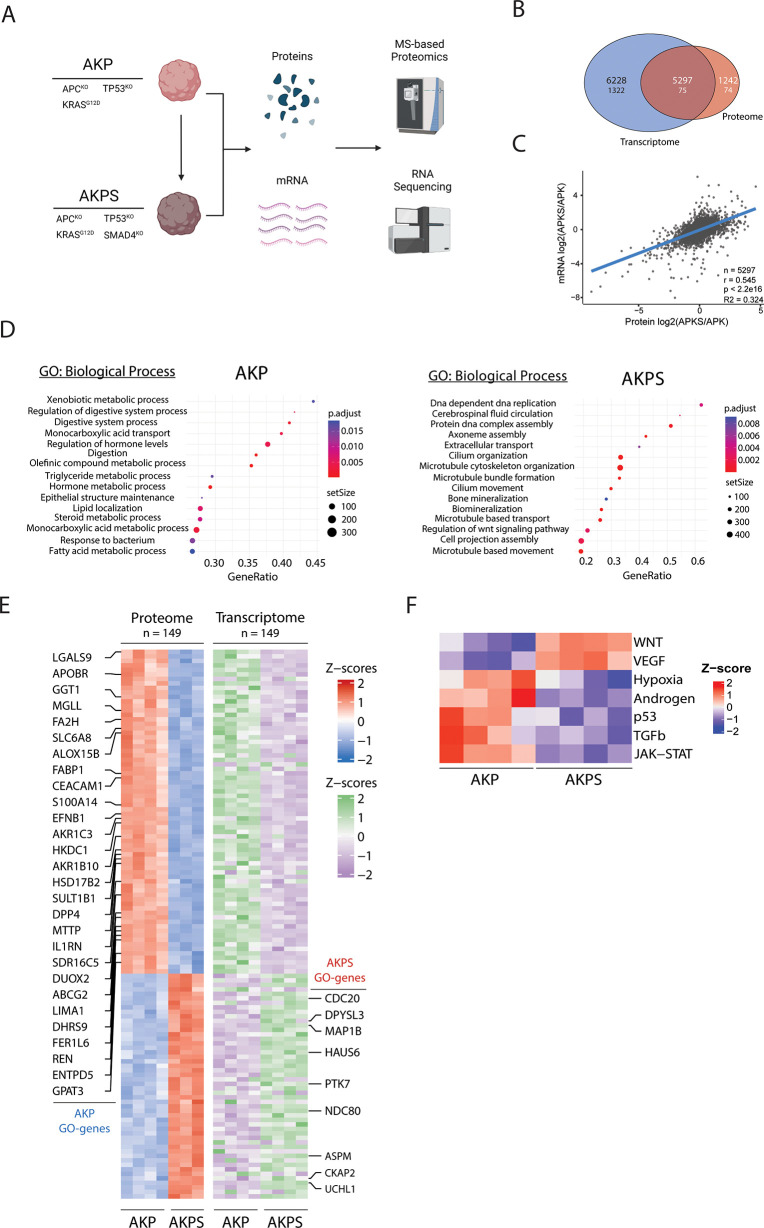
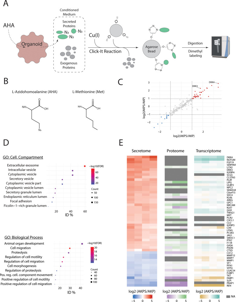
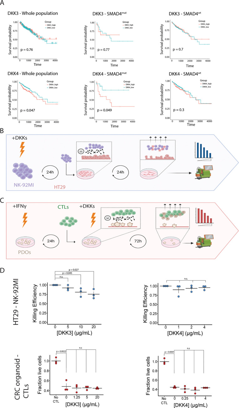
The development of metastasis severely reduces the life expectancy of patients with colorectal cancer (CRC). Although loss of SMAD4 is a key event in CRC progression, the resulting changes in biological processes in advanced disease and metastasis are not fully understood. Here, we applied a multiomics approach to a CRC organoid model that faithfully reflects the metastasis-supporting effects of SMAD4 inactivation. We show that loss of SMAD4 results in decreased differentiation and activation of pro-migratory and cell proliferation processes, which is accompanied by the disruption of several key oncogenic pathways, including the TGFβ, WNT, and VEGF pathways. In addition, SMAD4 inactivation leads to increased secretion of proteins that are known to be involved in a variety of pro-metastatic processes. Finally, we show that one of the factors that is specifically secreted by SMAD4-mutant organoids─DKK3─reduces the antitumor effects of natural killer cells (NK cells). Altogether, our data provide new insights into the role of SMAD4 perturbation in advanced CRC.

Keywords: SMAD4; cancer progression; colorectal cancer; multiomics; secretomics.


Conflict of interest statement

The authors declare no competing financial interest.

Figures

Figure 1
Figure 1
Transcriptomic and proteomic analysis of AKP and AKPS organoids. (A) Schematic overview of the used organoid lines and experimental procedures. (B) Venn diagram of detected and significant (FDR < 0.05 and FC > 2) transcriptomic and proteomic features. The upper number shows detected features, and the lower number shows significantly changing features. (C) Linear regression of relative transcriptomic and proteomic expression levels of AKP vs AKPS. n = 5297, r = 0.545, R2 = 0.324, p < 2.2 × 10⁻¹⁶. (D) Bubble plots showing the top 15 activated biological processes ranked on increasing FDR based on GSEA. All reported biological processes are over-represented with an FDR < 0.01. The left panel shows AKP-specific processes, and the right panel shows AKPS-specific processes. (E) Row-matched heatmaps showing the relative changes in protein and mRNA expression of the 149 significantly changing proteins shown in (B) (FDR < 0.05; FC > 1.5). Highlighted rows show significantly changing proteins that are associated with the biological processes in (D). (F) Heatmap showing significantly (FDR < 0.05) the relative activation of PROGENy pathways in AKP and AKPS organoids.
Figure 2
Figure 2
Secretomic analysis of AKP and AKPS. (A) Schematic overview of the application of BONCAT in secretomics. (B) Structure formulas of methionine (Met) and azidohomoalanine (AHA). (C) Dot plot showing changes in relative protein secretion levels between AKP and AKPS organoids. Proteins with an absolute mean FC > 2 were considered to be significantly differentially secreted. (D) Bubble plots showing the top 10 significantly (FDR < 0.01) enriched biological processes and cellular compartments ranked on increasing FDR based on ORA using all detected proteins. All reported biological processes and cellular compartments are over-represented with an FDR < 0.01. (E) Row-matched heatmaps showing the relative expression levels of secreted proteins (left), intracellular proteins (middle), and mRNA transcripts (right). All secreted proteins with a fold change >2 (colored dots in (C)) are shown.
Figure 3
Figure 3
DKK3 reduces the killing capacity of NK cells for CRC cells in vitro. (A) Kaplan–Meier plots showing the effect of DKK3 (top) and DKK4 (bottom) expression on survival probability in the TCGA-COAD cohort. Left panels include the whole cohort, middle panels show patients with mutations in SMAD4, and right panels show patients without SMAD4 mutations. (B) Schematic overview of killing assays using CRC cells and NK cells. NK-92MI cells are incubated for 24h with recombinant DKK-proteins after which they are co-cultured with HT29 cells. After 24h of coculture, non-adherent cells are removed and adherent cells are dissociated and counted by flow cytometry (C) Schematic overview of killing assays using patient-derived CRC tumor organoids and matched CTLs. Patient derived organoids (PDOs) are incubated with IFNγ for 24h after which they are cocultured with CTLs in combination with recombinant DKK proteins. After 72h, non-adherent cells are removed and adherent cells are dissociated and measured by flow cytometry (D) Dot plots showing the effect of DKK3 and DKK4 on the antitumor capacity of NK cells (top) and CTLs (bottom). No CTL indicates the control condition, where no CTLs are added to the tumor organoids; n = 3 independent replicates; error bars represent SEM. n.s: not significant.

Similar articles

Cited by

References

    1. Vogelstein B.; Papadopoulos N.; Velculescu V. E.; et al. Cancer Genome Landscapes. Science 2013, 339, 1546–1558. 10.1126/science.1235122. - DOI - PMC - PubMed
    1. Zhang L.; Shay J. W. Multiple Roles of APC and its Therapeutic Implications in Colorectal Cancer. J. Natl. Cancer Inst. 2017, 109, djw332.10.1093/jnci/djw332. - DOI - PMC - PubMed
    1. Ewing I.; Hurley J. J.; Josephides E.; Millar A. The molecular genetics of colorectal cancer. Frontline Gastroenterol. 2014, 5, 26–30. 10.1136/flgastro-2013-100329. - DOI - PMC - PubMed
    1. Drost J.; van Jaarsveld R. H.; Ponsioen B.; et al. Sequential cancer mutations in cultured human intestinal stem cells. Nature 2015, 521, 43–47. 10.1038/nature14415. - DOI - PubMed
    1. Welch D. R.; Hurst D. R. Defining the Hallmarks of Metastasis. Cancer Res. 2019, 79, 3011–3027. 10.1158/0008-5472.CAN-19-0458. - DOI - PMC - PubMed

Publication types