. 2022 Nov 29;41(9):111731.
doi: 10.1016/j.celrep.2022.111731.

Antigen-presenting T cells provide critical B7 co-stimulation for thymic iNKT cell development via CD28-dependent trogocytosis

Affiliations

Antigen-presenting T cells provide critical B7 co-stimulation for thymic iNKT cell development via CD28-dependent trogocytosis

Masashi Watanabe et al. Cell Rep. .

Abstract

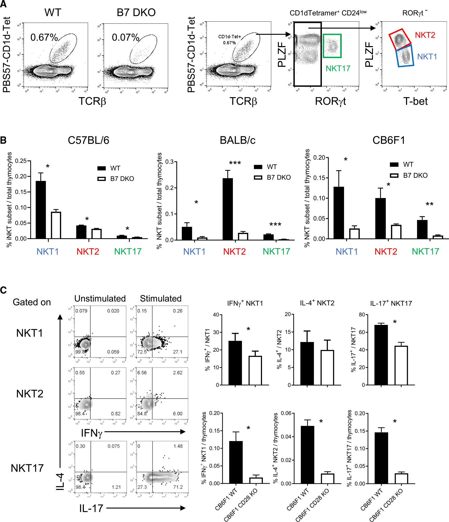
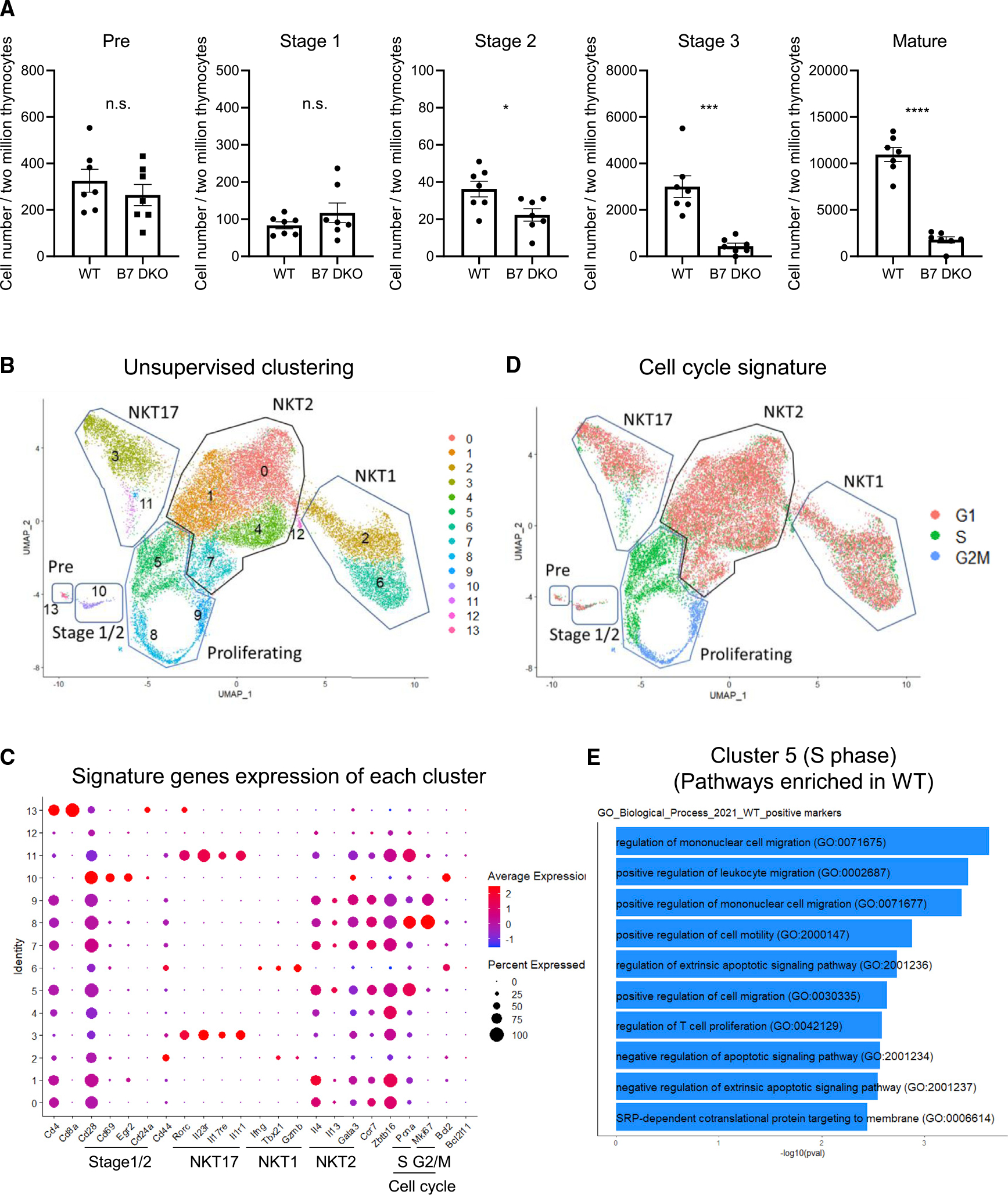
Invariant natural killer T (iNKT) cell development in the thymus depends on T cell receptor recognition of CD1d ligand on CD4/CD8 double-positive thymocytes. We previously reported that B7-CD28 co-stimulation is required for thymic iNKT cell development, but the cellular and molecular mechanisms underlying this co-stimulatory requirement are not understood. Here we report that CD28 expression on CD1d-expressing antigen-presenting T cells is required for thymic iNKT cell development. Mechanistically, antigen-presenting T cells provide co-stimulation through an unconventional mechanism, acquiring B7 molecules via CD28-dependent trogocytosis from B7-expressing thymic epithelial cells, dendritic cells, and B cells and providing critical B7 co-stimulation to developing iNKT cells. Thus, the present study demonstrates a mechanism of B7 co-stimulation in thymic T cell development by antigen-presenting T cells.

Keywords: B7-CD28 co-stimulation; CP: Immunology; antigen-presenting T cell; iNKT; thymic T cell development; trogocytosis.


Conflict of interest statement

Declaration of interests The authors declare no competing interests.

Figures

Figure 1.
Figure 1.. B7-CD28 co-stimulation is critical for all iNKT cell subset development
(A) Thymic iNKT cells were defined as B220−, PBS57-CD1d-tetramer binding TCRβ+ cell population. Gating strategy to define NKT1, NKT2, and NKT17 subsets within CD24low iNKT cells. (B) iNKT cell subset in C57BL/6 (B6), BALB/c, and CB6F1 strains. B6 WT = 6, B6 B7 DKO = 6, BALB/c WT n = 7, BALB/c B7 DKO n = 7, CB6F1 WT n = 4, CB6F1 B7 DKO n = 4. Data are pooled results of three independent experiments. (C) Cytokine production of each iNKT cell subset. Total CB6F1 thymocytes were stimulated with PMA and ionomycin, and intracellular cytokine staining was performed. Left panel: representative intracellular cytokine staining pattern of each iNKT cell subset defined as in (A). Upper right panel: frequency of indicated cytokine staining positive population per each iNKT cell subset. Lower right panel: frequency of indicated cytokine positive iNKT cell subset population per total thymocytes. CB6F1 WT n = 4, CB6F1 CD28 KO n = 4. Data are pooled results of three independent experiments. (B and C) Data are mean ± SEM. Statistical difference between groups were calculated with Student’s t test. *p < 0.05, **p < 0.01, ***p < 0.001. See also Figure S1.
Figure 2.
Figure 2.. Developmental iNKT cell stages and analysis of CD28 expression level
(A) Gating strategy to identify iNKT cell sub-populations based on CD44, CD24, and CD69 expression. (B) B6 Rag2-GFP expression level of each iNKT cell sub-population. n = 3. Data are representative of two independent experiments. (C) Defined developmental sequence of iNKT cell sub-populations. (D) CCR7 expression pattern at each developmental stage. n = 5. Data are pooled result of independent two experiments. (E) CD28 expression level (MFI) of each developmental stage in B6 WT, B7 DKO, and CD28 KO strains. (F) Difference of MFI (ΔMFI) between B7 DKO and WT was calculated at each stage. (E and F) Each group n = 3. Data are representative of three independent experiments. (B, D–F) Data are mean ± SEM. See also Figure S2.
Figure 3.
Figure 3.. Effect of absence of B7 co-stimulation on developing iNKT cell sub-populations
(A) Cell number at each developmental stage. BALB/c thymic iNKT cells were first enriched by magnetic-activated cell sorting with CD1d-tetramer and then analyzed by fluorescence-activated cell sorting (FACS). Data are expressed as cell numbers in each developmental stage within the tetramer-enriched iNKT cell population. Each group n = 7. Data are pooled results of three independent experiments. (B) Uniform manifold approximation and projection (UMAP) of six independent scRNA-seq data sets from BALB/c thymic iNKT cells (WT n = 3, B7 DKO n = 3) integrated using Seurat. (C) Scaled expression of selected signature genes for each cluster colored by average expression of each gene in each cluster. Dot size represents the percentage of cells in each cluster. (D) Cell cycle profiling of each cluster. (E) Gene ontology (GO) enrichment analysis. Differentially expressed genes between WT and B7 DKO for cluster 5 were identified using Wilcoxon rank-sum test. GO_Biological_Process_2021 database was used for GP enrichment analysis. (A) Data are mean ± SEM. Statistical differences between groups were calculated with Student’s t test. **p < 0.01, ***p < 0.001. See also Figure S3.
Figure 4.
Figure 4.. Thymocytes acquire and express B7 on cell surface in a CD28-dependent manner
(A) B7.1 and B7.2 expression on thymocytes in B6 WT, B7 DKO, and CD28 KO mice. Top panel: representative histogram results of at least three independent experiments. Bottom panel: quantification of pooled results for B7.1 and B7.2 MFI from four independent experiments. WT n = 5, CD28 KO n = 5, B7 DKO n = 3. (B) iNKT cell population in B6 DC, B, and/or TEC-specific B7 conditional KO mice generated by crosses with CD11c-Cre, CD19-Cre, and/or Foxn1-Cre respectively. B7flox n = 9, B7 DKO n = 4, TEC/DC/BC B7 cKO n = 3, TEC B7 cKO n = 5, DC B7 cKO n = 4, BC B7 cKO = 5. Data are pooled results of four independent experiments. (C) iNKT cell population in T cell-specific B7 conditional KO mice generated by cross with Lck-Cre. B7flox n = 24, B7 DKO n = 26, TEC/DC/BC B7 cKO n = 3, TC B7 cKO n = 5. Data are pooled result of at least three independent experiments. (D) Cell surface B7.1 expression on peripheral splenic CD4+ T cells in B6 B7 conditional KO strains. Red line indicates B7.1 staining of each cKO mouse. Black line indicates B7.1 staining of B7 DKO mice as negative staining control. Data are representative results of at least three independent experiments. (E) B7 expression on B6 B7 DKO T cells in mixed bone marrow chimeras with co-existence of B7 WT T cells. Red line indicates B7.1 staining of each cell type. Black line indicates B7.1 staining on each cell type from B7 DKO mice as negative staining control. (F) B7 expression on peripheral CD4+ T cells in B6 CD28 KO mice. B7.1 expression on splenic CD4+ T cells and CD11c+ dendritic cells is analyzed. Red line indicates B7.1 staining of each cell type. Black line indicates B7.1 staining of each cell type from B7 DKO mice as negative staining control. Data are representative results of at least three independent experiments. (G) B7 expression on DP thymocytes in B6 CD28-cytoplasmic-region-truncated mutant mice. Data are representative results of two independent experiments. Each group total n = 4. (A–C) Data are mean ± SEM. Statistical differences between groups were performed with one-way ANOVA followed by multiple comparison. *p < 0.05, **p < 0.01, ****p < 0.0001. See also Figure S4.
Figure 5.
Figure 5.. CD28 on antigen-presenting T cells is required for B7 co-stimulation in iNKT cell development
(A) CD28 expression on CD1d-expressing antigen-presenting T cells is required to co-stimulate iNKT cell development. To test the requirement for CD28 expression on antigen-presenting-cells for iNKT cell development, mixed bone marrow (BM) chimera mice on B6 background were generated. In groups 1, 2, and 3, BM1 was CD1dKO (CD45.1) and served as the indicator of iNKT cell development. BM2 (CD45.2) was CD1dKO/CD28WT (group 1), CD1dWT/CD28WT (group 2), or CD1d WT/CD28KO (group 3) as the source of CD1d-expressing APCs. In group 4, irradiated CD28 KO mice were reconstituted with CD28 KO BM to generate chimeras that were completely lacking CD28. 10 weeks after BM reconstitution, iNKT cells in BM1 compartment were analyzed. Group 1, n = 8; group 2, n = 6; group 3, n = 10; group 4 n = 7. Data are pooled results of at least three independent experiments. Data are mean ± SEM. Statistical comparisons between groups were performed with one-way ANOVA followed by multiple comparison. ***p < 0.001, (B) Solid red line represents B7.1 expression on CD1d+ DP thymocyte (BM2) in BM chimera mice in (A). Black dotted line is anti-B7.1 staining of B7 DKO DP as negative staining control. Data are representative result of at least three independent experiments. (C) B7 expression pattern on CD1d+ antigen-presenting cells and CD1d non-antigen-presenting-cells. See also Figure S5.

References

    1. Bendelac A, Rivera MN, Park SH, and Roark JH (1997). Mouse CD1-specific NK1 T cells: development, specificity, and function. Annu. Rev. Immunol. 15, 535–562. 10.1146/annurev.immunol.15.1.535. - DOI - PubMed
    1. Taniguchi M, Harada M, Kojo S, Nakayama T, and Wakao H (2003). The regulatory role of Valpha14 NKT cells in innate and acquired immune response. Annu. Rev. Immunol. 21, 483–513. 10.1146/annurev.immunol.21.120601.141057. - DOI - PubMed
    1. Crosby CM, and Kronenberg M (2018). Tissue-specific functions of invariant natural killer T cells. Nat. Rev. Immunol. 18, 559–574. 10.1038/s41577-018-0034-2. - DOI - PMC - PubMed
    1. Coles MC, and Raulet DH (2000). NK1.1+ T cells in the liver arise in the thymus and are selected by interactions with class I molecules on CD4+CD8+ cells. J. Immunol. 164, 2412–2418. 10.4049/jimmunol.164.5.2412. - DOI - PubMed
    1. Wei DG, Lee H, Park SH, Beaudoin L, Teyton L, Lehuen A, and Bendelac A (2005). Expansion and long-range differentiation of the NKT cell lineage in mice expressing CD1d exclusively on cortical thymocytes. J. Exp. Med. 202, 239–248. 10.1084/jem.20050413. - DOI - PMC - PubMed

Publication types