

Review
doi: 10.1183/16000617.0093-2022.
Print 2022 Dec 31.
Parasitic lung diseases
Affiliations
- PMID: 36450370
- PMCID: PMC9724914
- DOI: 10.1183/16000617.0093-2022
Item in Clipboard
Review
Parasitic lung diseases
Eur Respir Rev.
.
Abstract
Parasitic lung diseases are caused by a number of parasites as a result of transient passage in the lung or as a result of an immunologic reaction. The clinical presentation may be in the form of focal or cystic lesions, pleural effusion or diffuse pulmonary infiltrates. With increasing globalisation, it is important to consider parasitic infections in the differential diagnosis of lung diseases. This is particularly important since early identification and prompt therapy result in full cure of these conditions. In this review, we summarise the most common parasitic lung diseases.
Copyright ©The authors 2022.
Conflict of interest statement
Conflict of interest: All authors have no conflicts of interest to disclose
Figures
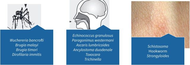
Route of transmission of common lung parasites.
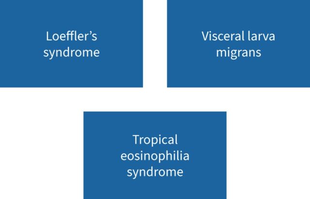
Common manifestations of parasitic eosinophilic lung disease.
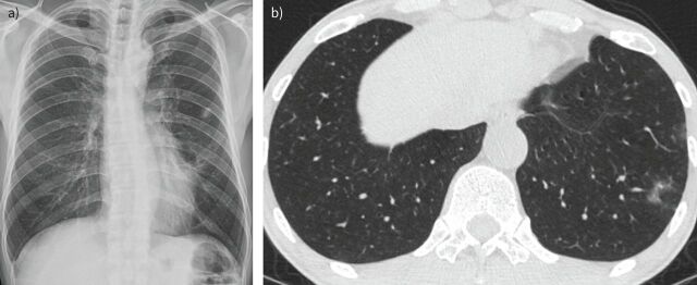
a) Nonspecific calcified nodules suggesting old Mycobacterium tuberculosis or other nontuberculous infection in the left upper lobe in an asymptomatic patient. b) Chest computed tomography (CT) scan of the same patient after 13 months, he was asymptomatic and the follow-up CT showed multifocal ill-defined ground-glass or solid nodular lesions in both lungs.
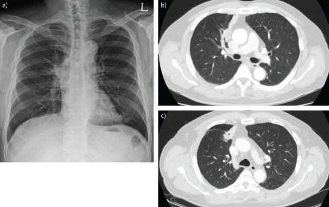
a) Posterior–anterior chest radiograph showing right hilar prominence in a patient with Paragonimus infection. b) Chest computed tomography (CT) scan showing a necrotic consolidation with bronchial cut-off at the right upper lobe anterior segment, suggesting infection/inflammation such as tuberculosis, actinomycosis, pneumonia and less likely a lung cancer in the same patient with Paragonimus infection. c) Follow-up chest CT scan after 8 months of therapy with antibiotics and antifungal therapy showing no improvements and cavitation.
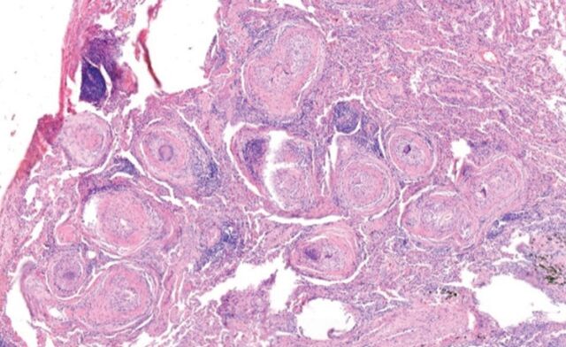
Histopathology of tissue showing chronic granulomatous inflammation with numerous parasitic eggs consistent with paragonimiasis.
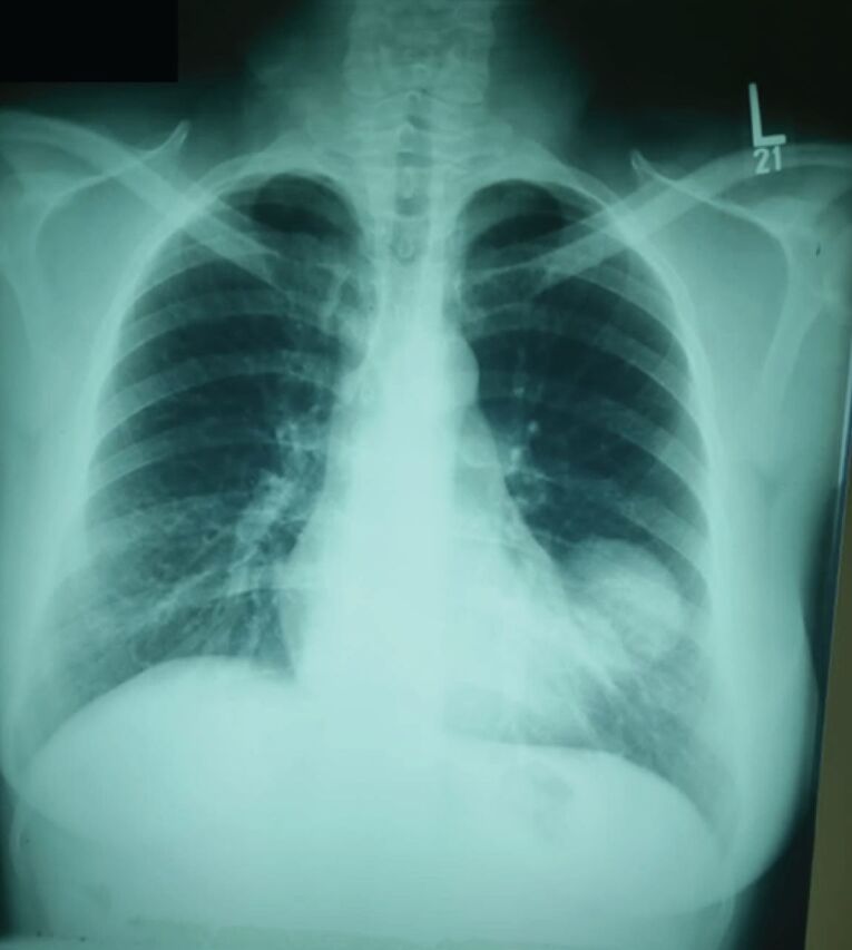
Plain chest radiograph showing a well-defined round radio-opacity in the left lower zone.
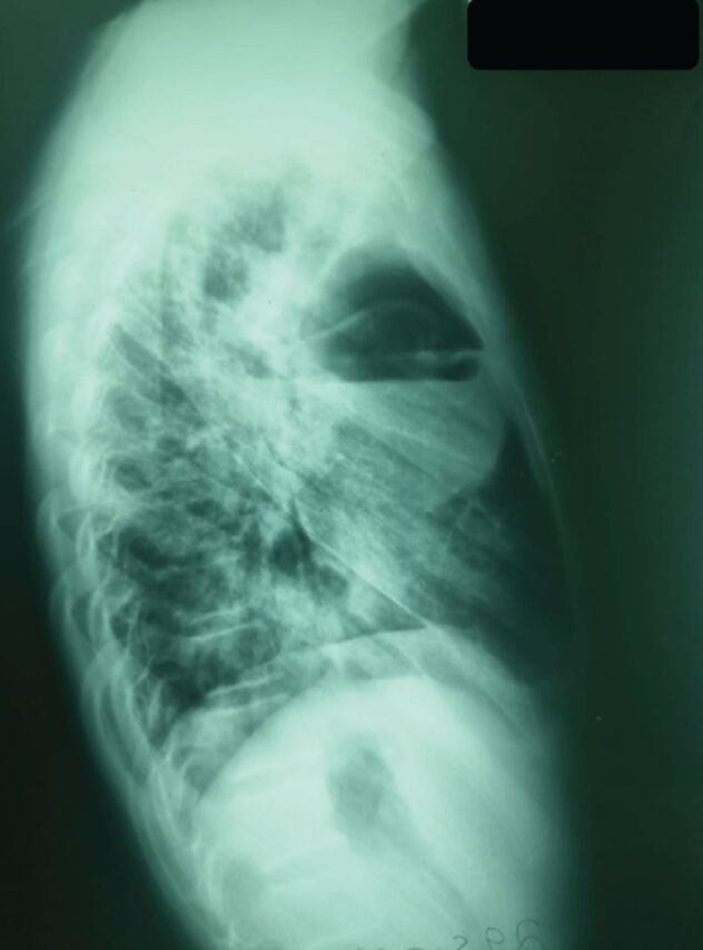
Lateral radiograph of the chest showing air fluid level within a pre-existing hydatid cyst suggesting superimposed infections.
Comment in
- doi: 10.1183/16000617.0150-2022
References
-
- Santeliz JV. Tropical pulmonary eosinophilia: an epidemiological and clinical review. Int J Respir Pulm Med 2019; 6: 102. doi:10.23937/2378-3516/1410102 - DOI
Publication types
MeSH terms
LinkOut - more resources
Full Text Sources